 |
| About Bioline | All Journals | Testimonials | Membership | News |
|
||||||
|
||||||
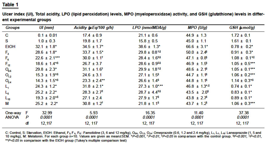
Indian Journal of Pharmacology, Vol. 36, No. 3, June, 2004, pp. 171-174 Research Paper Protective effect of increasing doses of famotidine, omeprazole, lansoprazole, and melatonin against ethanol-induced gastric damage in rats Sener Goksel , Paskaloglu K, Ayanoglu-dülger G Department of Pharmacology, School of Pharmacy, Marmara University, Istanbul, Code Number: ph04057 ABSTRACT OBJECTIVE: To study whether the increasing doses of omeprazole, lansoprazole and famotidine afford protection against ethanol-induced gastric damage and to compare their antioxidant effect with that of melatonin. MATERIAL AND METHODS: Mucosal damage was evaluated by macroscopic examination and by the measurement of lipid peroxidation (LPO), glutathione (GSH) levels and myeloperoxidase (MPO) activity. RESULTS: Ethanol administration-induced significant gastric damage, increased gastric acidity, and LPO and MPO activities, while tissue GSH levels decreased. The antiulcer drugs decreased the gastric acidity in a dose-dependent manner, whereas melatonin had no effect on this parameter. Biochemical parameters of oxidative damage, namely gastric LPO and GSH levels and MPO activities were reversed by both the antiulcer drugs and melatonin in a dose-dependent manner. CONCLUSION: These findings suggest that, parallel to increased acidity, reactive oxygen species have an important role in the pathogenesis of ethanol-induced gastric damage, and that melatonin, famotidine, lansoprazole and omeprazole are protective by their antioxidant property. However, according to our findings, inhibition of acid secretion is as important as the inhibition of oxidative damage in affording protection against ethanol-induced damage, and in this aspect melatonin seemed to be less efficient than the antiulcer drugs.INTRODUCTION Ethanol (EtOH) consumption is considered to be a risk factor in the development of gastroduodenal ulcers. Intragastrically administered EtOH rapidly penetrates the gastrointestinal mucosa, causing membrane damage, exfoliation of cells and erosion. The subsequent increase in mucosal permeability together with the release of vasoactive products from mast cells, macrophages and blood cells may lead to vascular injury, necrosis and ulcer formation.[1] Generation of free radicals has also been suggested as one of the mechanisms responsible for EtOH-induced gastroduodenal injury.[2] The stomach and upper gastrointestinal tract are the main sites of ethanol metabolism and the gastric mucosa is rich in xanthine oxidase that is capable of metabolizing acetaldehyde to acetate, accompanied by the generation of free radicals.[3] Lipid peroxidation mediated by oxygen free radicals is believed to be an important cause of the destruction of and damage to cell membranes, which has been demonstrated to play an important role in the pathogenesis of gastric mucosal injury induced by EtOH.[4] Omeprazole, lansoprazole, and famotidine are among the most widely used antiulcer drugs. Recently, these drugs have been demonstrated to possess antioxidant properties under various biochemical conditions.[5],[6] On the other hand, both in vivo and in vitro studies have demonstrated the antioxidant properties of the pineal hormone melatonin and it was shown to scavenge both hydroxyl and peroxyl radicals.[7] Several investigators have demonstrated that the damage-induced by EtOH may be prevented by treatment with melatonin.[8] The present study was undertaken to investigate the protective effect of increasing doses of the antiulcer drugs against EtOH-induced gastric damage in rats, to compare their antioxidant property with that of melatonin (a powerful antioxidant and free radical scavenger), and to study the contribution of their antisecretory effect in the protection against EtOH-induced damage. Material and Methods Animal treatment Studies were performed on seven groups of animals: 1-Control group (C), 2-Starvation group (S) which was deprived of food for 3 h and 48 h before sacrificing respectively, 3-Ethanol (EtOH) group (1 ml absolute ethanol, orally), 4-Famotidine (F) group (3, 6 and 12 mg/kg famotidine orally, 3 h before EtOH) 5-Omeprazole (O) group (0.6, 1.2 and 2.4 mg/kg omeprazole orally, 1 h before EtOH), 6-Lansoprazole (L) group (1, 5 and 10 mg/kg lansoprazole orally, 1 h before EtOH). 7-Melatonin (M) group (10 mg/kg melatonin dissolved in 0.2 ml of 0.25% ethanol:distilled water, i.p., 30 min before EtOH). All orally given drugs were administered by gavage through an intragastric tube and 10 animals were used for each group. Examination of gastric acidity Macroscopic analysis The gastric tissue samples were stored at -80°C until determination of gastric glutathione (GSH), and lipid peroxidation (LPO) levels, and tissue myeloperoxidase (MPO) activity. Determination of glutathione and lipid peroxide levels Tissue myeloperoxidase activity determination Statistical analysis Chemicals RESULTS Intragastric administration of 1ml EtOH consistently caused hemorrhagic lesions in the mucosa of the glandular stomach. Pretreatment of rats with higher doses of famotidine, lansoprazole or omeprazole prevented the gastric ulcerogenesis significantly and decreased the UI values. However, melatonin and low doses of the antiulcer drugs did not confer any significant protection against EtOH-induced gastric damage as reflected by the UI values [Table - 1]. Gastric acidity was found to be significantly higher in the EtOH group than in the control and the starvation groups. Only pretreatment with the highest dose of omeprazole caused a significant decrease in gastric acidity [Table - 1]. A significant correlation (r=0.94, P<0.0001) was observed between the acidity and the severity of the gastric damage (given as UI), demonstrating the importance of acidity in EtOH-induced ulcerogenesis. In the EtOH-treated group, gastric LPO was found to be significantly higher than in the control and the starvation groups. Famotidine, omeprazole, lansoprazole and melatonin pretreatment decreased it significantly. Lipid peroxidation was inhibited almost completely by melatonin and higher doses of lansoprazole and omeprazole [Table - 1]. There was a significant positive correlation (r=0.80, P<0.001) between the UI values and lipid peroxidation, indicating that lipid peroxidation may be one of the main causes of mucosal damage. The gastric MPO level of the EtOH group was significantly higher than that of the control and the starvation groups and all pretreatments decreased it significantly, indicating an inhibition of neutrophil infiltration [Table - 1]. In the EtOH treated group, gastric GSH was found to be significantly decreased. All drug-pretreatments prevented the EtOH-induced decrease in GSH levels except the lowest dose of famotidine. However, none of the drug-pretreatments was able to restore the GSH levels back to the control levels [Table - 1]. DISCUSSION Omeprazole, lansoprazole, and famotidine are widely used antiulcer drugs and have been demonstrated to bring about marked amelioration of gastric damage caused by EtOH in man and animals.[13],[14] However, the exact mechanism of this cytoprotection is speculative. We have recently demonstrated that antiulcer drugs are protective against acetylsalicylic acid-induced damage through their antioxidant property, as well as through their antisecretory effect.[15] The findings of the present study suggest that the same mechanisms of protection are also valid against EtOH-induced acute hemorrhagic gastric erosions. Studies suggest that the EtOH damage to the GI mucosa starts with microvascular injury, namely a disruption of the vascular endothelium resulting in increased vascular permeability, edema formation and epithelial lifting.[16] Findings of the present study are in agreement with these reports, since administration of 1 ml of absolute EtOH to pylorus-ligated rats caused severe gastric damage with hemorrhage. Parallel to ulcer formation, lipid peroxidation was observed to be significantly increased in this group of animals. The pineal hormone melatonin is known to possess powerful scavenging activity for both hydroxyl and peroxyl radicals, and its protective effect against the formation of acute gastric lesions induced by stress, ischemia-reperfusion or ethanol has been demonstrated previously.[7],[8] In parallel studies, the peroxidation of lipids and changes in the activities of glutathione peroxidase and myeloperoxidase were investigated as an index of gastric injury. Following melatonin pretreatment, these enzymes were observed to be decreased suggesting a marked protection against gastric injury, which could be due to its free radical scavenging activity and the ability to reduce neutrophil-induced toxicity.[17] Our present results are in accordance with the aforementioned studies. However, in the present study although melatonin caused 30% reduction in UI, it did not afford any morphological protection like the antiulcer drugs, and the UI values were not significantly different from that observed after EtOH alone. Though melatonin might have offered morphological protection at higher dose levels, in the present study only one dose (10 mg/kg) of melatonin was tried, and this dose is known to induce sufficient antioxidant effect. Pretreatment of the animals with the antiulcer drugs omeprazole, lansoprazole, and famotidine decreased the EtOH-induced gastric damage and at higher doses they were more efficient than melatonin in this respect. In several previous studies, these drugs have been demonstrated to have a powerful antioxidant effect under various conditions.[5],[6] Since one of the sources of oxygen radicals in gastric mucosal injury induced by EtOH in rats seems to be the neutrophils,[15] the role of the same was assessed by determining tissue-associated MPO activity, which was significantly high following EtOH. Melatonin and the antiulcer drugs inhibited the increase in MPO activity significantly, and restored it to control levels, suggesting that the neutrophil infiltration might have been suppressed by these drugs. The metabolism of EtOH generates superoxide radicals which may in turn promote lipid peroxidation.[1],[4] The stomach and the upper GI tract are the main sites of EtOH metabolism, and recent studies have implicated EtOH-generated free radicals, EtOH-induced lipid peroxidation and depression of glutathion as a mechanism of alcohol-induced gastric injury.[4],[16] Glutathione is an important constituent of the intracellular protective mechanism against various noxious stimuli including oxidative stress.[18] In humans a reduction in glutathione and cystein in the gastric body and antrum after ethanol consumption, was observed and glutathione pretreatment significantly decreased the EtOH-induced damage.[18] The decrease in glutathione following EtOH administration in the present study was accompanied by an increase in lipid peroxides. Omeprazole, lansoprazole, and famotidine inhibited glutathione depletion parallel to their efficacy as antioxidants, and omeprazole was observed to be as potent as melatonin in preserving glutathione levels. As can be seen from the results, EtOH increases gastric acidity significantly, even more than does acetylsalicylic acid.[15] With ASA, which is an acidic drug, this is not unexpected. On the other hand, according to our observation, EtOH-induced direct or indirect increase in acidity seems to be as important as the damage-induced by EtOH itself in promoting LPO and neutrophil infiltration, and the significant correlation between the suppression of acidity and the UI values demonstrate this quality. Thus, it can be suggested that treatment with agents that have both antioxidant and antisecretary properties (such as PPIs and H2 antagonists) affords the best protection against EtOH-induced gastric damage whereas melatonin, a strong antioxidant, was observed to be not as efficient. According to us, this is the most important observation of this study. In conclusion, melatonin and the antiulcer drugs omeprazole, lansoprazole and famotidine confer a dose-dependent protection against acute gastric mucosal injury-induced by EtOH, inhibit LPO and neutrophil infiltration significantly and prevent GSH depletion. At this dose level melatonin was as efficient an antioxidant as the higher doses of the antiulcer drugs, however, it was not as efficient in preventing the EtOH-induced gastric damage, apparently because it was not able to reduce the acidity. Thus, apart from their antioxidant property, inhibition of acid secretion by omeprazole, lansoprazole, and famotidine appears to contribute significantly to their protective effect against EtOH-induced oxidative gastric damage. REFERENCES
Copyright 2004 - Indian Journal of Pharmacology The following images related to this document are available:Photo images[ph04057t1.jpg] |
| |||||||||
