 |
| About Bioline | All Journals | Testimonials | Membership | News |
|
||||||
|
||||||
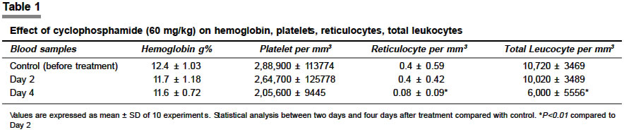
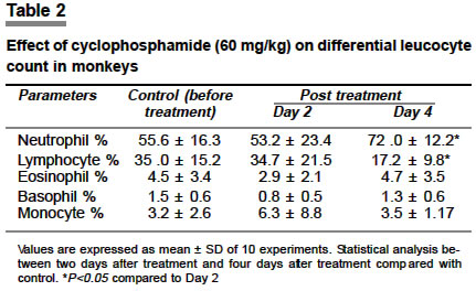
Indian Journal of Pharmacology, Vol. 36, No. 3, June, 2004, pp. 175-176 Research Letter Effect of cyclophosphamide pretreatment on hematological indices of Indian Bonnet monkeys Mythili MD, Nair SC, Gunasekaran S Departments of Physiology, Christian Medical College and Hospital, Vellore - 632 002 Code Number: ph04058 Sir, The leucocytes carried along with the transplanted tissue (passenger leucocytes) are important in graft rejection. It should be possible to greatly diminish the immunogenicity of a tissue graft if one can remove passenger leucocytes. Appropriate treatment of a tissue prior to transplantation can afford a marked reduction in its immunogenicity for the host. It is now possible to eliminate or at least reduce tissue immunogenicity by removal of leucocytes from the transplant prior to grafting. It therefore makes good theoretical and practical sense to attempt to alter tissue immunogenicity by treating the tissue to be grafted rather than the recipient.[1] Cyclophosphamide belongs to the nitrogen mustard subclass of alkylating agents. It is an immunosuppressant that alkylates DNA, thereby interfering with its synthesis and function, particularly in proliferating (also in non-proliferating) lymphocytes. Both B and T cells are affected by cyclophosphamide, although the toxicity produced is greater on B cells than T cells. Consequently, this drug exerts its greatest effect by suppressing humoral immunity. When cyclophosphamide is administered in very large doses, it can result in a specific tolerance to any new antigen to which it is simultaneously exposed.[2] Normal adult Indian Bonnet monkeys (Macaca radiata radiata) of both sexes, weighing between 2 and 6 kg were used in these experiments. After overnight fasting, about 1 ml of the blood was collected from saphenous vein and mixed thoroughly with the anticoagulant K2 EDTA. Cyclophosphamide (60 mg/kg) was administered intraperitoneally on day 0 and 2 after collecting a blood sample both the days. On day 4 the third blood sample was collected. The hematological indices such as hemoglobin, total leucocyte, differential leucocyte, platelet and reticulocyte were estimated. Hemoglobin was estimated by the Cyanmeth hemoglobin method with drabkins solution and a photoelectric calorimeter. Total leucocyte count and platelet counts were done manually by the bulk dilution method using Turk′s and platelet diluting fluid (formal citrate) respectively. Reticulocyte count was done manually by a supravital staining at 37°C for 20 min with Brilliant cresyl blue dye. Blood smears made were stained by the Leishman′s stain and differential leucocyte count done. All the blood samples were also duplicated on an automated cell counter with five-part differential capacity (coulter MAXM) and a good correlation was demonstrated. Totally, ten monkeys were used for this study. The statistical significance was determined using paired ′t′test and P<0.05 was considered significant. In [Table - 1], the change in the hemoglobin levels, platelet, reticulocyte and total leucocyte counts are shown. There was a slight but insignificant fall in the hemoglobin level and platelet count after two days and four days of treatment compared to pre-treatment values. There was no change in the reticulocyte count after two days of treatment but there was a significant fall four days after treatment. Cyclophosphamide is expected to have more effect on the lymphoid tissues. To find out this effect, total leucocyte and differential leucocyte count were done. There was an insignificant reduction in the total leucocyte count two days after treatment; on the other hand there was a significant marked fall four days after treatment when compared to pre-treatment and two days post-treatment values. There was a significant increase in the percentage of neutrophils four days after treatment. There was no change in the percentage of lymphocytes two days after treatment, whereas there was a significant fall four days after treatment. The percentage of and basophils had fallen two days after treatment, but came to the pretreated value four days after treatment. In the case of monocytes there was a slight increase after 2 days, but it came to the pretreated value four days after treatment [Table - 2]. In rats cyclophosphamide has an immunotoxic effect on lymphocytes in the spleen and blood. It has also been reported that cyclophosphamide pretreatment in rats sharply decreased the activity of all lymphoid cells, especially the CD4+ lymphocytes.[3] It was reported that both, destruction of donor antigen stimulated T cells in the periphery, and intrathymic clonal elimination of donor reactive T cells, were essential mechanisms of cyclophosphamide-induced tolerance.[4] Cyclophosphamide pretreatment reduces the lymphocytes in the spleen and in blood. The reduction in the activity of the lymphoid tissue by cyclophosphamide could be the reason for a decrease in the lymphocytes in the peripheral blood of the monkeys. ACKNOWLEDGEMENTS This work was supported by grants from Prof. M. Viswanathan Research Grant, Diabetes Research Center, Chennai, and FLUID Research Fund, CMCH. We gratefully acknowledge the help of Mr. Surander Singh and Miss. Rebecca for their help in counting and processing. We thank Mr. Y. Samuel for the maintenance of the monkeys. REFERENCES
Copyright 2004 - Indian Journal of Pharmacology The following images related to this document are available:Photo images[ph04058t2.jpg] [ph04058t1.jpg] |
| |||||||||
