 |
| About Bioline | All Journals | Testimonials | Membership | News |
|
||||||
|
||||||
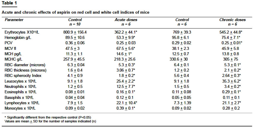
Indian Journal of Pharmacology, Vol. 36, No. 4, August, 2004, pp. 226-230 Research Paper Acute and chronic effects of aspirin on hematological parameters and hepatic ferritin expression in mice Merchant MA, Modi DeepakN Department of Zoology, Wilson College Chowpatty Mumbai - 400 007 Code Number: ph04075 ABSTRACT OBJECTIVE: To examine the acute and chronic effects of aspirin on peripheral blood and bone marrow counts and hepatic ferritin expression in mice. MATERIAL AND METHODS: Adult male albino mice were orally administered aspirin at a dose of 600 mg/kg thrice daily for 7 days or 150 mg/kg once daily for 6/7 days up to 25 weeks. At the end of the experiment the red and white blood cell counts, hemoglobin, and packed cell volume were estimated. Bone marrow films were studied to estimate the rate of erythropoiesis and leucopoiesis. Expression of liver ferritin was tested by immunohistochemistry. RESULTS: Acute or chronic doses of aspirin reduced the RBC count, hemoglobin and other red cell indices as compared to controls. The WBC counts were higher in the treated animals as compared to the untreated animals. Both the treatment regimens appeared to suppress the rate of erythropoiesis in the marrow, while the rate of leucopoiesis appeared to increase in the marrow of the treated animals. Aspirin treatment did not significantly affect the expression of ferritin in the liver. CONCLUSION: Aspirin in either acute or chronic doses induces anemia associated with leucocytosis in mice; the anemia does not seem to be induced due to alterations in iron metabolism. The drug appears to use multiple targets which affect red cell production and maturation processes. Keywords: Acetyl salicylic acid, hematological toxicity, red cell indices INTRODUCTION Aspirin, one of the widely used non steroidal antiinflammatory drugs, is probably the most highly consumed pharmaceutical product in the world. Recently, aspirin has gained greater importance not only as an analgesic but also as a cardio-protective drug. However, the use of aspirin is also associated with significant morbidity and mortality due to its adverse effects on multiple organ systems.[1] Long-term therapeutic consumption of aspirin is reportedly associated with the occurrence of gastrointestinal (GI) ulcerations, nephrotoxicity, hepatotoxicity and even renal cell cancer.[1],[2] Numerous clinical observations have associated the use of aspirin with blood disorders like anemia and cytopenias.[3] While the relative risk of occurrence of blood disorders with the use of aspirin is considered to be low, significant mortality rates have been reported due to blood disorders caused by the use of aspirin.[3] Although blood disorders with the use of aspirin have been well documented clinically, relatively few experimental studies have been conducted to clarify and confirm the association. It has been shown that oral administration of low doses of aspirin significantly reduces circulatory erythrocyte and leukocyte counts suggesting the inhibitory action of this drug on bone marrow hemopoiesis.[4] However, in that study only peripheral blood was investigated and no attempt was made to study the bone marrow, a speculated target site of aspirin action causing blood toxicity.[5],[6] Furthermore, the animals in the study[4] were dosed continuously in drinking water, a pattern of drug intake not encountered clinically. Aspirin is known to cause GI tract erosion resulting in occult bleeding; it is also reported to reduce iron uptake from it resulting in iron deficiency.[7],[8] To confirm or rule out this possibility we investigated the expression of hepatic ferritin, a major form of iron stores in the body as reduction in ferritin from liver and bone marrow is a characteristic feature of iron deficiency anemia.[9] In the present communication we report the effects of acute and chronic doses of aspirin administration on blood and bone marrow indices, and hepatic ferritin of mice. MATERIAL AND METHODS The present study was approved by the local ethics committee. Reagents: All the chemicals used in this study were purchased from Sigma (USA) unless otherwise stated. NaCl, NaH2PO4, Na2HPO4, KCl and MgCl2 were purchased from Qualigens (India). Adult Swiss albino mice were purchased from Haffkins Biopharmaceutical Corporation Limited (Parel Mumbai) and were allowed to breed in the animal house of the department. 12-week-old albino male mice were used in the present study. Mice were housed 2-3 per cage and were allowed free access to food and water throughout the experiment. Aspirin was suspended in normal saline and administered orally by gastric intubation in the dose regimes as described below.
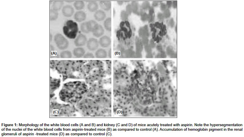
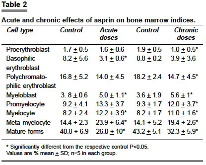
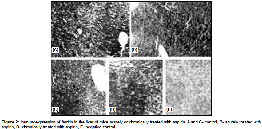
The control animals in both the groups received equivalent amounts of saline only. The doses used in the present study may be extremely high for consumption in humans if calculated on weight-by-weight basis, however preliminary experiments had shown that this high dose is necessary to maintain the plasma salicylate levels similar to those found in humans during various regimens with aspirin (data not shown). This is because the clearance rate of aspirin in mice is several folds higher as compared to humans.[12] At the end of each trial, the animals were immobilized by ether anesthesia and the blood was drawn from the tail vein using EDTA as an anticoagulant. Total erythrocyte count, leucocyte count and hemoglobin were estimated using standard methods; packed cell volume was measured using a microhematocrit method. All absolute values were calculated according to the instructions of Dacie and Lewis.[9] Marrow smears were made from the tibia or femoral bones and air-dried. Blood and marrow films were fixed in methanol, stained with Leishman or Giemsa stain and 200 WBCs were counted to obtain the differential counts. Absolute WBC counts were estimated by extrapolating the percent differential counts with that of total leukocyte counts.[9] For measurement of erythrocyte diameter a drop of fresh blood was mounted under a cover slip and a diameter of 200 erythrocytes was measured using a precaliberated occulometer. RBC thickness was estimated as described previously[9] and the sphericity index was obtained as a ratio of RBC thickness to the diameter. Liver sections (n=5 in each group) were used for localization of ferritin using conventional immunohistochemistry.[13] Briefly, the sections were deparaffinized and incubated in 1% H2O2 for 30 min at room temperature and then blocked in blocking solution of 1% sheep serum containing 0.1% TritonX-100 in 0.01M phosphate buffer saline (PBS) for 3 h. The sections were then incubated overnight at 4°C with a polyclonal ferritin antibody (1:100 dilution, Sigma, USA) prepared in the blocking solution. The sections were then washed with 0.05% Triton X-100 in 0.01M PBS thrice to remove excess antibody and subsequently incubated with peroxidase conjugated secondary antibody for 5 h at room temperature. The color was then developed using diaminobenzedene (DAB, Sigma USA) as substrate for 5 min. The sections were washed, dehydrated and mounted in DPX [p-xylene-bis-(N-pyridinium bromide)]. 5-micron thick paraffin sections of kidney from control and treated animals were stained with hematoxylin, counterstained in eosin and observed under a microscope. The data are presented as mean ± SD and the statistical significance was analysed using Student′s ′t′test, P<0.05 was considered significant. RESULTS Animals treated with aspirin in acute or chronic doses had significantly lower erythrocyte (RBC) count, hemoglobin (Hb) and packed cell volume (PCV) as compared to the controls [Table - 1]. The mean cell hemoglobin (MCH) and the mean cell volume (MCV) were significantly greater than controls; while the mean cell hemoglobin concentration (MCHC) was similar in both the groups [Table - 1]. The erythrocyte diameter was significantly lower whereas the thickness was higher in the treated animals as compared to controls [Table - 1]. The total leucocyte (WBC) count, absolute neutrophil count, eosinophil count and lymphocyte count were higher in mice treated with acute or chronic doses of aspirin as compared to controls. The monocyte count was higher than the controls in the acutely treated animals while no significant difference was noted in the monocyte counts of chronically treated animals and controls [Table - 1]. Morphologically, the neutrophils of the acutely treated animals had hypersegmentation of the nuclei with coarse or clumped chromatin. Occasionally, the nuclei appeared fragmented in some of the acutely treated animals [Figure - 1]A and B. No morphological alterations were detected in the chronically treated animals. Irrespective of the treatment regimen, the erythroid count in the bone marrow was significantly lower in the treated groups than controls; the myeloid counts were higher in the treated animals as compared to controls [Table - 2]. There was a reduction in the RBC precursors in the marrow; the proerythroblasts and polychromatophilic erythroblasts were reduced in the chronically treated animals, whereas only the percentage of basophilic erythroblasts was significantly reduced in the acutely treated animals as compared to controls [Table - 2]. All the myeloid precursors were significantly increased in the treated animals as compared to controls; the matured forms were however reduced in the treated animals as compared to untreated animals. This was anticipated, as the matured forms in circulation were higher in treated animals as compared to controls [Table - 1] and [Table - 2]. Generally, the marrow of the chronically treated animals was hypocellular and no morphological alterations in this group. However, the myeloid elements in the marrow of the acutely treated animals showed marked morphologic alterations such as nuclear fragmentation and occasional necrosis. Ferritin was immunolocalized in the cytoplasm of the hepatocytes throughout the hepatic parenchyma of the control animals; the expression was panlobular in most cases [Figure - 2]. As is evident from [Figure - 2] the pattern of expression was not altered due to treatment with either low or high doses of aspirin. No immunoreactivity was evident when the primary antibody was excluded from the reaction [Figure - 2]E, indicative of the specificity of the reaction. Histologically, the kidney sections of animals treated with acute doses of aspirin had hemoglobin pigment deposited in the glomeruli and some of the tubules; no such pigmentation was seen in the control animals [Figure - 1]B and C. The kidneys of the chronically treated animals were similar to controls (data not shown). DISCUSSION Hematological disorders as a result of aspirin ingestion are well documented clinically, and the results of the present study further confirm the association, as anemia accompanied by marked leucocytosis was observed in the mice treated with aspirin in acute and chronic doses. Prolongation of bleeding time was one of the first clinically recognized hematological side effects of aspirin administration[14]; anemia, thrombocytopenia, agranulocytosis and leucopoenia are some of the most frequently reported adverse effects of aspirin.[3] In the present study, aspirin treatment significantly reduced the circulating red cell counts, hemoglobin and packed cell volume, and the MCV and MCH were significantly higher than controls. These results are in agreement with the published reports in rats.[4] Aspirin in vivo is reported to alter the iron uptake from the GI tract.[8] This effect coupled with acute or chronic blood loss due to GI tract erosion induced by aspirin is believed to cause iron deficiency anemia in humans.[15],[7] To rule out the possibility of iron deficiency in mice treated with aspirin, we investigated the expression of ferritin in the liver of treated and control animals. Since ferritin is the first form of stored iron to be utilized in the event of iron deficiency, its levels in blood, bone marrow and the liver are indicative of the iron status of the individual.[9] However, the expression of ferritin in the liver of treated animals was similar to that of controls. Therefore iron deficiency does not appear to be a possible cause of the anemia observed in the treated animals. Acetylation of bone marrow macromolecules by aspirin has been suggested as a possible mechanism causing blood disorders.[5],[8] Based on reduced circulating erythrocyte and leucocyte counts in rats treated with aspirin, an inhibitory effect of aspirin on bone marrow hemopoiesis has been proposed.[4] Although in the present study too we observed low erythroid count in the circulation of the treated animals, there was a marked increase in the circulating leucocyte count in the drugged animals as compared to controls. At present we do not have any obvious explanation for the contradiction observed in the WBC count in the present study as compared to the previous study;[4] our observations on the circulating levels of the two cell types were confirmed by the studies on the marrow which showed enhanced rates of leucopoiesis and depressed erythropoiesis. High doses of aspirin may be toxic as morphologic alterations were noted in the myeloid elements of the bone marrow and peripheral blood of acutely treated mice. However, the erythroid elements in the marrow of both acute and chronic groups were morphologically normal. Apart from reduced counts in the circulation and the bone marrow, the erythrocytes of the treated animals had altered size and volume. The erythrocytes of both acutely and chronically treated animals had lower diameter than the controls; the thickness and the volume were significantly higher in the treated animals as compared to controls, which is indicative of spherocytosis. At the biochemical level, aspirin alters red cell membrane functions by inhibiting cholinesterase and ion-dependent ATPase activity and by altering the ion permeability across the cell wall.[11], [16], [17] With these defects along with altered cell shape, it is reasonable to assume that the cells may be more susceptible to intravascular hemolysis. Although, in the present study, no specific test was performed to confirm the occurrence of hemolysis in vivo, hemoglobin pigment was detected in some of the renal elements and free hemoglobin was also detected in the plasma of some of the acutely treated mice (data not shown). Both these observations are reported as indicators of hemolysis in vivo.[9] Thus, it appears that intravascular hemolysis may be a major contributing factor for the reduced red cell counts observed in the present study. Hemolytic anemia has been reported with the use of aspirin in humans, especially in cases with certain hemoglobinopathies.[3], [6] At the cellular level, aspirin uncouples mitochondrial oxidative phosphorylation [8], [11] and alters the activity of several respiratory enzymes in different tissues (Modi and Merchant unpublished data). These metabolic defects in the aspirin-treated animals coupled with anemia would render the tissues hypoxic and could be expected to stimulate erythropoiesis by increasing the erythropoietin (Ep) production in vivo.[18] Contrary to this expectation, the rate of erythropoiesis in the treated animals appeared depressed, probably indicating Ep deficiency. This speculation is supported by the fact that since there is a direct relationship between the renal prostaglandin levels and Ep production,[8] inhibition of prostaglandin production by aspirin (through inhibition of cyclooxygenase-2) [19] may reduce renal erythropoietin production. It is possible that the reduction in Ep production may further aggravate the anemia already induced by aspirin in the treated animals. In conclusion, aspirin in either acute or chronic doses induces significant blood dyscrasias in mice; the drug appears to use multiple targets which affect red cell production and maturation processes. ACKNOWLEDGEMENTS We express our gratitude to Mrs Anuradha for her help in differential WBC counts. The kind guidance provided by Dr GS Gazdar is gratefully acknowledged. REFERENCES
Copyright 2004 - Indian Journal of Pharmacology The following images related to this document are available:Photo images[ph04075t2.jpg] [ph04075f1.jpg] [ph04075t1.jpg] [ph04075f2.jpg] |
| |||||||||
