 |
| About Bioline | All Journals | Testimonials | Membership | News |
|
||||||
|
||||||
Indian Journal of Pharmacology, Vol. 36, No. 6, November-December, 2004, pp. 355-359 Research Paper Pyrogallol: A novel tool for screening immunomodulators Joharapurkar AA, Wanjari MM, Dixit PV, Zambad SP, Umathe SN Department of Pharmaceutical Sciences, University Campus, Nagpur University, Nagpur - 440 033 Code Number: ph04127 ABSTRACT OBJECTIVE:To induce immunosuppression in rats by pyrogallol and to develop a novel model to screen the immunomodulatory activity of a known agent.
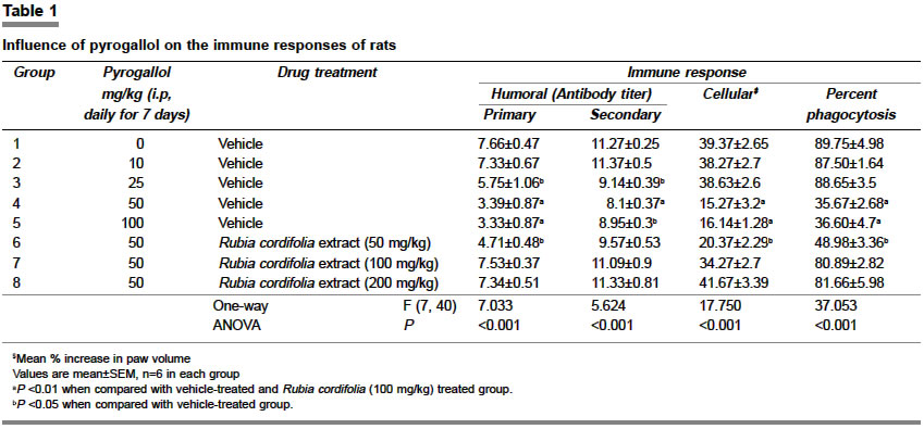
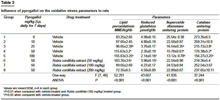
KEY WORDS: Immunosuppression, oxidative stress, Rubia cordifolia INTRODUCTION While screening the immunomodulatory activity, most of the studies employ agents like cisplatin, cyclophosphamide, or corticosteroids in order to induce the immunosuppression in the experimental animals.[1] These agents are also known to generate free radicals in the biological system and thereby cause oxidative stress.[1],[2],[3] Whether the impairment in immune responses is subsequent to their ability to generate oxidative stress is not clear. However, several workers have demonstrated that prooxidants suppress the immune responses in experimental animals.[3] In addition, it has been documented that the immunomodulators isolated from plant sources possess antioxidant activity.[4] It is known that pyrogallol is a strong generator of free radicals[5] and it is evidenced that it can suppress the proliferation of mouse lymphocytes in vitro.[6] In view of these evidences, it was proposed to investigate whether a strong prooxidant like pyrogallol can induce immunosuppression in rats and to test the utility of such a method to screen the immunomodulatory activity of a known agent like the alcoholic extract of Rubia cordifolia (RC).[7] In order to investigate the involvement of free radicals in the influence of pyrogallol on the immune system, the markers of oxidative stress such as the lipid peroxidation (LPO) levels, reduced glutathione (GSH) content, superoxide dismutase (SOD) and catalase (CAT) activities were assessed in blood. MATERIAL AND METHODS Chemicals Alcoholic extract of RC Animals Treatments Immunological responses 1. Humoral immune response[7] On Day 13 and 20, blood was withdrawn from the retro-orbital plexus of all antigenically challenged rats. Twenty-five µl of serum was serially diluted with 25 µl of phosphate-buffered saline. SRBC (0.025 x 10⁹ cells) were added to each of these dilutions and incubated at 37°C for one hour. The rank of minimum dilution that exhibited hemagglutination was considered as an antibody titer. The level of antibody titer on Day 13 of the experiment was considered as the primary humoral immune response and the one on Day 20 of the experiment was considered as the secondary humoral immune response. 2. Cellular immune response[7] This was assayed by the footpad reaction method. The edema was induced in the right paw of rats by injecting SRBC (0.025 x 10⁹ cells) in the subplantar region on Day 20. The increase in the paw volume in 48 h, i.e. on Day 22 was assessed on digital plethysmometer (UGO Basile-7150). The mean percentage increase in paw volume was considered as delayed type of hypersensitivity and as an index of cell-mediated immunity. The volume of the left hind paw, injected similarly with phosphate-buffered saline, served as a control. 3. Phagocytosis[8] On Day 22, 3.0 ml of Hank′s balanced salt solution (HBSS), containing 10% bovine serum albumin (BSA), was put into the peritoneal cavity of the animal and the same was recovered by gentle aspiration. The macrophages (3 x 10⁵/600µl), present in the aliquots, were incubated on glass plates at 37°C for 30 min in a humidified chamber. The cells, adhered to the glass, were incubated with live cells of Candida albicans, previously opsonized in autologous plasma (2 x 10⁶/250µl), at 37°C for 30 min, then washed with HBSS, again incubated for 30 min with autologous plasma and finally stained with Wright′s dye. A total of 300 cells were counted under the microscope and the results are reported as phagocytosis percentage. Oxidative stress parameters They were assessed in the blood of the rats, which was withdrawn from the retro-orbital plexus, on Day 22 of the experiment. Lipid peroxidation (LPO)[9] To 2.0 ml of the 5% suspension of RBC in 0.1 M phosphate-buffered saline, 2.0 ml of 28% trichloroacetic acid was added and centrifuged. 1.0 ml of 1% thiobarbituric acid was added to the supernatant, heated in boiling water for 60 min and then cooled. The absorbance was measured at 532 nm (UV-1601, Schimadzu). Lipid peroxidation was calculated based on the molar extinction coefficient of malondialdehyde (MDA) (1.56 x 10⁵), and expressed in terms of nanomoles of MDA/g Hb. Superoxide dismutase (SOD)[10] It was estimated in the erythrocyte lysate prepared from the 5% RBC suspension. To 50 µl of the lysate, 75 mM of Tris-HCl buffer (pH 8.2), 30 mM EDTA and 2mM of pyrogallol were added. An increase in absorbance was recorded at 420 nm for 3 min in spectrophotometer (Schimadzu 1601). One unit of enzyme activity is 50% inhibition of the rate of autooxidation of pyrogallol as determined by change in absorbance/min at 420 nm. The protein content of lysate was estimated by Lowry′s method[11] and the activity of SOD is expressed as units/mg protein. Catalase (CAT)[12] Catalase activity was determined in erythrocyte lysate using Aebi′s method with some modifications. The erythrocyte lysate (50 µl) was added to a cuvette containing 2.0 ml of phosphate buffer (pH 7.0) and 1.0 ml of 30 mM H2O2. Catalase activity was measured at 240 nm for 1 min using spectrophotometer. The molar extinction coefficient of H2O2, 43.6 M cm-1 was used to determine the catalase activity. One unit of activity is equal to one millimole of H2O2 degraded per minute and is expressed as units per milligram of protein. Reduced glutathione (GSH)[13] Blood glutathione was measured by addition of 0.2 ml of whole blood to 1.8 ml distilled water followed by 3.0 ml of precipitating mixture (1.67 g metaphosphoric acid, 0.2 g EDTA and 30 g NaCl to make 100 ml of solution). It was centrifuged at 5000 x g for 5 min and 1.0 ml of the filtrate was added to 1.5 ml of the phosphate solution, followed by the addition of 0.5 ml of DTNB reagent. The optical density was measured at 412 nm using a spectrophotometer. Statistical analysis All data were analyzed with one-way ANOVA followed by Dunnett′s multiple comparisons. The intergroup difference was considered significant at P<0.05. Using Pearson correlation analysis, the changes in the LPO levels were first correlated with the changes in GSH, SOD and CAT. Then, the changes in the LPO levels were correlated with the changes in the immunological parameters. RESULTS [Table - 1] indicates the effect of pyrogallol treatment on the immunological responses. The daily treatment with pyrogallol for seven days significantly (P<0.01) impaired the primary (F=7.033) and secondary (F=5.624) humoral immune responses, cell-mediated immune response (F=17.750) and phagocytosis (F=37.053), at 50 and 100 mg/kg dose whereas at 25 mg/kg dose it was only effective to suppress the humoral immune response (P<0.05) in rats. The immunosuppressant effects of the 50 and 100 mg/kg doses of pyrogallol were found to be quantitatively similar. [Table - 2] indicates the influence of pyrogallol on the marker parameters of oxidative stress in rats. Pyrogallol dose-dependently elevated the LPO levels (F=52.291), which were maximum at 100 mg/kg dose (P<0.01). Pyrogallol treatment decreased GSH content (F=43.657) and reduced SOD (F=41.035) and CAT (F=37.244) activities. [Table - 1] and [Table - 2] further indicate that a 100 mg/kg dose of RC significantly prevented the pyrogallol (50 mg/kg)-induced changes in the oxidative stress parameters and the immunosuppression (P<0.01). The effect of a higher dose of RC (200 mg/kg) was quantitatively similar with that of 100 mg/kg dose whereas the lower dose (50 mg/kg) was effective in preventing the influence of pyrogallol only on secondary immune response, SOD and CAT activities (P<0.05). DISCUSSION The present investigation has revealed that pyrogallol at 50 and 100 mg/kg dose, produced significant impairment of humoral as well as cell-mediated immune responses and inhibited phagocytosis. At a 25 mg/kg dose, pyrogallol could only suppress the humoral immune response with no effect at 10 mg/kg dose. In addition, it was observed that the influences of higher doses of pyrogallol (50 and 100 mg/kg) were quantitatively similar. Therefore, the minimum required dose of pyrogallol to impair the immune responses appears to be 50 mg/kg daily for 7 days. In the later part of the study, it was observed that daily oral administration of alcoholic extract of RC (100 mg/kg) significantly prevented the influence of pyrogallol on immune responses. This suggests that the immunomodulatory effect of RC can be screened by the method in which the immunosuppression was induced by pyrogallol. The observed immunomodulatory activity of RC is well in accordance with earlier reports[7] in which the immunosuppression was induced by a method other than pyrogallol. This is the first attempt at using pyrogallol for inducing experimental immunosuppression and literature has not adequately recorded the immunotoxicity of pyrogallol. There is in vitro evidence to show that pyrogallol can suppress the mouse lymphocyte proliferation.[6] In fact, pyrogallol is toxic to the biological system and its toxicity is attributed to its ability to generate free radicals.[5] The preliminary toxicity studies (data not shown) revealed that pyrogallol did not produce any mortality up to 100 mg/kg dose, and there was no apparent symptom of any sickness. However, in a dose range of 100 to 200 mg/kg, pyrogallol produced marked hematuria, and 25% mortality at 200 mg/kg dose. Thus, in view of the proximity of the 100 mg/kg dose to the toxic dose range and the fact that 50 mg/kg could induce significant immunosuppression, it appears that 50 mg/kg of pyrogallol dose (i.p., daily for 7 days) can be recommended as a safe and dependable tool to induce immunosuppression. The literature has documented several evidences of the vulnerability of the immune system to the free radical-induced oxidative stress, which indicate that the cellular and humoral components of the immune system are particularly sensitive to increased levels of reactive oxygen species, which may cause premature immunosenescence.[1],[2],[3],[4] The endogenous antioxidant system prevents the deleterious influence of the free radicals on the immune cells and preserves their normal function.[3],[4] Circumstances such as chronic inflammatory diseases, exposure to toxic chemicals, environmental pollutants, radiation, alcohol, and high fat diet, which are known to impair the immune system, are also known to generate free radicals.[2],[4] Impairment in these conditions may thus be subsequent to over-utilization of endogenous antioxidants.[2],[3],[4] In view of this, it appears that pyrogallol, which is a strong generator of superoxide radicals,[10] might impair the immune response through oxidative stress. Such a possibility is substantiated by an increase in lipid peroxidation and decrease in antioxidant defense, after pyrogallol treatment. This suggests a strong causal relationship between pyrogallol-induced oxidative stress and immunosuppression. This has been further substantiated by the fact that the Pearson correlation analysis revealed a significant correlation between the changes in LPO levels, a representative marker of oxidative stress, and the changes in the immunological parameters. This analysis has shown that the level of LPO is inversely correlated with the changes in endogenous antioxidant defense [for GSH (r=-0.886), for SOD (r=-0.820), and catalase activity (r=-0.874), all at P<0.01]. Hence, the level of LPO was considered as a representative of oxidative stress and then correlated with the individual immunological parameters. This has revealed that the pyrogallol-induced immunosuppression is also inversely correlated to the LPO level (r=-0.861, for primary humoral immune response; r=-0.733, for secondary humoral immune response; r=-0.832, for cellular immune response; and r=-0.809, for phagocytosis, all at P<0.05). It is known that the peroxidative toxicity of pyrogallol in vivo may also be through its effect on iron release from ferritin,[14] which can induce lipid peroxidation via Fenton or Haber-Weiss reaction.[15] In addition, the alcoholic extract of RC, which is known for its antioxidant activity, not only attenuated the influence of pyrogallol on the immune system but was also found to prevent the changes in the oxidative stress parameters, which were induced by pyragallol. It is known that RC contains a significant concentration of anthraquinone, which can chelate iron and thereby exhibit antioxidant activity.[16] As the present method of inducing reproducible immunosuppression by 50 mg/kg of pyrogallol was without any mortality and the same could screen the well-proven immunomodulatory activity of Rubia cordifolia, it can be proposed as a novel method for screening an immunomodulatory agent. REFERENCES
Copyright 2004 - Indian Journal of Pharmacology The following images related to this document are available:Photo images[ph04127t2.jpg] [ph04127t1.jpg] |
| |||||||||
