 |
| About Bioline | All Journals | Testimonials | Membership | News |
|
||||||
|
||||||
Indian Journal of Pharmacology, Vol. 37, No. 1, January-February, 2005, pp. 38-40 Research Letter Effect of Vitex negundo on oxidative stress Tandon Vishal, Gupta RK Postgraduate Department of Pharmacology and Therapeutics, GMC, Jammu - 180 001
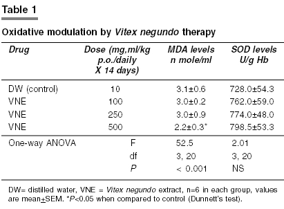
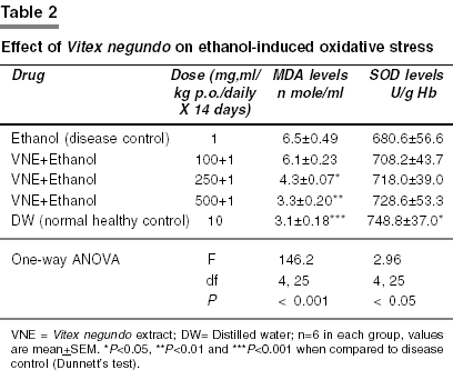
Correspondence Address:Postgraduate Department of Pharmacology and Therapeutics, GMC, Jammu - 180 001 Code Number: ph05010 Sir, In vitro, the antiradical potential of the freeze-dried root extract of Vitex negundo (VN) was investigated by Munasinghe et al (2001)[1] by determining their abilities to scavenge DPPH(1,1-diphenyl-2-picrylhrazyl) free radical and to inhibit hydroxyl radical-mediated damage to deoxyribose. They however did not study the in vivo effects of VN on lipid peroxidation or endogenous antioxidant enzymes like superoxide dismutase (SOD). TThe leaves of VN are known to possess various antioxidant chemical constituents like flavonoids,[2] vitamin C and carotene[3] which may have a modulatory effect on oxidative stress or endogenous antioxidants. Moreover, no one has as yet investigated the in vivo effects of VN leaf extract on oxidative stress. Therefore, the present study was undertaken to investigate the effect of VN leaf extract on oxidative stress in albino rats. The plant was collected from the local area of Sevagram (MS), and was authenticated by an expert. The fresh leaves of VN were shade-dried and powdered. The powder was macerated for 24 h in 70% v/v ethanol. Then, it was subjected to percolation using 70% v/v ethanol as solvent. The menstrum collected was again shade-dried. The final viscous yield (9.5%), was then suspended in 1% gum acacia and dissolved in distilled water to prepare suspension in desired concentrations just before use. Albino rats (125-180 g) of either sex (Wistar strain) were used. Animals were housed and fed as per standard guidelines of CPCSEA. The animals were given a weeks time to get acclimatized to laboratory conditions and were fasted overnight before the experiment. The project was approved by the institutional ethics committee. The animals were divided into four groups, each consisting of six rats. Group I served as control and received distilled water. The VN leaf extract was fed orally for 14 days in a volume of 10 ml/kg body weight in doses of 100 mg/kg/day (Group II), 250 mg/kg/day (Group III) and 500 mg/kg/day (Group IV) for assessment of the oxidative modulatory effect of VN. Dose selection was based on preliminary trials in our laboratory and estimated oral LD50 dose of VN leaf extract (7.58 g/kg, b.w.) in one of our previous reported study.[4] For studying the effect of VN on ethanol-induced oxidative stress,[5],[6] another set of animals were divided into five groups consisting of six rats each. Group I served as a disease control in which 1 ml/kg/day of 20%v/v ethanol was instilled intra-gastrically for 14 days. Groups II, III and IV were given the doses of VN extract orally for 14 days as mentioned earlier. After 30 minutes of this pre-treatment, each group received 1 ml/kg/day of 20%v/v ethanol orally for 14 days. Group V served as healthy control. After completion of 14-day treatment, 3-5 ml of blood was collected from the inner canthus of the eye from each animal under light ether anesthesia using capillary tube, in a vial containing EDTA as anti-coagulant. Plasma was separated by centrifugation at 3000 rpm for 10 minutes. It was stored at -20oC and used to estimate malondialdehyde levels. Buffy coat was removed; remaining erythrocytes were washed three times in cold 0.9% saline and hemolysed by adding the same volume of ice-cold water to yield a 50% hemolysate. The hemolysate was stored in 500 µl aliquots at -20oC. It was used to estimate the activity of the enzyme SOD. The estimation of malondialdehyde (MDA)[7] was carried out by the TBARS method. Thiobarbituric acid (TBA) reacts with MDA, one of the aldehyde products of lipid peroxidation, to give a colored product which was extracted in butanol and absorbance measured spectrophotometrically at 530 nm. MDA (Sigma chemicals) was dissolved in 0.05 M sulphuric acid to prepare 10 µM stock solution. By diluting the stock solution, different concentrations (1 to 5 nm/ml) of MDA were obtained for preparing a standard curve. To 0.5 ml each of plasma and MDA standards, 2.5 ml of 20% TCA was added and centrifuged at 3000 rpm for 10 minutes. The supernatant was decanted and the precipitate was washed once with 0.05M sulphuric acid and 3 ml of 0.2 g/dl TBA reagent (Loba chemie, India) was added to the precipitate. The mixture was heated in a boiling water bath for 30 minutes. After cooling, the resultant chromogen was extracted with 4 ml of n-butyl alcohol and the separation of the organic phase was done by centrifugation at 3000 rpm for 10 min. Absorbance of the butyl alcohol extract of samples was measured at 530 nm against distilled water as blank. The standard curve was plotted and the concentration of the sample was calculated and expressed as nmol/ml malondialdehyde. An elevated level of MDA was taken as the index of oxidative stress. The estimation of superoxide dismutase (SOD) activity in erythrocytes was carried out by the method of Marklund and Marklund (1974)[8] utilizing the inhibition of auto-oxidation of pyrogallol by SOD enzyme. The final assay mixture contained 3 ml of triscacodylate buffer (50 mM, pH 8.2) containing air equilibrated 0.2 mM pyrogallol [E merck, India], 1 mM EDTA, 1 mM DTPA(diethylene triamine penta acetic acid) and 100 ml of enzyme source. The amount of hemoglobin in the hemolysate was estimated by Drabkin′s method.[9] Hemoglobin was then precipitated and SOD was extracted in Tsuchihasi extract containing chloroform and ethanol (1.5:1). This was used to estimate the enzyme activity. Three sets of reaction mixture were prepared. Standard: Known amounts of SOD (Sigma St. Louis, USA) from pure bovine source, in different concentrations were added to the assay mixture to achieve 30% to 50% inhibition of pyrogallol auto-oxidation. Test: The clear supernatant from Tsuchihasi extract was added instead of standard SOD. Control: Neither test sample, nor standard was added to assay mixture. The crude extract was replaced by 50 mM of phosphate buffer (0.1 Mm, pH 5.6) to obtain uninhibited auto-oxidation of pyrogallol. In all sets, the reaction was started by the addition of pyrogallol (0.2 mM) and the change in optical density was recorded for 4 min at 420 nm. The initial 10 sec were considered as the induction period of the enzyme. Change of absorbance/minute ("A) was calculated from the reading. The %inhibition of the test system was calculated from the standard graph. The SOD activity was expressed in units/g hemoglobin. One unit of SOD activity being defined as the amount of enzyme required to cause 50% inhibition of pyrogallol auto-oxidation. The raised levels of SOD, an endogenous antioxidant was taken as an indicator of the antioxidant property of the test drug. The data were analyzed using one-way (ANOVA) followed by Dunnett′s test. P <0.05 was considered significant. The data are represented as mean ± SEM. The results are shown in [Table - 1] and [Table - 2]. Vitex negundo in the present study produced significant (P<0.05) reduction in MDA levels after 14-day treatment in only the higher dose (500 mg/kg/po). Although a marginal rise of SOD in this dose was observed it was not significant. Usually, induction or activation of enzymes leads to a several-fold rise in enzymatic activity. From this marginal rise, it is difficult to infer whether the enzyme activity is really altered. Moreover, serum MDA levels in control rats seem slightly towards the higher side, which may probably be due to the fact that blood was collected under light ether anesthesia. Therefore, it is difficult to comment that continuous VN therapy can reduce oxidative stress. Whereas, in the ethanol-induced oxidative stress model, it significantly (P<0.05 and P<0.01) reduced only MDA levels in both moderate and higher doses and the effects on SOD were non-significant. Moreover, in the lower doses it failed to show any modulatory effect on lipid peroxidation or SOD enzyme activity. Ethanol-induced lipid peroxidation and reduction of SOD activities are well-known facts.[5] Although a report contrary to this is also available showing a significant increase in SOD activity.[6] In our study however, ethanol treatment has produced greater alteration in lipid peroxidation in comparison to SOD activity. The findings of the present study suggest that VN leaf extract can produce reduction of oxidative stress by reducing lipid peroxidation whereas it has failed to modulate endogenous antioxidant enzyme (SOD) activity. However, these findings are contrary to the findings of Munasinghe et al (2001).[1] Their study did not show anti-radical activity of VN. On the contrary they observed pro-oxidant role of VN. This discrepancy might be due to a different experimental set-up in the form of different methods used, different part of the plant used and different method of extraction used in their study. The leaves of VN are known to possess various antioxidant chemical constituents like flavonoids,[2] vitamin C and carotene,[3] which might possibly be responsible for the reduction of lipid peroxidation produced by it in the present study. As there are several limitations in the measurement of MDA by the TBA method, GSH, GPx, GR, GST and catalase along with MDA and SOD are needed to be evaluated in future studies to prove the effect of Vitex negundo on oxidative stress. The limitation of the present study was that no positive control was taken. This is the first report which has indicated that VN can produce reduction of oxidative stress mainly by reducing lipid peroxidation, which needs to be substantiated by a detailed study. Acknowledgements We are extremely thankful to Dr. B. C. Harinath, Director (J.B.T.D.R.C) and Head, Department of Biochemistry, M.G.I.M.S, Sevagram (Wardha) for allowing us to do biochemical investigations in the biochemistry department. We also extend our sincere thanks to the staff for their full cooperation and technical assistance. The authors also wish to acknowledge the central drug store, M.G.I.M.S, Sevagram for providing chemicals. References
Copyright 2005 - Indian Journal of Pharmacology The following images related to this document are available:Photo images[ph05010t2.jpg] [ph05010t1.jpg] |
| |||||||||
