 |
| About Bioline | All Journals | Testimonials | Membership | News |
|
||||||
|
||||||
Indian Journal of Pharmacology, Vol. 37, No. 6, November-December, 2005, pp. 397-398 Research Letter In vitro cytotoxic properties of Ipomoea aquatica leaf Prasada KN, Ashokb G, Raghub C, Shivamurthya GR, Vijayan P, Aradhyac SM Department of Studies in Botany, Manasagangotri, Mysore-6,
Jss College of Pharmacy, Rocklands, Ootacamund-643001 and Department of Fruit
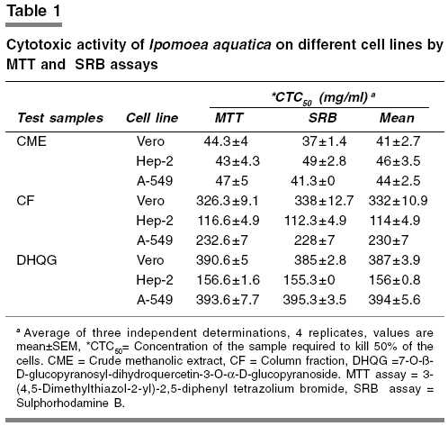
and Vegetable Technology,CFRTI, Mysore-570 020 Code Number: ph05104 Efforts are being made to develop anticancer agents from natural sources by many workers. In the past, plants have provided anticancer compounds like vincristine and taxol. Several members of the species Ipomoea ( Convolvulaceae ) are being used traditionally for the treatment of a large number of disease conditions. Among them Ipomoea bahiensis is reported to posses anticancer activity.[1] Ipomoea aquatica leaf is being used as a green leafy vegetable and also in traditional medicine for nose bleed and high blood pressure with high antioxidant properties.[2] No investigation has been carried out for its anticancer property till now. In view of these reports and ethnomedical uses of Ipomoea aquatica leaf, we studied the in vitro cytotoxic properties of its crude methanolic extract (CME), its column fraction (CF) and purified bioactive compound i.e., 7-O-β-D-glucopyranosyl-dihydroquercetin-3-O-α-D-glucopyranoside (DHQG) isolated from it. The leaves of Ipomoea aquatica were collected from Mysore, Karnataka, India, authenticated by a taxonomist and a specimen preserved (specimen no: 9/1) at the Department of Botany, University of Mysore, Mysore. The leaves (628 g) were oven dried at 48°C for 36 h to prevent enzyme action,[3] powdered and extracted with methanol (2.5 l) in a Soxhlet extractor for 8 h. The extract was concentrated under reduced pressure and controlled temperature to yield CME (146 g, 23.3%). This extract (60 g) was purified partially by using silica column chromatography to obtain the CF. This fraction was in turn purified by using XAD-4 resin column chromatography to obtain a pure compound (1.7 g). This 100% pure compound based on UV, NMR and GC-MS studies was identified as DHQG.[2] Each test sample (CME, CF and DHQG) were separately dissolved in 1% dimethyl sulphoxide[4] (DMSO) and the volume was made up to 10 ml with Dulbecco′s modified Eagles medium (DMEM), pH 7.4 supplemented with 2% heat inactivated newborn calf serum (Maintenance medium, PAA Laboratories, Austria), to obtain a stock solution of 1 mg/ml concentration, sterilized by filtration and stored at -20°C till use. The CME, CF and DHQG were investigated for cytotoxic properties against normal and cancer cell lines. Vero (normal African green monkey kidney) and Hep-2 (human larynx epithelial carcinoma) cell lines were obtained from Pasteur Institute of India, Coonor. A-549 (human small cell lung carcinoma) was obtained from National Center for Cell Sciences, Pune. The cultures were maintained in DMEM, pH 7.4 supplemented with 10% heat inactivated newborn calf serum,[5] penicillin (100 IU/ml), streptomycin (100 µg/ml) and amphoterecin-B (5 µg/ml) and were grown in 25 cm2 tissue culture flasks (Tarsons Products [P] Ltd., Kolkata, India) until confluent and used for cytotoxicity assays. Cell lines in exponential phase were washed with serum free DMEM medium, trypsinised using 0.2% trypsin and re-suspended in DMEM medium with 10% heat inactivated newborn calf serum.[5] Cells were plated at 10,000 cells/well in 96 well microtiter plates (Tarsons Products [P] Ltd., Kolkata, India) and incubated for 24 h at 37°C, 5% CO2 in a humidified atmosphere, during which period a partial monolayer was formed. The cells were then exposed to different concentrations (15.6, 31.2, 62.5, 125, 250, 500 and 1000 mg/ml, prepared by serial two-fold dilutions using maintenance medium from the stock solution) of test sample in quadruplicate. Control wells received only maintenance media. The cells were incubated at 37°C in a humidified incubator with 5% CO2 for a period of 72 h according to method of Brown[4] et al ., (2004). Morphological changes were examined using an inverted tissue phase-contrast microscope (Olympus Optical Co., Ltd., Japan, Model 1X70) at 24 h time intervals and compared with the control. At the end of 72 h, cellular viability was determined using standard 3-(4,5-Dimethylthiazol-2-yl)-2,5-diphenyl tetrazolium bromide (MTT)[6] and Sulphorhodamine B (SRB)[7] assays and the CTC50 value (concentration of the sample required to kill 50% of the cells) was calculated. DHQG showed cytotoxicity towards cell cultures with CTC50 values of 387 mg/ml against normal Vero cell line, where as 156 and 394 mg/ml, against Hep-2 and A-549 cell lines respectively. The CME and CF gave CTC50 values ranging from 41-332 mg/ml in Vero, 46 - 114 mg/ml in Hep-2 and 44 - 230 mg/ml in A-549 cell lines. [Table - 1] The CME was more potent than that of DHQG probably due to synergistic effects resulting from the combination of anthocyanins and other phenolic compounds.[8] Presence of sugar moiety in DHQG may be attributed to its low activity, probably due to steric hindrance by addition of sugar moieties.[9] The results of CTC50 values thus obtained indicate that DHQG showed cytotoxicity towards cancer cell lines tested. Hence further investigations are required to prove the toxicity of DHQG in vivo . References
Copyright 2005 - Indian Journal of Pharmacology The following images related to this document are available:Photo images[ph05104t1.jpg] |
| |||||||||
