 |
| About Bioline | All Journals | Testimonials | Membership | News |
|
||||||
|
||||||
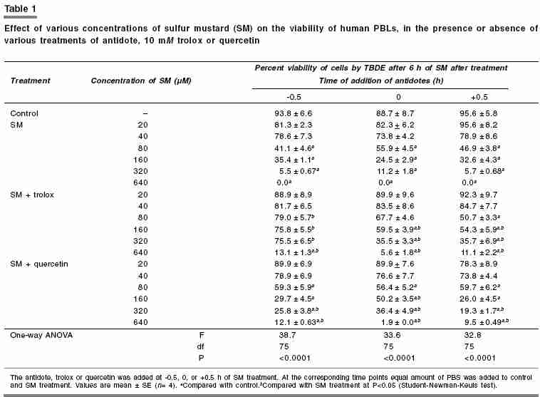
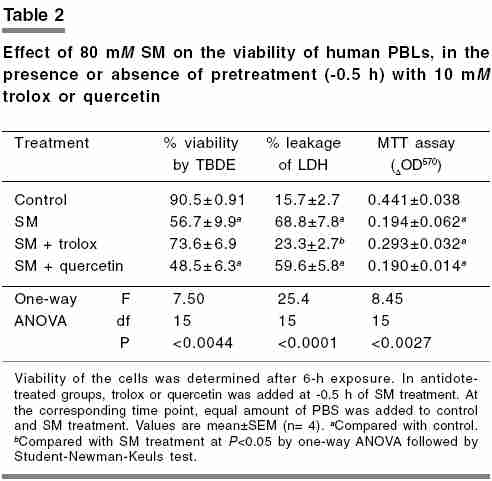
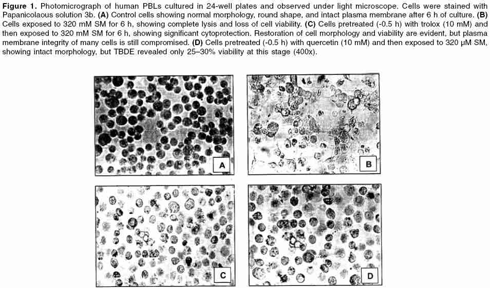
Indian Journal of Pharmacology, Vol. 38, No. 1, January-February, 2006, pp. 38-42 Research Paper Effect of trolox and quercetin on sulfur mustard-induced cytotoxicity in human peripheral blood lymphocytes Bhattacharya R, Tulsawani R K, Vijayaraghavan R Division of Pharmacology and Toxicology, Defence Research and Development Establishment, Jhansi Road, Gwalior-474002, Madhya Pradesh, India Code Number: ph06007 Abstract Objective: To evaluate the protective activity of antioxidants, viz. trolox and quercetin, against sulfur mustard (SM)-induced cytotoxicity.Materials and Methods: Cytotoxicity of various concentrations (20-640 µM) of SM, in the presence or absence of 10 µM trolox or quercetin (-0.5, 0, or +0.5 h) was determined in human peripheral blood lymphocytes after 6-h exposure. Cell viability was measured by Trypan blue dye exclusion (TBDE). Further, a cytotoxic concentration of SM (80 µM) was challenged by the two antidotes (-0.5 h) and cell viability was measured by TBDE and leakage of intracellular lactate dehydrogenase (LDH). Mitochondrial integrity and peroxide levels were measured by 3-4,5-dimethyl thiazol- Z -yl)-2,5-diphenyltetrazolium bromide and 2',7'-dichlorofluoroscin diacetate assay, respectively. Morphological changes of cells exposed to 320 µM SM (with or without antidotes) were also visualized under light microscope. Results: On the basis of TBDE , SM caused cell death of approximately 50% at 80 µM and 100% at 640 µM, respectively. Pretreatment of trolox conferred significant protection compared with quercetin. Also, pretreatment of trolox significantly reduced cell death and LDH leakage caused by 80 µM SM but did not prevent the loss of mitochondrial integrity. Trolox significantly reduced the levels of peroxides generated by SM. The better protection offered by trolox was evidenced in cell morphology studies too. Conclusion: Pretreatment (-0.5 h) of trolox afforded significant protection against SM-induced cytotoxicity in human lymphocytes. The protection was related to the antioxidant property of trolox, a water soluble analog of a-tocopherol. Keywords: Alkylating agent, antioxidant, cytotprotective, WBC Introduction Although India is a signatory to the Chemical Weapons Convention, threat from unscrupulous use of sulfur mustard (SM; bis [2-chloroethyl] sulfide), a potential chemical warfare agent still persists.[1],[2] SM is a highly reactive bifunctional alkylating agent that causes blisters and several other injuries on exposure.[3],[4],[5] SM forms sulfonium ion in the body and alkylates DNA, leading to strand breaks and cell death.[3],[5] Owing to the high electrophilic property of the sulfonium ion, SM binds to several cell components and causes various toxic effects on different cell types or tissues.[3], [4], [6] A variety of substances such as free radical scavengers, inhibitors of cell death, promoters of cell survival, radioprotectors, and numerous other pharmacological agents of diverse action have been shown to attenuate SM toxicity in vivo and in vitro . [7],[8],[9],[10],[11],[12] Protective effects of various antioxidants, viz. vitamin E, glutathione, and thiol reagents such as dithiothreitol and N -acetylcysteine (NAC) have also been reported against toxicity of SM or its analogs.[13], [14] However, none of the agents have been approved for human use so far. The best protection against SM poisoning is avoidance of contact and in the event of contact, rapid decontamination or detoxification of the contaminated area should be done. Only a few chemical decontaminants for human use have shown very good efficacy.[15], [16] The possible strategies to counter SM poisoning include (i) prevention of SM-induced alkylation of macromolecules, (ii) reversal of alkylation, and (iii) prevention or reversal of cascade of biochemical events following alkylation. Clinical manifestation of SM exposure is delayed and often has an extended latent phase of injury.[6] In view of this, many researchers have proposed prophylaxis as a more pragmatic approach for SM poisoning,[7], [10], [12], [14] more so because of the risks involved in the destruction of SM or during inspection by the Organization for Prohibition of Chemical Weapons. In the present study, the effect of trolox (a water-soluble analog of α-tocopherol; vitamin E) and quercetin (a polyphenolic natural bioflavonoid) was evaluated against SM-induced cytotoxicity in human peripheral blood lymphocytes (PBL). Protective efficacy of trolox and quercetin was studied by pretreatment (-0.5 h), simultaneous treatment (0 h), or post-treatment (+0.5 h) to SM exposure. Materials and Methods Chemicals Lymphocyte preparation Treatments Leakage of LDH MTT assay Measurement of peroxides Photomicrography Statistical analysis Results [Table - 1] shows the cell viability determined by TBDE after 6 h of SM exposure, in the presence or absence of trolox or quercetin treatment. Cytotoxicity caused by SM was concentration-dependent and viability of the control cells was significantly lost at 80 mM onwards. Almost 50% cell death was caused by 80 mM SM, whereas 100% cell death was observed at 640 mM. Pretreatment of trolox conferred significant protection at all the concentrations except 640 mM. Also, simultaneous treatment and post-treatment of trolox protected the cells, but the effects were not statistically significant as compared with control. Treatment with quercetin offered only marginal protection. Further study was carried out with only 80 mM SM, which was challenged by pretreatment with trolox or quercetin [Table - 2]. Significant reduction in cell viability accompanied by leakage of intracellular LDH and decrease in mitochondrial succinate dehydrogenase activity (MTT assay) were observed following treatment with 80 mM SM. Both cell viability and LDH leakage was significantly prevented by pretreatment with trolox. Although MTT assay showed attenuation, the protection by trolox was not significant. Pretreatment of quercetin did not afford protection on any of the end points evaluated. The percent peroxide levels generated by SM, in the presence or absence of trolox or quercetin, was also quantified after 6-h exposure (data not presented). The peroxide level (percent of control) was significantly elevated to 198.6±12.9 by 80 m M by SM, which was reduced to 112.9±14.6 and 156.9±16.9 by pretreatment of trolox and quercetin, respectively. Although both the agents reduced the levels of peroxide, the effect of trolox was significant. [Figure - 1] shows the photomicrographs of lymphocytes treated with a cytotoxic concentration (320 mM) of SM in the presence or absence of pretreatment with trolox or quercetin. Compared with the normal control cells [panel (A)], the SM-treated cells were completely lysed without any intact morphology or plasma membrane integrity [panel (B)]. However, cells pretreated with trolox showed near-normal morphology of the cells with intact plasma membrane [panel (C)]. Although morphology of the cells protected with quercetin [panel (D)] was almost similar to those treated with trolox, the TBDE of the cells revealed only 25-30% cell viability.Discussion Various in vitro models have been used to delineate SM toxicity or its antagonism, but human lymphocyte culture is a model with many advantages.[23] Previous study from this laboratory revealed that pretreatment of trolox, but not quercetin, was cytoprotective against cyanide poisoning in vitro .[19] In view of the oxidative injury reported with SM toxicity, we evaluated the effects of pretreatment, simultaneous treatment, or post-treatment of trolox or quercetin against the same in human lymphocytes in vitro. SM causes cytotoxicity through lipid peroxidation, which is mediated by depletion of reduced glutathione (GSH).[3] Flavonoids (gossypin and hydroxyethyl rutaside) and vitamin E have been shown to decrease SM-induced lipid peroxidation in mice, and this protection is attributed to their possible antioxidant and free-radical scavenging properties.[7] In the present study, the dose-response of SM was similar to that observed previously in liver slice culture, but the severity of toxicity was more.[12] Pretreatment with trolox generally protected against the cell membrane-damaging effects (TBDE, LDH release) of SM, but did not protect the mitochondrial activity, which is more sensitive to free radicals. Perhaps, the diminished mitochondrial activity was sufficient to sustain the cell viability and would have restored to normalcy at a later stage. Trolox, however, protects cyanide-induced DNA fragmentation and mitochondrial and nuclear dysfunction by attenuating the peroxide levels in thymocytes in vitro.[19] Reactive oxygen species mediated cytotoxicity and DNA damage is usually accompanied by depletion of intracellular GSH.[24] Trolox is known to penetrate biomembranes and protect mammalian cells from oxidative damage and DNA fragmentation, and it is considered to be more potent than vitamin E.[25] Generation of free radicals is considered to be one of the earliest events preceding cell death.[24] Because of this reason cells pretreated with the antioxidants could only show significant protection as compared with simultaneous or post-treatment. Lymphocytes are known to lack the ability to synthesize cysteine, a primary component for GSH, and its intracellular glutathione level depends largely on the extracellular cysteine. Therefore, GSH-mediated protection by trolox cannot be anticipated as observed in case of NAC, a precursor to glutathione.[26] Antioxidant properties of quercetin is ascribed to its ability to interrupt membrane lipid peroxidation rather than scavenging the free radicals, which is excessively generated in SM toxicity. Perhaps for this reason we did not observe appreciable protection by quercetin. Also, in our previous study we did not observe notable protection by quercetin.[19] The present study also indicates that trolox was better than quercetin in terms of reducing the levels of peroxide generated by SM. This also suggests strong antioxidant property of trolox, which has been widely recognized for many pathological conditions.[25] In view of the present findings, prophylactic implications of trolox alone or with other agents cannot be overlooked against SM poisoning. Detailed animal studies would further validate its scope. Acknowledgment The authors thank Er. K. Sekhar, Director, DRDE, Gwalior, for providing keen support and encouragement in this work.References
Copyright 2006 - Indian Journal of Pharmacology The following images related to this document are available:Photo images[ph06007t2.jpg] [ph06007f1.jpg] [ph06007t1.jpg] |
| |||||||||
