 |
| About Bioline | All Journals | Testimonials | Membership | News |
|
||||||
|
||||||
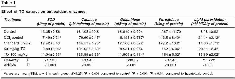
Indian Journal of Pharmacology, Vol. 38, No. 1, January-February, 2006, pp. 54-55 Research Letter In vivo antioxidant activity of hydroalcoholic extract of Taraxacum officinale roots in rats Sumanth Meera, Rana* A.C Al Ameen college of Pharmacy, Hosur Road, Opp. Lalbagh Main Gate, Bangalore 560 027, India. *Department of Pharmacological Sciences, Dr. H. S. Gour University, Sagar 470 003, Madhya Pradesh, India. Code Number: ph06010 In the traditional system of medicine, there are a number of plants which are used in the treatment of liver disorders. Their extract, fractions, and active constituents exhibit marked hepatoprotective action, which has been related to their antioxidant properties.[1] Traditionally, Taraxacum officinale , WEBER, family Compositae, commonly known as "Dandelion" has been used as a remedy for jaundice, other disorders of the liver and gall bladder, and to counteract water retention. Free radicals are reactive molecules involved in many physiological processes and human diseases, such as cancer, aging, arthritis, Parkinson′s syndrome, ischemia and liver injury. The elevation of free radical levels seen during the liver damage is owing to enhanced production of free radicals and decreased scavenging potential of the cells. A variety of intrinsic antioxidants (reduced glutathione, superoxide dismutase (SOD), catalase and peroxidase) are present in the organism, which protect them from oxidative stress, thereby forming the first line of defence.[2] The present study was undertaken to evaluate antioxidant activity of 70% hydroalcoholic extract of roots of T. officinale (TO) in rats. The plant TO was collected from the Gandhi Krishi Vigyan Kendra (GKVK), Bangalore, in the month of December 2002 and was authenticated by Dr. Yoganarsimhan, Scientist, Regional Research Centre [Ay], Bangalore. The dry powder of roots of TO was extracted with 70% alcohol in a soxhlet apparatus for 24 h at approximately 60°C. The extract was then concentrated by distilling the solvent below 60°C and dried in a dessicator. A suspension was prepared by using equal volumes of the extract and gum acacia for administration to rats using oral gague. The chemicals used for investigation of antioxidant activity were CCl4 (Quality Fine Chemicals Ltd. India), Liv-52 (Himalaya, India), hydroxylamine hydrochloride (Sigma, India), nitro-blue tetrazoleum (NBT) (Sigma, India), hydrogen peroxide, EDTA, and Ellman′s reagent (Sigma, India). All other chemicals obtained from local sources were of analytical grade. Albino rats of Wistar strain, weighing 100-150 g, maintained on normal diet (Amrut Laboratory Animal Feeds, Bangalore) and water ad libitum , were divided into five groups of six animals each. Before starting the experiment, permission from the Institutional Animal Ethics Committee was obtained. Group-I animals served as normal control, treated with distilled water. Group-II animals served as hepatotoxic control, treated with CCl4 in a single dose of 1.5 ml/kg, i.p., to produce acute hepatotoxicity. Group III served as a standard group, and was administered Liv-52 in a dose of 56 mg/kg, p.o. Group-IV and -V animals were treated with daily doses of 50 and 100 mg/kg, p.o., respectively, of TO extract for 7 days. The animals of Groups III-V were given single dose of CCl4, 1.5 ml/kg, i.p., 6 h after the last treatment. On day 8 the rats were sacrificed by carotid bleeding and liver was rapidly excised, rinsed in ice-cold saline, and a 10% w/v homogenate was prepared using 0.15 M KCI, centrifuged at 800 g for 10 min at 4°C. The supernatant obtained was used for the estimation of catalase, peroxidase, and lipid peroxidation. Further, the homogenate was centrifuged at 1000 g for 20 min at 4°C and the supernatant was used for estimation of SOD and glutathione Estimation of SOD was done by autoxidation of hydroxylamine at pH 10.2, which was accompanied by reduction of NBT, and the nitrite produced in the presence of EDTA was detected colorimetrically.[3] One enzymatic unit of SOD is the amount in the form of proteins present in 100 µl of 10% liver homogenate required to inhibit the reduction of 24 mM NBT by 50% and is expressed as units per milligram of protein. Catalase activity was estimated by determining the decomposition of H2O2 at 240 nm in an assay mixture containing phosphate buffer.[4] One international unit of catalase utilized is that amount that catalyzes the decomposition of 1 m M H2O2/min/mg of protein at 37°C. Catalase activity was calculated using the millimolar extinction coefficient of 0.07 and expressed in terms of micromole per minute per milligram of protein. Glutathione was estimated using Ellman′s reagent (5,5¢-dithiobis-(2-nitrobenzoic acid) [DTNB]). The sulphydryl groups present in glutathione forms a colored complex with DTNB, which was measured colorimetrically at 412 nm.[5] The amount of glutathione was determined using its molar extinction coefficient of 13600/m/cm and expressed in terms of µmol/mg of protein. Peroxidase estimation is based on periodide formation. Periodide can be spectrophotometrically determined at 353 nm, and this is directly proportional to the peroxidase concentration in the reaction mixture containing approximate amounts of H2O2 and enzyme.[6] One unit of peroxidase activity is defined as the change in absorbance per minute and expressed in terms of units per milligram of protein. Malondialdehyde (MDA), a secondary product of lipid peroxidation , reacts with thiobarbituric acid at pH 3.5. The red pigment produced was extracted in n -butanol-pyridine mixture, and estimated by measuring the absorbance at 532 nm.[7] Results were subjected to one-way ANOVA. P< 0.05 was considered significant. The post hoc analysis was carried out by Dunnet′s multiple comparison test. As shown in [Table - 1], CCl4 treatment decreased SOD, catalase, glutathione, and peroxidase and increased lipid peroxidation. Pretreatment with 100 mg/kg (p.o.) of TO extract improved the SOD, catalase, glutathione, and peroxidase levels significantly and reduced lipid peroxidation. SOD is a ubiquitous cellular enzyme that dismutates superoxide radical to H2O2 and oxygen and is one of the chief cellular defence mechanisms. The H2O2 formed by SOD and other processes is scavenged by catalase that catalyzes the dismutation of H2O2 into water and molecular oxygen. Thus, the antioxidant enzyme catalase is responsible for detoxification of H2O2. Glutathione is a tripeptide of glycine, glutamic acid, and cysteine. Glutathione is an important naturally occurring antioxidant as it prevents the hydrogen of sulfhydryl group to be abstracted instead of methylene hydrogen of unsaturated lipids. Therefore, levels of glutathione are of critical importance in tissue injury caused by toxic substances. The antioxidant enzymes and glutathione form the first line of defence against free radical-induced damage, offer protection against free radicals, and thereby maintain low levels of lipid peroxide.[3] Peroxidase is an enzyme that catalyzes the reduction of hydroperoxides, including hydrogen peroxides, and functions to protect the cell from peroxidative damage. As the TO extract, in the dose of 100 mg/kg, p.o., has improved the SOD, catalase, glutathione, and peroxidase levels significantly, which were comparable with Liv 52.[2] We conclude that the hydroalcoholic extract from the root of TO possesses antioxidant activity, confirming the traditional use of the plant in treatment of liver disorders. References
Copyright 2006 - Indian Journal of Pharmacology The following images related to this document are available:Photo images[ph06010t1.jpg] |
| |||||||||
