 |
| About Bioline | All Journals | Testimonials | Membership | News |
|
||||||
|
||||||
Indian Journal of Pharmacology, Vol. 38, No. 2, March-April, 2006, pp. 107-110 Research Paper In vitro prevention by ACE inhibitors of cataract induced by glucose D. G. Langade, G. Rao*, R. C. Girme**, P. S. Patki***, P. M. Bulakh** Department of Pharmacology, Grant Medical College, Mumbai, *Ranbaxy Laboratories Ltd.,**Department of Biochemistry, B.J. Medical College, Pune,
***Department of Pharmacology, B.J. College, Pune.Medical Code Number: ph06027 Abstract Objectives: To study, the anticataract activity of lisinopril and enalapril on cataract induced by glucose, in goat lenses.Materials and Methods: Goat lenses were incubated in artificial aqueous humor containing 55 mM glucose (cataractogenesis) with lisinopril or enalapril in different concentrations at room temperature for 72 h. Biochemical parameters studied in the lens were electrolytes (Na+ , K+ ), Na+-K+ -ATPase activity, malondialdehyde (MDA) and proteins. Results: Glucose induced opacification of goat lens began 8-10 hrs after incubation and was complete in 72-80 hrs. Cataractous lenses showed higher Na+ , MDA (P<0.001), lower Na+ -K+ -ATPase activity, and water-soluble protein content. Lenses treated with lisinopril or enalapril in concentrations of 1, 5, and 10 ng/ml showed higher protein (total and water soluble proteins) content and prevented formation and progress of cataract by glucose, as evidenced by biochemical parameters. Conclusion: The anticataract activity of lisinopril and enalapril may be because of the antioxidant and free radical scavenging activity, as evidenced by a decrease in MDA in treated lenses. Further in-vitro and in-vivo studies in various experimental models and long term clinical trials are required to validate the anticataract activity of ACE-inhibitors. Keywords: Antioxidant, enalapril, lisinopril Introduction Cataract is the opacification of lens often associated with old age and is a major complication of diabetes mellitus because higher glycosylated hemoglobin levels are significantly associated with increased risk of cataract.[1] Although many cataractogenic factors have been identified, the biochemical background of cataractogenesis is still unknown. The lens Na+ -K+ -ATPase activity plays an important role in maintaining lens transparency, and its alteration is one of the major events leading to the cataract formation.[2] Oxidative damage by the free radicals is also implicated in the pathology of cataractogenesis.[3] Although a number of agents have been tried for prevention and therapy of cataract, none have proved useful.[4] ACE inhibitors have been found to afford protection from free radical damage in many experimental conditions. [5],[6],[7],[8] Therefore, this study was conducted to find the efficacy of lisinopril and enalapril in the prevention of experimental cataract induced by glucose.Materials and Methods Various in vivo or in vitro experimental models in rats, mice, and rabbits have been utilised to study cataractogenesis. In this study, goat lenses were used as they were easily available. The study was approved by the institutional ethics committee. Lens culture Study drugs and groups A total of 80 lenses were divided into following categories (n=10 in each category): Group I : Normal lens [Control (Glucose 5.5mM)] Homogenate preparation The homogenate was centrifuged at 10,000 G at 4°C for 1 hour and the supernatant used for estimation of biochemical parameters. For estimation of water-soluble proteins, homogenate was prepared in sodium phosphate buffer (pH 7.4). Biochemical estimation
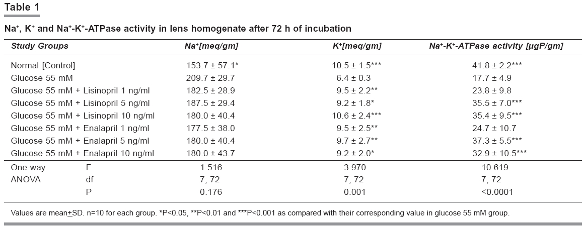
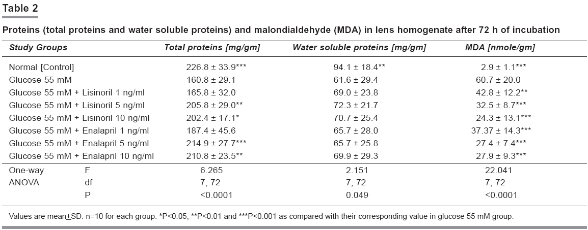
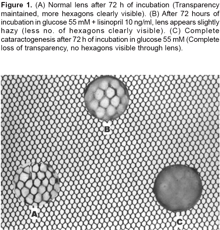
Photographic evaluation Statistical analysis Results Incubation of lenses with glucose 55 mM showed opacification starting after 8 hrs at the periphery, on the posterior surface of the lens. This progressively increased towards the centre, with complete opacification at the end of 72 hrs. Biochemical changes Glucose 55 mM treated lenses also showed significantly low concentrations of proteins (total and water soluble proteins) in the lens homogenate (P< 0.001) and very high MDA (P< 0.001) compared with control group having normal lenses. [Table - 2] Both lisinopril and enalapril groups had significantly higher concentrations of total lens proteins at 5 and 10 ng/ml concentrations (P< 0.05), compared with glucose 55 mM group. At the same time, they had higher water-soluble proteins at all three concentrations of lisinopril and enalapril, compared with glucose 55 mM group. However, the difference was not significant statistically. [Table - 2] MDA levels were found to be very high in glucose 55 mM treated lenses, compared with normal lenses (60.7 Vs 2.9). Lenses treated with lisinopril and enalapril had significantly reduced MDA content (P< 0.01) at all the three concentrations, compared with glucose group. [Table - 2] Photographic evaluation [Figure
- 1] Discussion In cataractogenesis, the parameters commonly considered are electrolytes (Na+ and K+ ), malondialdehyde (MDA) and proteins (total proteins and water soluble proteins). Incubation in the media containing high glucose (55 mM) concentration has shown to cause considerable drop in Na+ -K+ -ATPase activity, with progression of opacity.[10] This study, is in agreement with this finding. Na+ -K+ -ATPase is important in maintaining the ionic equilibrium in the lens, and its impairment causes accumulation of Na+ and loss of K+ with hydration and swelling of the lens fibers leading to cataractogenesis.[13] This alteration in the Na+ -K+ ratio alters the protein content of the lens, leading to a decrease in water-soluble proteins′content and increase in insoluble proteins. This causes lens opacification.[14] This study showed higher Na+ -K+ -ATPase activity, total and water-soluble proteins and K+ ions whereas lower concentrations of Na+ ions with lisinopril and enalapril treated groups. Therefore, these ACE inhibitors seem to prevent the alteration of Na+ and K+ imbalance, which may be due to a direct effect on lens membrane Na+ -K+ -ATPase or indirect effect through their free radical scavenging activity. Oxidative stress may also be implicated in the cataract induced by glucose, due to the formation of superoxide (O 2.-) radicals and H 2 O 2 . High glucose (55 mM) has shown to induce antioxidant enzymes, suggesting oxidative stress in the cells.[15] In this study MDA levels were significantly higher in high glucose (55 mM) group, compared with normal control group. The MDA levels were significantly less in the lisinopril and enalapril treated groups at all concentrations. These results are in agreement with those of Bhuyan KC, et al . [16] They found significant reduction in the rate of superoxide (O 2.-) production in animals treated with captopril, in cataract model induced by diquat in rabbits. Noda Y, et al .[17] demonstrated scavenging activity of lisinopril on nitric oxide. Lisinopril and enalapril have also been shown to increase the content of water-soluble proteins, retarding the process of cataractogenesis initiated by high glucose concentration. Incubation in presence of high glucose (55 mM) concentration simulates a state of clinical diabetes where ACE inhibitors are commonly used in these patients to treat associated cardiovascular disorders. A preventive role of lisinopril and enalapril as seen in this in vitro model may, to some extent, suggest an additional utility of ACE inhibitors in the form of preventing and/or retarding the progression of diabetic cataracts. The concentrations of lisinopril and enalapril used in this study ranged between 1 to 10 ng/ml. However, higher concentrations upto 20 ng/ml may show better anticataract activity, and further evaluation with higher concentrations is required. This in vitro study may not directly correlate with the in vivo conditions. Therefore, in vivo studies in different animal models are required for further elucidation of the role of ACE inhibitors in preventing cataract. Conclusion Lisinopril and enalapril demonstrated in vitro anticataract activity in experimental cataract induced by high glucose. Further in vitro and in vivo studies to elucidate the exact mechanism of ACE inhibitors in prevention of cataractogenesis are needed. Clinical evaluation of patients already receiving ACE inhibitors may be followed to identify the presence of the additional benefit of cataract prevention/progression.References
Copyright 2006 - Indian Journal of Pharmacology The following images related to this document are available:Photo images[ph06027t1.jpg] [ph06027t2.jpg] [ph06027f1.jpg] |
| |||||||||
