 |
| About Bioline | All Journals | Testimonials | Membership | News |
|
||||||
|
||||||
Indian Journal of Pharmacology, Vol. 38, No. 2, March-April, 2006, pp. 120-124 Research Paper Studies on Dalbergia sissoo (Roxb.) leaves: Possible mechanism(s) of action in infectious diarrhoea S. Brijesha , P. G. Daswania , P. Tetalib , N. H. Antiaa, c , T. J. Birdia aThe Foundation for Medical Research, 84A, RG Thadani Marg, Worli, Mumbai - 400018, Maharashtra, India. Code Number: ph06030 Abstract Objective : Several medicinal plants have been evaluated for their antidiarrhoeal activity. Most studies evaluated their effect on intestinal motility and antimicrobial activity and, therefore, did not take into account the pathogenesis of infectious diarrhoea. Features of infectious diarrhoea like abdominal pain, cramps, inflammation, and passage of blood/mucus in the stools are the combined effect of one or more virulence factors of the infecting organism. The effect of medicinal plants on the microbial virulent features can serve as marker(s) for testing their efficacy. In this study, we evaluated the effect of a decoction of dried leaves of Dalbergia sissoo on aspects of pathogenicity, that is, colonisation to intestinal epithelial cells and production/action of enterotoxins. This was done to define its possible mechanism(s) of action in infectious diarrhoea.Materials and Methods : Antibacterial, antiprotozoal, and antiviral activities of the plant decoction were checked by agar dilution method, tube dilution method, and neutral red uptake assay, respectively. Cholera toxin (CT) and Escherichia coli labile toxin (LT) were assayed by ganglioside monosialic acid receptor ELISA. Suckling mouse assay was used to assess E. coli stable toxin (ST). As a measure of colonisation, the effect against adherence of E. coli and invasion of E. coli and Shigella flexneri to HEp-2 cells were studied. Results: The decoction had no antibacterial, antiprotozoal, and antiviral activity. It reduced the production and the binding of CT and bacterial adherence and invasion. Conclusion : This study showed that D . sissoo is antidiarrhoeal as it affects bacterial virulence. However, it has no antimicrobial activity. Keywords: Gastrointestinal infection, Indian rosewood, plant antimicrobial Introduction Infectious diarrhoea is the most common infectious disease worldwide.[1] Gastrointestinal infections kill 1.8 million people globally each year, mainly children in developing countries.[2] Acute, watery, bloody diarrhoea may be due to a variety of pathogens- bacterial (e.g., Escherichia coli , Vibrio cholerae , Shigella flexneri , and Campylobacter jejuni ), protozoal (e.g., Giardia lamblia , Entamoeba histolytica and Cryptosporidium parvum ) and viral (e.g. rotavirus, astrovirus, and adenovirus) agents. These organisms disrupt intestinal functions and cause diarrhoea through several mechanisms. These include microbial attachment to the intestinal epithelium and localised effacement, production of toxin(s), and penetration and invasion of intestinal epithelial cells that result in alteration of absorption due to the rearrangement in cytoskeletal structure.[1] Dalbergia sissoo Roxb. (Fabaceae), known as Indian Rosewood, is reported to be useful in many conditions including fever, ulcers, digestive disorders, and skin diseases.[3],[4] It is also known to be effective against diarrhoea and dysentery.[3],[5] Furthermore, this plant had the highest frequency of quote (5.2%) in an ethnobotanical survey carried out by us (unpublished observation). To the best of our knowledge, no experimental evidence is available on its antidiarrhoeal activity. This work was, therefore, undertaken to assess the antidiarrhoeal activity of the dried leaves of D. sissoo on antimicrobial (antibacterial, antiprotozoal, and antiviral) activity and bacterial virulence parameters, such as, colonisation, production and action of toxins. Materials and Methods The study design Cell cultures, media, and reagents The bacterial growth media and MEM were purchased from Himedia laboratory, Mumbai, India. DMEM and FCS were procured from GibcoBRL, UK. The constituents of the Diamond′s medium for G. lamblia and the antibiotics (penicillin, streptomycin, gentamicin, and metronidazole) were procured from local manufacturer. Trypan blue, neutral red, polymyxin B sulphate, anticholera toxin, and bovine serum albumin were purchased from Sigma, USA, and swine anti-rabbit immunoglobulin (Ig) was obtained from Dako, Denmark. The 96-well ELISA plates were purchased from Nunclon, Denmark, and the ELISA plate reader was purchased from Labsystems, Finland. Preparation of plant extract Phytochemical analysis Microorganisms used Antimicrobial activity The antiprotozoal activity was assayed by incubating a 24 h culture of G. lamblia trophozoites without (control) and with different dilutions of the decoction for 24 h. The number of viable trophozoites was counted in a haemocytometer with trypan blue.[8] The antigiardial drug metronidazole (100 µg/ml) was used as the positive control. The antiviral activity was determined by assaying the entry and the subsequent survival of rotavirus in MA-104 cells by the neutral red uptake assay.[9] Briefly, a 72 h culture of MA-104 cells was infected with rotavirus and incubated without (control) and with different dilutions of the decoction for 90 min. The culture was further incubated for 72 h after removal of the decoction and the unabsorbed virus. Thereafter, the cells were incubated with neutral red dye for 30 min. The intracellular dye was released with 1:1 solution of 100 mM acetic acid and ethanol. The released dye was measured at 540 nm (reference 630 nm) in an ELISA plate reader. Effect on toxins The assays were based on two protocols: i) Preincubation: Bacterial strains were grown without (control)
and with different dilutions of the decoction in casein hydrolysate yeast
extract broth (CAYE), and the LT/CT produced by the respective bacterial
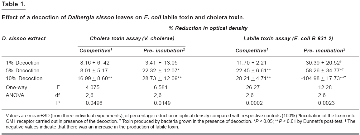
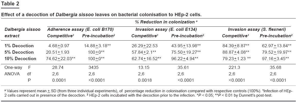
strains was assayed using the GM1-ELISA. Stable toxin (ST), which is an exotoxin, obtained as a culture supernatant of E. coli TX1 was assayed by the method originally described by Gianella.[12] Briefly, the toxin was inoculated intra-gastrically into 2-3 day-old Swiss albino suckling mice. Following an incubation of 3 h at room temperature, the pups were sacrificed and the ratio of gut weight to that of the remaining carcass weight was calculated. Ratio of > 0.083 was considered as positive. The assays were based on two protocols: Effect on colonisation to HEp-2 cell line The effect of the decoction on invasion by E. coli E134 and S. flexneri was studied by a method described by Vesikari et al .[14] Briefly, a 48 h culture of HEp-2 cells grown in a 24-well tissue culture plate was infected with log phase culture (10 8 cells/ml) of the bacterial grown in BHI and incubated for 2 h. The culture was further incubated with gentamicin (100 µg/ml) for 3 h to kill the uninvaded bacteria. The epithelial cells were then lysed by cold shock, and the released bacteria were counted by plating on NA. The assays were based on two protocols: Statistical analysis Results Phytochemistry Antimicrobial activity Effect on toxins Effect on colonisation Discussion According to the World Health Report 2004, diarrhoea is the cause of 3.3% of all deaths.[2] The past decade has witnessed several attempts towards the management of diarrhoea. These include improved formulations of oral rehydration solution (ORS) and the development of a feasible vaccine. Although ORS has contributed to reduction in diarrhoeal mortality rates, it is often less efficient in high stool output state. In addition, response to vaccines in developing countries is not encouraging.[15] With the threat of drug resistance, a definite niche exists for the development of an alternative approach to treat infectious diarrhoea. Medicinal plants can fill this niche. This study was an attempt to explore the antidiarrhoeal activity of crude decoction of D. sissoo leaves. The decoction did not have antibacterial activity against the strains tested nor did it have antigiardial or antirotaviral activity. It was observed that although the decoction did not arrest the growth of V. cholerae , it prevented the production of CT indicating that the reduction in the production of CT was metabolic and not due to reduction in bacterial counts. There was a two-fold increase in the production of LT in the presence of the decoction, but its binding to the receptor was reduced. It is known that LT and CT are closely related structurally, functionally, biologically, and immunogenically.[16] Therefore, the reduction in binding of LT and CT to GM1 receptor implies that the decoction may contain chemical(s) that either bind(s) directly to the receptor or to the common antigenic moiety of the toxins. The decoction also affected colonisation. It inhibited the adherence of E. coli B170 and invasion by E. coli E134 and S. flexneri . The decrease in colonisation was observed in both the protocols suggesting that D. sissoo modifies/affects receptors on HEp-2 cells in a way that restricts bacterial attachment and entry. This is especially true because the decoction did not affect the morphology of the HEp-2 cells and, as mentioned earlier, had no antibacterial activity. The findings of the biological assays are indicative of the selective antidiarrhoeal action of D. sissoo leaves. The results suggest that the leaves may not be active against diarrhoea induced by LT and ST or those caused by protozoa and virus. However, it appears to be most efficacious against cholera and diarrhoeal episodes caused by enteropathogenic and enteroinvasive bacterial strains. To conclude, this study besides describing the possible mechanisms of antidiarrhoeal action of D. sissoo leaves also highlights the necessity of including multiple parameters for judging the efficacy of medicinal plants. Assaying bacterial virulent features as a marker for demonstrating the antidiarrhoeal efficacy of a plant, has been previously reported by us using two indigenous plants viz. Cyperus rotundus [17] and Holarrhena antidysenterica .[18] This is especially important in the absence of antimicrobial activity, which in most of the studies reported earlier[19],[20],[21],[22] has been considered the marker for antidiarrhoeal activity. Acknowledgments We are thankful to Dr. P. D′Mello and Mr. Yogesh Palav, Department of Pharmacognosy, Principal, K. M. Kundanani, College of Pharmacy, Mumbai, India, for their assistance in carrying out the phytochemical studies. We are also thankful to Dr. N. F. Mistry, Foundation for Medical Research and Foundation for Research in Community Health (FRCH), for her critical suggestions and Mr. S. Jangam and Mr. A. Gurav, the field workers of FRCH, for collection of plant material. This work has been supported by the Department of Science and Technology, Ministry of Science and Technology, Government of India through grant number 91283. References
Copyright 2006 - Indian Journal of Pharmacology The following images related to this document are available:Photo images[ph06030t1.jpg] [ph06030t2.jpg] |
| |||||||||
