 |
| About Bioline | All Journals | Testimonials | Membership | News |
|
||||||
|
||||||
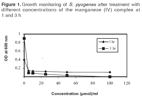
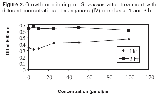
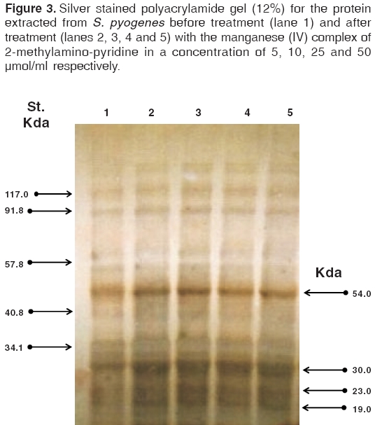
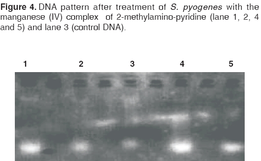
Indian Journal of Pharmacology, Vol. 38, No. 3, May-June, 2006, pp. 177-180 Research Paper Bactericidal activity of manganese (IV) complex of 2-methylamino-pyridine against Streptococcus pyogenes and Staphylococcus aureus El-Masry S, El-Sayed IH, Lotfy Mahmoud, El-Shahat M, Ali SA, Nasif WA Molecular and Cellular Biology Department, Genetic Engineering and Biotechnology Research Institute, Minufiya University, Sadat City, Minufiya Code Number: ph06043 Abstract Objective: To investigate the bactericidal activity of manganese (IV) complex of 2-methylamino-pyridine against Streptococcus pyogenes ( S. pyogenes ) and Staphylococcus aureus ( S. aureus ).Materials and Methods: The inhibitory effect of the complex was studied on the molecular level and by turbidity measurement. Treatment of bacteria was carried out using 5, 10, 25, 50 and 100 µmol of the complex per ml of culture media. Results: The results showed that the growth of S. pyogenes rapidly decreased with increasing concentrations of the complex. In contrast, the complex caused no significant decrease in the growth rate of S. aureus . The molecular level studies showed that four protein bands, with apparent molecular weights of 19, 23, 30 and 54 Kda, respectively, increased in the protein pattern of the S. pyogenes extract after the complex treatment using silver stained polyacrylamide gels, under reducing condition . However, there was no detectable change in the protein pattern of the S. aureus extract after the complex treatment . No DNA damage was detected while using agarose gel electrophoresis and ethidium bromide staining in both types of bacteria. Conclusion: Manganese (IV) complex of 2-methylamino-pyridine showed an apparent antibacterial inhibitory effect against S. pyogenes, but S. aureus was apparently resistant. Keywords: Free radicals, gram-positive cocci, metal complex, SOD. Introduction Gram-positive cocci are a heterogeneous collection of approximately 21 genera that colonise humans. Among these, Streptococcus and Staphylococcus are important pathogens in humans.[1] S. pyogenes has demonstrated the ability to develop drug resistance, particularly in patients with mixed infections that involve S. aureus . The drug resistance in staphylococci is due to penicillinase and acquisition of the mec A gene, which codes for a novel penicillin-binding protein, PBP2 .0[1],[2] Free radicals and, in particular, superoxide radical (O- 2) cause cellular disruption due to peroxidation of membrane lipids. Superoxide dismutase (SOD) is believed to be involved in all oxygen-metabolising cells.[3] Four different types of SOD have been found, two of which have been found in eukaryotic cells. A copper and zinc containing form is located in the cytosol; and a manganese containing form is located in the mitochondria.[4] Its wide distribution among aerobic organisms[3] suggests that superoxide is formed inside all cells that grow in air and is toxic. Cells devoid of cytosolic SOD suffer enzyme inactivation, growth deficiencies and DNA damage. It has been suggested that the scant superoxide, generated by aerobic metabolism, harms even cells that contain abundant SOD. The vulnerability of bacteria to increased intracellular superoxide explains the widespread use of superoxide-producing drugs as bactericidal weapons.[5] Extensive studies were conducted to address the antibacterial activity for many compounds and metal complexes that were found to have redox-cycling or pro-oxidative activity.[6],[7],[8],[9] In a previous study, we found an antitumour activity of some metal complexes having SOD-like activity on Ehrlich ascites carcinoma cells.[10] The present study aims to examine the antibacterial activity of one of these complexes [Manganese (IV) complex of 2-methylamino-pyridine, (MnL 2 O) 2 Cl 4 .2H 2 O where L is 2-methylamino-pyridine] on S. aureus and S. pyogenes . The inhibitory effect of this complex was studied on the molecular level and by turbidity measurement. Materials and Methods All chemicals used in this study were of analytical grade and were purchased from Sigma Chemical Co. (St. Louis, Mo, USA), unless mentioned otherwise. Manganese (IV) complex of 2-methyl-aminopyridine was synthesised and supplied by Prof. AM Ramadan, Chemistry Department, Faculty of Education, Kafer El-Shiekh, Tanta University, Egypt. The SOD-like activity of the complex was ascertained by the method of Dechatelet, et al[11] and has been reported earlier.[10]
Cultures Bacterial DNA isolation and agarose gel electrophoresis SDS-polyacrylamide gel electrophoresis (SDS-PAGE)
Statistical analysis Results The results showed that there was a decrease in growth of S. pyogenes [Figure - 1], but not S. aureus [Figure - 2] after 1 and 3 h of the complex treatment. The protein patterns of the S. aureus and S. pyogenes extracts were separated on silver stained polyacrylamide gels (12%), under reducing condition. The results showed no change in the protein profile of the S. aureus extract after the complex treatment. But in the S. pyogenes extract, there was a change in the protein profile at low molecular weights after the complex treatment. The intensity of four protein bands with apparent molecular weights of 19, 23, 30 and 54 Kda, respectively, increased as revealed by the silver stained SDS-PAGE gels after the complex treatment. [Figure - 3] No apparent DNA damage was produced upon treatment of S. pyogenes and S. aureus with different complex concentrations as revealed by agarose gel electrophoresis and ethidium bromide staining. [Figure - 4] Discussion Oxygen is known to form highly reactive free radicals (reactive oxygen species: ROS) such as superoxide ions, hydroxyl radicals and hydrogen peroxide in prokaryotic and eukaryotic cells. Free radicals, by the possession of unpaired electrons, make them very reactive as they urgently seek to gain or lose electrons in order to reach a more stable configuration.[17] Most facultative and aerobic organisms contain a high concentration of superoxide dismutase enzyme (SOD). This enzyme converts the superoxide anion into ground state oxygen and hydrogen peroxide or both, to eliminate H 2 O 2 and convert it into water and ground state oxygen.[18] We reported a pronounced antitumour effect of manganese (IV) complex of 2-methylamino-pyridine, with SOD-like activity due to the production of H 2 O 2 , earlier.[10] In this context, the antibacterial activity of this complex on S. aureus and S. pyogenes was examined and the possible mechanism of action was explained. The obtained data showed that a pronounced antibacterial activity of manganese (IV) complex of 2-methylamino-pyridine with different concentrations was found against S. pyogenes. Further, the antibacterial activity of the complex was not observed at any degree in the case of S. aureus. Moreover, in the S. pyogenes extract there was a change in the protein profile at low molecular weights after the complex treatment. The intensity of four protein bands with apparent molecular weights of 19, 23, 30 and 54 Kda, respectively, increased as revealed by the silver stained SDS-PAGE gels after the complex treatment. These bands may be related to the enzyme system of the bacteria that deal with free radicals utilisation. Aronovitch, et al [13] reported that the bacterial activity of epinephrine-Cu (II) complex caused little killing of E. coli , but rapid killing was induced by the addition of 0.5 mM H 2 O 2. The H 2 O 2 forms a recycling redox system to damage the cytoplasmic membrane of E. coli , which is apparently the main reason for its bacterial activity. They suggested that with the addition of H 2 O 2 , the epinephrine-Cu (II) complex binds to the cell surface to induce oxidative membrane damage. Hoshino, et al [7] proposed that catechin-Cu (II) on the cell surface reacts locally with molecular oxygen to produce H 2 O 2 . The H 2 O 2 generated on the surface can enter into the cell easily and cause damage to the cytoplasmic membrane. The apparently low concentrations of H 2 O 2 observed in the bulk suspension, will not reflect the local concentration of H 2 O 2 generated on the cell surface. Therefore, they proposed that Cu (I) and its redox reactions, involving catechins and H 2 O 2 on the cell surface, must be involved in the killing of bacteria. It was found that the DNA double strand did not break in the process of killing, indicating that the antibacterial activity of manganese (IV) complex of 2-methylamino-pyridine is derived from the damage of the cytoplasmic membrane. Hoshino, et al [7] treated the E. coli with catechin-copper (II) complexes and obtained the same result. Gram-negative bacteria such as E. coli have negatively charged lipopolysaccharide on their cell surface. [12] Sonohara, et al [19] reported that gram-positive bacteria such as S. aureus have a positively charged surface and Tichy[20] reported that lipopolysaccharides had an ability to bind Cu (II). The binding of copper ions to S. aureus cells was found to decrease with an increase in the concentration of epigallocatechin (EGC). Indeed, the percentage of copper ions bound to S. aureus cells was only 2.4% in the presence of 100 µmol/L EGC. Therefore, it is possible that copper ions, complex with EGC and attracted by the negative charge of lipopolysaccharides of E. coli , bind to it tightly and generate H 2 O 2 locally on the surface of E. coli cells. On the other hand, copper ions, complexed with EGC in the bulk buffer, cannot bind to the S. aureus cells. H 2 O 2 was not generated on the surface of S. aureus cells but in the bulk buffer the EGC-Cu (II) complex does not generate active hydrogen species from H 2 O 2 . This may be the reason why the bactericidal activity of the catechin-Cu (II) complex is low against S. aureus . From studies on S. aureus and E. coli , it was concluded that the binding of copper ions to the cell surface plays an important role in the bactericidal activity of catechin Cu (II) complexes.[6] The explanation of Hoshino, et al [6] is not applicable in our situation. Our study is concerned with two bacterial species which are both gram-positive cocci carrying the same charge. Although the antibacterial activity of the complex is apparent in S. pyogenes, it is not so in S. aureus . S. pyogens lacks the enzyme catalase, which utilises H 2 O 2 . However, S. aureus has this enzyme, which has the ability to convert H 2 O 2 and protect the bacterial cells from the SOD-like activity of such a complex. The induction of endogenous catalase in the cells increased their ability to resist being killed by a combination of catechins and Cu (II). [7] In conclusion, our results suggest that the metal complex (manganese (IV) complex of 2-methylamino-pyridine) with SOD-like activity exerts its antibacterial activity by increasing H 2 O 2 production and possibly damaging the cytoplasmic membrane. References
Copyright 2006 - Indian Journal of Pharmacology The following images related to this document are available:Photo images[ph06043f2.jpg] [ph06043f1.jpg] [ph06043f4.jpg] [ph06043f3.jpg] |
| |||||||||
