 |
| About Bioline | All Journals | Testimonials | Membership | News |
|
||||||
|
||||||
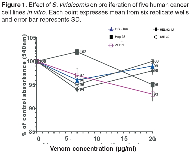
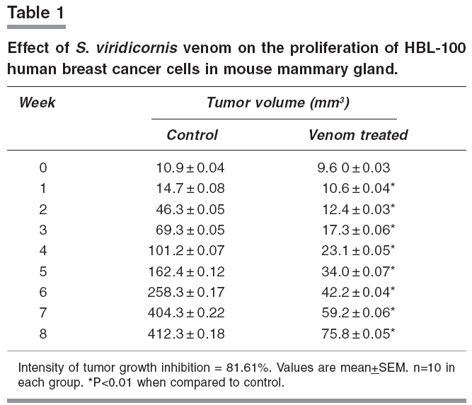
Indian Journal of Pharmacology, Vol. 38, No. 4, July-August, 2006, pp. 291-292 Research Letter Venom of a hill centipede Scolopendra viridicornis inhibits growth of human breast tumor in mice T. Bhagirath, B. Chingtham, Y. Mohen* Department of Life Sciences, Manipur University, Canchipur, Imphal – 795003. *Department of Pathology, Regional Institute of Medical Sciences, Lamphel, Imphal – 795001. E-mail: bhagirath_thounaojam@yahoo.com Code Number: ph06078 A hill centipede Scolopendra viridicornis (Arthropoda, Chilopoda, Scolopendridae) found in north-east India (Indo-Burma region) features extensively in tribal folk medicine. Dipping the insect in colorless local wine brewed from rice containing about 30% alcohol leads to oozing of the venom from the tips of the poison claws. Oral ingestion of the alcoholic solution is a popular treatment for various diseases proliferating among tribals who display symptoms like chronic headache, persistent uterine bleeding, chronic loss of appetite, blood in stool, blood and pus in urine, and white vaginal discharge. This treatment is now popularly used in Manipur for cancer patients as a last resort. Use of this venom in traditional medicine and the reports of antitumor activity of snake venom disintegrins[1] led us to examine similar activity of the venom. Turbidimetric test of the venom using human thrombocytes showed positive disintegrin activity (unpublished observation) and this further led us to examine anticancer properties of the venom. Since an antitumor agent may be cytotoxic[3] or noncytotoxic[7] in vitro MTT [3-(4,5-Dimethylthiazol-2-yl) -2,5-diphenyltetrazolium Bromide] assay to detect cytotoxic antitumor property and in vivo test using tumor mouse model to detect noncytotoxic antitumor property were used. The following cell lines were purchased from the National Centre for Cell Science, Pune University Campus, Pune: Hep 3B (ATCC NO. HB8064) - hepacellular carcinoma, HBL-100 (ICLC NO. HTL 00004)- breast myoepithelial tumor, IMR-32 (ATCC NO. CCL-127) - neuroblastoma, HEL 92.1.7 (ATCC NO. TIB 180) - leukemia, ACHN (ATCC NO. CRL - 1611) - renal cell adenocarcinoma. Inbred female Swiss albino virgin mice, 15 weeks old, 25-30 g in weight were used. Due to the involvement of surgical procedure in the experiment, the mice were kept in well maintained formalin fumigated cages and room. Scolopendra viridicornis was collected from the hills. The specimens were identified following the key to family Scolopendridae. The venom was obtained by electrical stimulation of 2000 live centipedes as described for scorpion milking.[2] MTT assay, was used for in vitro cytotoxicity test and was performed as per the method of Alley et al .[3] Cells were harvested from experimental-phase maintenance cultures. Four hundred cells were counted by trypan blue exclusion and dispensed within triplicate 96-well culture plates in 100 µl volumes for each venom concentration. The assay at each concentration was repeated twice. Following a 24 h incubation at 37°C, 5% CO 2 , 100% RH (Shellab 2123 CO 2 incubator) 100 ml each of culture medium, venom solution and venom carrier (PBS) were dispensed (carrier control group n=6, each drug group n=3). Peripheral wells of each plate (lacking cells) were utilized for venom blank (n=2) and medium/tetrazolium reagent blank (n=6) background determination. Venom concentrations tested were 1.75 µg and 5 µg in 250 µl of cell culture. Working venom solutions were prepared at 7 µg/ml and 20 µg/ml of PBS without calcium and magnesium. These venom concentrations were selected based on the concentrations of snake venom disintegrin that were tested for cancer cell cytotoxicity.[1] Culture plates were incubated for 6 days. On day 7, 50 µl of MTT working solution (1 mg/ml of PBS) was added to each well. After 4 h incubation at 37°C, culture plates were centrifuged at 2000 rpm for 5 min. Supernatant was removed from wells by slow aspiration through a blunt 18-gauze needle leaving about 20-30 µl in each well. Then 150 µl of DMSO (Sigma, St.Louis) was added to each well. After thorough mixing with a mechanical plate shaker, absorbance at 540 nm was measured with a Multiskan MS microwell plate reader. Cell growth inhibition was expressed in terms of percentage of control absorbance (±1 SD% ) following subtraction of mean background absorbance. For in vivo antitumor test in animals, approval of the University Ethics Committee was obtained and the guidelines in accordance with CPCSEA were followed. Among the five human cancer cell lines used here, breast tumor cell line HBL-100 was tumorigenic in mouse.[6] Human breast tumor model in mouse established with HBL-100 was, therefore, used for in vivo test of the venom. The heterologous tumor mouse model was established according to Price et al.[5] Mice (n=20) were anaesthetized by ether (BDH, Mumbai) in a closed chamber. A 1 cm longitudinal incision was made on the lateral side of the second nipple from the rostral side where the mammary fat pad beneath the skin was carefully exposed. HBL-100 cells (5 x 10[5]) suspended in 0.1 ml PBS (Glaxo, Mumbai) were injected into the mammary fat pad. The wound was then sutured. Palpable tumor masses appeared on day 16 after injection with tumor cells in 14 of the 20 mice and on day 30 in the remaining 6 mice. On the day palpable tumor mass appeared (zero week), mice were randomized into control and treatment groups (10 each). Daily infiltrative injections into the established tumor masses were carried out with 5 µg (maximum dose of cytotoxicity test) of venom in 0.1 ml of 0.15 M NaCl over a period of seven weeks. The control animals received corresponding volume of venom carrier (NaCl solution). Tumor mass in each animal was a single lump that remained inseparable into composites throughout the experimental period. Diameters of the tumor masses (sub-cutaneous) were measured weekly beginning from zero week with a caliper and tumor sizes were calculated from the formula: volume (mm 3 ) = (width) 2 x length x 0.5. Intensity of inhibition = (Mean control volume - mean treatment volume) X 100 ¸ Mean control volume. Significance of difference in tumor size between treatment and control was tested by student t-test. P< 0.05 was considered significant. The venom at 7 µg/ml and 20 µg/ml did not cause significant reduction in light absorbance of MTT reaction mixtures of all the cancer cell lines tested indicating that the venom did not specifically kill cancer cells leaving the normal cells unharmed. [Figure - 1] Daily injection of venom 5 µg into the tumors over a seven week period showed that mean growth of tumors was substantially lower than that of control. [P< 0.01; [Table - 1]] Mean growth of control tumors (n=10) was 37 times the initial volume, compared to venom treated tumors (n=10) which was only seven times the initial volume. Intensity of tumor growth inhibition by the venom was 81.61%. During venom treatment, the appearance of tumor area, bleeding or oozing of body fluid from the venom injection site, food intake were monitored daily, and body weights were measured weekly. No abnormality was observed. MTT assay originally developed for cell viability testing is now widely used in cancer cell cytotoxicity testing.[3] Negative results in MTT assay of S. viridicornis venom against five human cancer cell lines show that the venom is not cytotoxic to the cancer cell lines at the doses tested. Although a cancer cell cytotoxic agent may not be effective against certain cancer cell lines considering the number and diversity of the cell lines tested, the venom is unlikely to be cytotoxic to other cancer cell lines. Inspite of noncytotoxicity the venom substantially inhibited cancer progression in an in vivo model indicating that it interacts with cancer progression factors rather than acting directly on cancer cells. Because of limited supply of venom the test was conducted using only one tumor model albeit an antitumor testing protocol used by others as well.[4] As in the case of present study, use of tumor models established in immuno-competent mice for antitumor test has been reported.[4] Tumorigenicity of HBL-100 cell line in mouse has already been demonstrated.[6] Cytotoxic antitumor agents selectively kill cancer cells through various mechanisms and exhibit positive results in both the in vitro and the in vivo antitumor tests.[4] On the other hand noncytotoxic antitumor agents inhibit tumor progression through suppression of processes associated with tumorigenesis displaying positive results only in the in vivo antitumor test.[7] Considering these present and earlier observations, it is evincing that S. viridicornis venom has noncytotoxic antitumor property although further test should corroborate it. Acknowledgment We are grateful to the Department of Biotechnology, Government of India for financial support for this workReferences
Copyright 2006 - Indian Journal of Pharmacology The following images related to this document are available:Photo images[ph06078t1.jpg] [ph06078f1.jpg] |
| |||||||||
