 |
| About Bioline | All Journals | Testimonials | Membership | News |
|
||||||
|
||||||
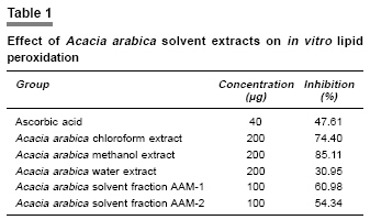
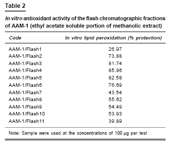
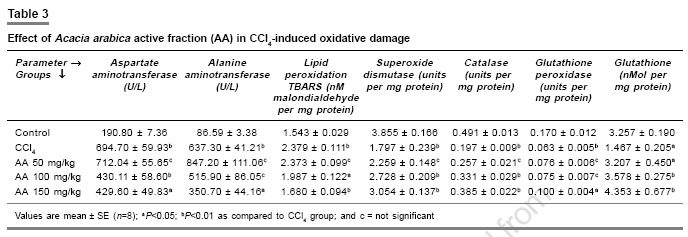
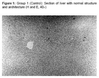
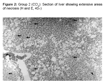
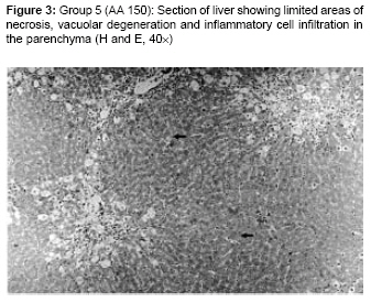
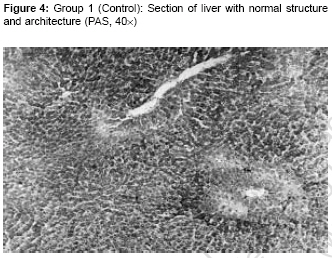
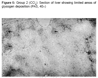
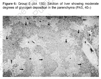
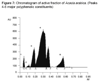
Indian Journal of Pharmacology, Vol. 39, No. 1, January-February, 2007, pp. 33-38 Research Paper Antioxidant activity of ethyl acetate soluble fraction of Acacia arabica bark in rats Sundaram R, Mitra SK R and D Center, The Himalaya Drug Company, Bangalore-562 123 Date of Submission: 21-Oct-2005 Code Number: ph07008 Abstract Objective: To study the antioxidant activity of various extracts and fractions of Acacia arabica by in vitro and in vivo experimental models.Materials and Methods: Various solvent extracts were prepared by Soxhlet extraction. Extract fractionations were done by solvent-solvent extraction and flash chromatographic separation. In vitro lipid peroxidation was carried out by tertiary butyl hydroperoxide -induced lipid peroxidation. The most active fractions were identified and standardized by thin layer chromatography (TLC). In vivo experiments on the most active fraction were carried out with 50, 100, and 150 mg/kg, p.o. doses, in carbon tetrachloride (CCl 4 )-induced hepatotoxicity, in rats. Various biochemical parameters like serum aspartate aminotransferase (AST), serum alanine aminotransferase (ALT), superoxide dismutase (SOD), catalase, glutathione peroxidase (GSH-Px), glutathione (GSH), and lipid peroxidation were estimated. Results: Flash chromatographic fractions 2-6 of ethyl acetate extract exhibited maximum activity with in vitro lipid peroxidation. In vivo evaluation of this active fraction (AA) in CCl4-induced hepatotoxicity for 19 days at a dose of 150 mg/kg offered marked liver protection, which was evident by significant changes in lipid peroxidation, glutathione, superoxide dismutase and catalase ( P <0.01). The treatment also showed significant changes in AST, ALT, and GSH-Px levels ( P <0.05). At lower doses, the protection was not consistent. Conclusion: The polyphenol rich active fraction of Acacia arabica is a potent free radical scavenger and hepatoprotective and protects TBH-induced lipid peroxidation and CCl 4 -induced hepatic damage. Keywords: Antioxidant, free radical scavenger, hepatoprotective, mimosaceae, plant extract. Introduction Free-radical reactions have been implicated in the pathology of many human diseases/disease conditions like atherosclerosis, ischemic heart disease, aging process, inflammation, diabetes, immunosupression, neurodegenerative diseases etc.[1],[2],[3] Radicals and other reactive oxygen species are formed constantly in the human body and are removed by the enzymic and non-enzymic antioxidant defense systems.[4] The disturbance in ′redox homeostasis′ occurring when antioxidant defenses are inadequate can damage lipids, proteins, carbohydrates, and DNA. Drugs with multiple protective mechanisms, including antioxidant activity, may be one way of minimizing tissue injury.[5] A number of plants and plant isolates have been reported to protect free radical-induced damage in various experimental models.[6] In the present study, Acacia arabica bark (Mimosaceae), an ingredient of various traditional preparations, used in the treatment of asthma, bronchitis, diabetes, dysentery, diarrhea and skin diseases[7] has been selected. The bark is reported to contain (+) catechin, (-) epicatechin, (+) dicatechin, quercetin, gallic acid, (+) leucocyanidin gallate, sucrose and (+) catechin-5-gallate.[8] The purpose of the present study was to evaluate, whether various extracts and fractions of Acacia arabica bark have antioxidant property against TBH-induced free radical damage and the most potent fraction on carbon tetrachloride (CCl 4 )-induced hepatic damage. Both the selected models are known to implicate free radicals. Tertiary butyl hydroperoxide (TBH) is a short-chain lipid peroxide analogue used to induce acute oxidative stress in rat hepatocytes and the in vivo rat liver. Two distinctive pathways are involved in the metabolism of TBH in hepatocytes. The first employs the microsomal cytochrome P-450 system leading to the production of reactive oxygen species (ROS) such as peroxyl and alkoxyl radicals that initiate lipid peroxidation while the second involves the conversion of GSH-Px to t -butanol and oxidized glutathione disulphide (GSSG). Glutathione disulphide is then reduced to GSH by GSH reductase, resulting in nicotinamide adenine dinucleotide phosphate (NADPH) oxidation. Decreased GSH and oxidized NADPH contribute to altered Ca 2+ homeostasis, which is considered a major event in TBH-induced toxicity.[9] CCl 4 induces liver damage by producing free-radical intermediates.[10] CCl 4 is converted to trichloromethyl radical (CCl 3 ) by the cytochrome P-450 system, which in turn is converted to peroxy radical (CCl 3 O 2 ), which causes the damage.[11] Materials and Methods Plant material Acacia arabica bark was collected from the outskirts of Bangalore and was identified by Dr. Kannan, Botanist of The Himalaya Drug Company, Bangalore. A specimen of the same was deposited in the herbarium of our R and D Center. Extraction The dried raw material was coarsely powdered by a pulverizer and packed in a 5-litre capacity Soxhlet (continuous extraction) apparatus. The temperature for chloroform, methanol, and water extraction were 60, 70, and 100oC, respectively. The extraction was monitored continuously and at each stage the completion was confirmed by color, optical density and thin layer chromatography. The extracts were then filtered through Whatman filter paper No. 1 and concentrated by distillation. The concentrated extracts (solid content about 50%) were taken in china dishes and evaporated in a boiling water bath to remove the solvents. The extracts were then weighed to calculate the yield. Solvent fractionation About 20 gram of methanolic extract of Acacia arabica was dispersed uniformly in 50% aqueous methanol. This mixture was taken in a separating funnel and extracted repeatedly with ethyl acetate till the ethyl acetate layer became colorless. The ethyl acetate layer was concentrated using a rotary evaporator. The concentrated extract was dried completely on a water bath to obtain the fraction AAM-1. Similarly, the residue left after ethyl acetate extraction was also dried to obtain the fraction AAM-2. Flash chromatography Flash chromatographic separation was carried out using ISCO Combiflash system consisting of glass columns packed with 25 gram of silica gel. The column was equilibrated with chloroform and about 2 gram of the extract was packed in the column. The column was eluted with the gradient of chloroform and methanol starting from 100 to 25% chloroform. The elutes were collected in test tubes at the rate of one tube per 30 sec using Foxy 200 fraction collector. A total of 260 elutes were collected. Ten microliters of each fraction was spotted on Silica gel precoated TLC plates (Merck) and developed in the solvent chloroform: methanol (9:1) or benzene: ethyl acetate: acetone (10:2:10). Fractions were prepared by mixing elute showing similar color, TLC pattern and/or intensity of the bands and were evaporated on a water bath. A total of 11 fractions were obtained and were evaluated in vitro . Lipid peroxidation of solvent extracts and fractions The extracts were prepared at the concentration of 1 mg/mL in 5% dimethyl sulphoxide (DMSO). Lipid peroxidation was carried out by the following method.[12] The liver of normal rat, after ether anesthesia, was perfused in vitro with 0.9% ice-cold sodium chloride and the tissue was homogenized at a concentration of 10% w/v in 0.15M of potassium chloride using a glass homogenizer. The homogenate was centrifuged at 800 g and the supernatant was used for the study. About 200 µL of various extracts or fractions (equivalent to 100-200 µg) was mixed with 1 mL of the homogenate and the lipid peroxidation was induced using 0.35% solution of TBH (200 µL) in DMSO. Ascorbic acid was used as a standard reference. The mixture was incubated at 37°C for 20 minutes and the reaction was stopped by adding 40 µL of 10% butylated hydroxy toluene reagent in alcohol. Then 2 mL 0.8% thiobarbituric acid reagent in water and 2.5 mL of 1% phosphoric acid were added. After heating at 80°C for 30 min, Thiobarbituric Acid Reactive Substances (TBARS) were extracted with 5 mL of butanol by gentle vortexing. The absorbance of butanol layer was measured Spectrophotometrically (Shimadzu UV-1700 pharmaspec) at 532 nm against butanol as blank. The samples were taken in duplicates and the percentage inhibition of lipid peroxidation was calculated with respect to the positive control. Lipid peroxidation of flash chromatographic fractions The lipid peroxidation was carried out for the flash chromatographic fractions by the same procedure as mentioned above except that the sample used was 100 µg. In vivo experiments Inbred Wistar male rats (220-250 gram) housed under standard conditions of temperature (22±2°C), relative humidity (30-60%), and 12 hour light/dark cycle. They were fed with standard pellet diet and water ad libitum . The Institutional Animal Ethics Committee approved the experimental protocol. All the animals received humane care according to the criteria outlined in the ′Guide for the Care and Use of Laboratory Animals′ prepared by the ′National Academy of Sciences,′ and published by the ′National Institutes of Health.′ Active fraction of Acacia arabica for in vivo study was prepared by combining Flash Chromatographic Fractions 2-6 and coded as AA. The content of polyphenols in this fraction was determined as gallic acid equivalents (GAE). The fraction was also standardised by TLC fingerprinting technique using the method described above under the section ′Flash chromatography.′ CCl 4 -induced hepatotoxicity in rats About 40 rats were divided into five groups of eight animals each. Group I served as a control. Group II rats served as CCl 4 control and groups III, IV, and V were administered with active fraction of Acacia arabica (AA) at doses of 50, 100, and 150 mg/kg b.wt. p.o. respectively for 19 days. On days 17 and 19, groups II-V received CCl 4 at the dose of 1 mL/kg b.wt. p.o. in liquid paraffin (1:1). After 24 hours of the second dose of CCl 4 the rats were killed by decapitation, since many of the biochemical and histological changes are known to manifest after 24 hours of CCl 4 administration.[13] Blood was collected and serum was separated for estimation of alanine aminotransferase (ALT)[14] and aspartate aminotransferase (AST).[14] The liver was dissected out, immediately washed in ice-cold saline and a 5% homogenate prepared in 0.15m potassium chloride. The homogenate was used for the assay of lipid peroxidation, estimated in terms of malondialdehyde or thiobarbituric acid reactive substances.[15] A portion of liver was homogenized in Tris-HCl sucrose buffer for the estimation of antioxidant enzymes. Superoxide dismutase was estimated by the method of Kono[16] and the result was expressed as units of SOD activity per mg protein. Catalase was estimated by the method of Beers and Sizer[17] and the results were expressed as units of catalase activity per mg protein. Glutathione was estimated by the method of Ellman[18] and the GSH content was expressed as mmole/mg protein. Glutathione peroxidase was estimated by the method of Paglia and Valentine[19] and expressed as units per mg of protein. Protein was estimated by using folin phenol reagent.[20] A portion of the liver was preserved in 10% neutral buffered formalin for histopathological evaluation. Histopathological evaluation was done by Hematoxylin and Eosin (H and E) staining as well as Periodate-Schiff (PAS) staining. Statistical analysis All the values were expressed as mean ± SEM. Statistical analysis was carried out using one-way anova followed by Dunnett′s multiple comparison post-hoc test. The minimum level of significance was fixed at 95% confidence limit. Graphpad Prism (Version 4.0) software package was used for statistical analysis. Results In vitro lipid peroxidation The effect of Acacia arabica solvent extracts and ascorbic acid on TBH-induced lipid peroxidation is summarized in [Table - 1]. As the methanolic extract showed maximum activity, it was taken for solvent fractionation. The ethyl acetate soluble portion of methanolic extract (AAM-1) showed better protection than the residue (AAM-2). The fraction AAM-1 was subjected for flash chromatographic separation and the results are presented in [Table - 2]. The fractions AAM-1/Flash 2 (the code given to the fraction) to AAM-1/Flash 6 showed maximum activity in in-vitro lipid peroxidation assay [Table - 2]. CCl 4 -induced hepatotoxicity Serum ALT, AST and liver lipid peroxidation were significantly elevated in CCl 4 -intoxiciated rats compared to the control group [Table - 3]. These parameters were restored to normalcy following administration of active fraction AA. AST was significant ( P < 0.01, P < 0.05) at 100 and 150 mg/kg, while ALT was significant ( P < 0.05) only at 150 mg/kg. CCl 4 treatment resulted in a significant increase in malondialdehyde or TBARS. Treatment with AA significantly prevented the increase in TBARS (100 mg/kg [ P < 0.05] and 150 mg/kg [ P < 0.01]) and significantly ( P < 0.01) ameliorated the CCl 4 -induced changes in SOD and catalase. However, GSH-Px level increased significantly with the treatment of AA only with 150 mg/kg dose ( P < 0.05). Carbon tetrachloride intoxication resulted in a significant depletion of GSH. AA treatment (50, 100 and 150 mg/kg) significantly elevated the GSH levels. Histopathological evaluation In CCl 4 -treated group, liver showed extensive areas of necrosis, vacuolar degeneration, and inflammatory cell infiltration. Treatment with AA (100 and 150 mg/kg) caused reversal of these changes [Figure - 1],[Figure - 2],[Figure - 3]. PAS stained sections of CCl 4 -treated group showed severe glycogen depletion. A reversal trend was observed with the treatment of AA (100 and 150 mg/kg [Figure - 4],[Figure - 5],). [Figure - 6]Discussion Acacia Arabica bark is used traditionally in disorders implicated with oxidative stress and free radical damage, like diabetes.[7] In the present study, activity guided fractionation was adapted to identify the active fraction of this drug, responsible for the antioxidant activity. A three-step fractionation, namely solvent extraction, solvent-solvent fractionation, and flash chromatography of Acacia Arabica bark, was undertaken. After each fractionation, the antioxidant potential was studied by in vitro lipid peroxidation. In vitro lipid peroxidation assay showed that the methanolic extract was more potent, compared to the other two (aqueous and chloroform) extracts. The methanolic extract was subjected to solvent fractionation using ethyl acetate. The ethyl acetate soluble portion (AAM-1) was found to be more potent when compared to the residue (AAM-2). After this step, AAM-1 was subjected to flash chromatography. The fractions obtained by flash chromatographic separation were evaluated for activity. It was observed that the fractions 2-6 had maximum activity. The major chemical constituents of these fractions were found to be identical by TLC fingerprinting technique [Figure - 7]. Hence, the fractions 2-6 were combined as active fraction of Acacia arabica and coded as AA. The fraction AA was found to be rich in polyphenols by various chemical tests and the total polyphenol was about 40% in gallic acid equivalents (GAE).[9] This active fraction was evaluated for antioxidant activity by CCl 4 -induced oxidative damage. Extensive evidence in rodent models, using structurally and mechanistically diverse hepatotoxicants such as acetaminophen (APAP), carbon tetrachloride (CCl 4 ), chloroform (CHCl 3 ), thioacetamide (TA), trichloroethylene (TCE) and allyl alcohol (AA), it has been demonstrated, that tissue repair plays a critical role in determining the final outcome of toxicity.[21] However multiple doses of CCl 4 ensures complete hepatic damage. Significant changes in the biochemical parameters in selected dose level of CCl 4 control reflect that the possibility of dynamic compensatory cell proliferation and tissue repair is very remote. The hepatic injury induced by CCl 4 results in an increase in serum AST and ALT levels due to the leakage of cellular enzymes into circulation. The decrease in the elevation of serum enzymes following treatment with AA may be due to the reduction in cell membrane disturbances. Superoxide dismutase, the enzyme responsible for dismutation of superoxide radicals, is significantly depleted with CCl 4 treatment. Treatment with AA significantly increased the superoxide dismutase levels. Catalase, the enzyme responsible for detoxification of hydrogen peroxide was also significantly increased with AA treatment. Glutathione is responsible for maintaining the intracellular redox milieu, replenishing a number of antioxidant compounds like vitamin E and C. Glutathione is the substrate for H 2 O 2 catalyzing enzymes like GSH-Px and glutathione-S-transferase. The significant increase in GSH and GSH-Px levels with the AA treatment suggests that it has direct free-radical scavenging property and also strengthens the enzymatic antioxidant defense system. A single dose of CCl 4 can lead to centrizonal necrosis and steatosis.[22] The histological changes observed after CCl 4 administration include necrosis, inflammatory cell infiltration, vacuolar degeneration and glycogen depletion. Administration of AA prevented these histological changes in liver in a dose dependent manner. The antioxidant property of polyphenolic compounds is well documented.[23] Polyphenolic compounds exert antioxidant property by free radical scavenging and metal chelating properties. Many phenolic antioxidants that are widely distributed in plant-based diets have shown free radical scavenging property when assessed by the Trolox equivalent antioxidant capacity (TEAC), the ferric reducing antioxidant power (FRAP), the hypochlorite scavenging capacity, the deoxyribose method and the copper-phenanthroline- dependent DNA oxidation assays.[24] The polyphenolic constituents have ideal structural chemistry for free radical-scavenging activities and have been shown to be more effective antioxidants in vitro than vitamin E and C.[25] The active fraction was found to contain about 40% of total polyphenols indicating that they could be responsible for the pharmacological activity. Other possible mechanisms for the amelioration of CCl 4 -induced damage by AA may include, altered toxicokinetics of CCl 4 ; like inhibition of the metabolizing enzyme CYP2E1, diminished bio-activation of CCl 4 through interference with binding sites in liver, alteration of kinetic profile (lowering plasma t 1/2 , increased volume of distribution and increased plasma clearance). Though there are reports of CYP2E1 inhibition by polyphenols,[26] the other possibilities need further investigations. Identification of the exact chemical nature of these constituents would further enlighten the mechanism of action. With these findings, it may be concluded that the ethyl acetate fraction of Acacia arabica bark is a free radical scavenger and hepatoprotective. Acknowledgement The authors acknowledge Dr. B.G. Madhumathi, Histopathologist, R and D Center, The Himalaya Drug Company, Bangalore, towards her contribution for this study. References
Copyright 2007 - Indian Journal of Pharmacology The following images related to this document are available:Photo images[ph07008f4.jpg] [ph07008t3.jpg] [ph07008f1.jpg] [ph07008f2.jpg] [ph07008f3.jpg] [ph07008t2.jpg] [ph07008f5.jpg] [ph07008t1.jpg] [ph07008f7.jpg] [ph07008f6.jpg] |
| |||||||||
