 |
| About Bioline | All Journals | Testimonials | Membership | News |
|
||||||
|
||||||
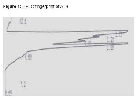
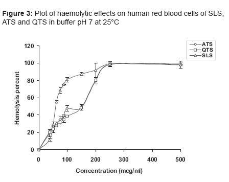
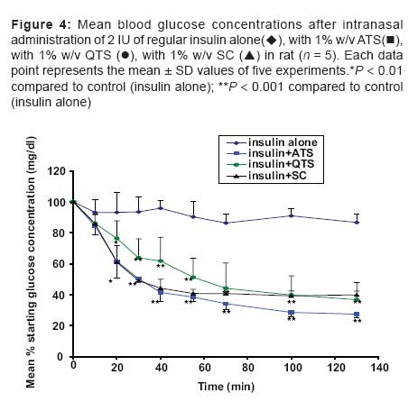
Indian Journal of Pharmacology, Vol. 39, No. 5, September-October, 2007, pp. 226-230 Research Paper Isolation, characterization and study of enhancing effects on nasal absorption of insulin in rat of the total saponin from Acanthophyllum squarrosum S.A. Sajadi Tabassi, H. Hosseinzadeh*, M. Ramezani*, E. Moghimipour*, S.A. Mohajeri* Pharmacological Research Center of Medicinal Plants and School of Pharmacy Correspondence Address:Pharmacological Research Center of Medicinal Plants and School of Pharmacy, Mashhad University of Medical Sciences, P O Box 1365-91775, Mashhad Date of Submission: 18-Jan-2005 Code Number: ph07057 Abstract Objective: Isolation of the total saponins from Acanthophyllum squarrosum Boiss. and investigation of its surface activity, haemolytic effects on human erythrocytes, as well as enhancing potentials on intranasal insulin absorption in rat as compared to two other enhancers, i.e, Quillaja total saponin (QTS) and sodium cholate (SC). Keywords: Absorption enhancers, acanthophyllum, insulin, saponin Saponins are a ubiquitous group of amorphous glycosides that are widely distributed in the higher plants. They dissolve in water to form colloidal solutions that foam upon shaking and can be distinguished from other glycosides by their surface active properties. [1] Most saponins have shown haemolytic activity and are toxic to most cold-blooded animals. [2] In view of the vital role of insulin and the daily need for injections (the only available dosage form of insulin) in type I diabetes patients, there is pressing need to identify other possible administration routes, e.g., the intranasal pathway; however, an answer to this problem has eluded pharmaceutical researchers over the recent decades. [3] Studies which have been carried out on nasal drug delivery for systemic purposes in recent years have shown that the nasal route can be exploited for the systemic delivery of drugs such as small molecular weight polar drugs as well as peptides and proteins. [4] Small lipophilic molecules are generally well absorbed through the nasal mucosa. [4] However, the same is not applicable for high molecular weight polypeptides such as insulin. To overcome the problem of poor nasal absorption of polypeptides and proteins, different strategies have been adopted, including the use of chemical absorption enhancers. [5],[6] Various substances have been studied for use as absorption enhancers of insulin through nasal mucosa, including bile salts; [7] polyoxyethylene-9-lauryl ether [laureth-9]; [3],[8] anionic surfactants, e.g, sodium lauryl sulfate (SLS) and potassium lauryl sulfate; [9] l-α-lysophosphatidyl choline, [10] chitosan [11],[12] and Quillaja saponins. [13] Pillion and coworkers studied the enhancing effects of Quillaja total saponin (QTS) and its derivatives on nasal and ocular delivery of insulin. [13] However, no report was found in the literature regarding the absorption-enhancing effects of Acanthophyllum total saponin (ATS). The aim of the present study was to isolate and characterize ATS and investigate its enhancing effects on intranasal insulin absorption in comparison with two other enhancers, i.e., QTS and SC. Materials and Methods Chemicals Chemicals and their suppliers were as follows: QTS (Sigma, USA); sodium cholate (SC) (Merck, Germany); carboxy methyl cellulose (CMC) (Alborz Co. Iran); glucometer strips (Apex Biotechnology Corp. USA); regular insulin solution 100 IU/ml (NOVO, Denmark); heparin 5000 IU/ml (B. Braun Medicals S.A, Germany). All chemicals were of analytical grade and used as received. Extraction of saponin ATS was extracted from the roots of Acanthophyllum squarrusom using the method described earlier. [14] Briefly, the roots were collected from around the city of Tabas, north of Yazd Province, Iran and identified by Fersdowsi University of Mashhad. Voucher samples were preserved for reference in the herbarium of the Department of Pharmacognosy, School of Pharmacy, Mashhad (Voucher no. 003-0119-1). The roots of the plant were dried, powdered and defatted with petroleum ether in a Soxhlet. The air-dried powder was extracted with methanol and after evaporation yielded a syrupy brown residue. It was then extracted with water-saturated n -butanol. The residue was dissolved in the least amount of methanol and precipitated by the addition of five volumes of diethyl ether. After filtration, the residue was dissolved again in methanol and precipitated by adding diethyl ether twice. Finally, the total saponin was freeze-dried (Freeze Dryer 3, LABCONCO) and stored in an amber, air tight glass container at room temperature for further studies. Characterization of saponin High performance liquid chromatography (HPLC) : HPLC fingerprint of the isolated ATS was taken (Waters 600E, Waters Co, UK) with a Licrosphere C 18 column (5µ , 125 x 4 mm). The samples were eluted with water: methanol: methyl cyanide at a flow rate of 1.0 ml/min and detected by a UV detector at the wavelength of 210 nm. Surface activity and critical micelle concentration (cmc) : Surface activity of the aqueous solutions of saponin was determined using Wilhelmy plate apparatus (Model K 12 , Kruss Processor Tensiometer). Approximately, 20 ml of saponin solution was placed in a clean beaker in the instrument chamber. After temperature equilibrium was attained, a vertical plate of platinum-iridium, attached to a balance, was immersed in the solution and the force due to wetting was determined by the instrument. Surface tension of a series of concentrations of saponin was measured and the concentration at which an abrupt change in the surface tension took place was regarded as the cmc. Hemolytic effects of ATS : Preparation of red blood cell (RBC) suspension: Approximately 10 ml of blood was taken from a healthy volunteer into a heparinized tube and put in ice. The plasma and buffy coat were removed by centrifuging blood samples at 2200 g (Hermle Z 230A) for 10 min and the erythrocytes were washed three times with at least five times their volume of McIlvaine′s buffer, pH 7.0. The erythrocytes were adjusted to approximately 12% haematocrit by resuspending them in the same buffer to 3.33 times their original weight. The erythrocyte suspension was stored on ice at 4° C and was used within 48 h of collection. Haemolysis experiments : 200 µl of RBC suspension was incubated for 30 min with an equal volume of the saponin solution, prepared in McIlvaine′s buffer, at 37° C. After incubation, the mixtures were spun in a microcentrifuge (Hettich 2415) at 3200 g for 15 s and 200 μl of the resulting supernatant was added to 3 ml Drabkin′s reagent to assay for the amount of haemoglobin released. Positive controls consisted of 200 μl samples taken from uncentrifuged mixtures of erythrocyte suspensions (200 μl) and buffer (200 μl), which were added to 3 ml Drabkin′s reagent to obtain complete hemolysis. A negative control was also included to assess the levels of spontaneous hemolysis; it comprised 200 μl buffer mixed with 200 μl erythrocyte suspension. The absorbance at 540 nm of all test and control samples was determined spectrophotometrically using a UV-visible spectrophotometer (Shimadzu 160-A, Japan) and the values, expressed as percentage hemolysis, were calculated by comparing the absorbance of test samples with that of positive control. Haemolysis caused by negative controls was always less than 1%. In vivo animal studies Preparation of control and test solutions : All solutions were prepared freshly on the day of experiment and refrigerated until use. Control solutions were prepared by dissolving 40 mg carboxymethylcellulose (CMC) in 2 ml PBS, pH 7.0 (1% w/v final concentration) by gentle heating and adding 2 ml regular insulin (100 IU/ml) after it cooled. Test solutions were made by the addition of 40 mg each of ATS, QTS and SC to 2 ml of the cooled CMC solution and mixing with 2 ml regular insulin to obtain 1% w/v enhancer concentration and 50 IU/ml of insulin in the final solution. Animal experiments : Male Wistar rats weighing 240-300 gm were bred in the animal house of the Department of Pharmacology, School of Pharmacy of Mashhad. The rats were treated surgically according to the method described earlier [11] with slight modifications. After anesthesia with intraperitoneal injection of urethane (1.3 g/kg), an incision was made at the neck of the rat to expose the trachea. A polyethylene tube was inserted about 1.5 cm into the trachea to maintain respiration during the experiment. Body temperature was fixed at 37 ± 0.5°C. The esophagus was also cannulated with another similar polyethylene tube, which was closed at the end with an adhesive agent and inserted toward the posterior nasal cavity. The nasopalatine was sealed with an adhesive agent to prevent drainage of the drug solution from the nasal cavity into the mouth. The left carotid and right external jugular veins were cannulated for blood sampling and fluid replacement, respectively. Insulin absorption studies : Groups of five animals were used for the nasal administration of control and test solutions. The animals were manually restrained in a supine position while the solution was instilled into the nostril. Test solutions were always administered through the rat′s right nostril and the nostril was then closed immediately with an adhesive agent. At time points 0 and 5 min, 20 μl of insulin solution containing 1 IU insulin was instilled into the rat′s nostril. Thus, each rat received 2 IU of insulin. The mean blood glucose concentration of three samplings at 5 min intervals before drug administration was measured and taken as the basal blood glucose concentration. Blood glucose concentration was determined using a glucometer (Sensorex, Metertech Inc, USA) with special glucometer strips (Apex Biotechnology Corp.) and the value was taken as an indicator of the systemic absorption of insulin via the nose. Blood samples were withdrawn from the jugular vein and replaced by the same volume of normal saline. Samples were taken at time points 10, 20, 30, 40, 55, 70, 100 and 130 min after drug administration and blood glucose concentrations were determined as described above. The blood glucose level was reported as a percentage of basal blood glucose concentration for each sample. Statistical analysis Analysis of data was done using the Minitab software. The data were expressed as mean values ± SD and tested for significance by one-way analysis of variance (ANOVA) followed by the multiple comparison test of Tukey-Kramer. P < 0.05 was regarded as indicating statistical significance. Results HPLC analysis Figure 1 shows the HPLC fingerprint of the total saponin isolated from A. squarrosum . Surface tension studies : According to the data obtained in this study, the average surface tension of pure water at room temperature (21° C) was found to be 72.62 ± 0.67 mN/m. Critical micelle concentration (cmc) was taken from the region where the surface tension vs concentration curve reached a plateau and it was found to be 120, 50 and 160 µg/ml for SLS, ATS and QTS, respectively [Figure 2]. Hemolysis studies : Figure 3 shows the haemolysis vs concentration profiles for SLS, ATS and QTS. In general, the maximum hemolytic effect occurs at concentrations above cmc. There was a sharp increase in hemolysis with increasing concentrations of the three compounds. As Figure 3 indicates, among the compounds tested, SLS showed the maximum efficacy in terms of disruption of red blood cell membranes. The hemolysis profiles of ATS and QTS are almost the same and both of them caused complete hemolysis at a concentration of about 250 μg/ml. Nasal absorption studies In this study, nasal insulin absorption in the presence of three absorption enhancers was investigated by determination of the decrease in blood glucose levels in rat. As Figure 4 shows, ATS at a concentration of 1% w/v decreased blood glucose significantly ( P < 0.01 or P < 0.001) as compared to the control at all time points after nasal administration of insulin except at the time point of 10 min. Figure 4 also shows the effect of a 1% w/v concentration of both QTS and SC on nasal absorption of insulin. It is evident that both compounds decreased blood glucose significantly as compared to the control. At 130 min post-instillation of insulin in the presence of the enhancers blood glucose was found to be 27.46%, 36.79% and 39.95% that of basal values for ATS, QTS and SC, respectively. Although the effect of ATS on blood glucose and insulin absorption was found to be more pronounced than that of the other two enhancers Figure 4, the difference was not statistically significant ( P > 0.05). Discussion Surface tension studies showed that ATS was able to lower the surface tension of water in a concentration-dependent manner. SLS, which is an ionic surfactant, induced a sharp decrease in surface tension. QTS was also shown to decrease the surface tension of water in a similar manner, though it was less effective than SLS. In a study carried out by Shuman and coworkers [15] on the micellar properties of Quillaja saponins, the cmc of saponins was reported as 130-510 µg/ml, depending on the purity and source of the saponin. [15] Our study showed the cmc of ATS as being around 80-120 µg/ml, which is in logical agreement with the above results if we take into account the difference in the plant species and also the degree of purity of ATS as shown in the HPLC chromatogram. Saponins have the ability to rupture erythrocytes and this has led to the development of the hemolytic assays for detecting the presence of saponins in plant extracts. [16] Pillion et al. , found that saponins isolated from Quillaja saponaria bark hemolysed sheep red blood cells at fairly low concentrations. [13] As the results of the present study show, the hemolysis profile of the ATS and QTS were almost identical and complete hemolysis was observed for both compounds at the concentration of 250 µg/ml. Since this concentration is above the CMC of ATS, membrane disruption could be attributed to the formation of mixed micelles and extraction of membrane components by saponin micelles. In vivo animal studies demonstrated that intranasal administration of solutions containing insulin with QTS, SC and ATS significantly ( P < 0.05) reduced blood glucose compared to the basal values in rat. This indicates that the tested compounds possess an enhancing effect on intranasal absorption of insulin. The extent of the effect of these three enhancers was not found to be different statistically ( P > 0.05). QTS and cholates are known absorption enhancers and have been studied by other researchers as well. Chandler et al. , studied the effect of sodium taurodihydrofusidate (STDHF; a bile derivative), L-α lysophosphatidyl choline (LPC), polyoxyethylene-9-lauryl-ether (laureth 9) and diethylamine ethyl-dextran (DEAE-dextran) on nasal absorption of insulin in rat. [17] They found that all these enhancers increased insulin absorption with their ranking order being as follows: Laureth 9 > LPC = STDHF > DEAE-dextran ≥ no enhancer. [17] The effect of sodium deoxycholate (SDC) on intranasal absorption of insulin was studied and the results showed that when insulin (0.5 IU/kg) in saline (pH 7.4) was administered without any absorption enhancer, no change in blood glucose level and insulin concentration was observed; however, blood glucose reduced in 10-20 min when 1% SDC was added to the insulin solution. [18] The potency of bile salts in enhancing nasal absorption of insulin in human volunteers was ranked as deoxycholate > chenodeoxycholate > cholate > ursodeoxycholate. [7] It has been reported that bile salts such as cholate, glycocholate and taurocholate produce smaller effects than nonionic, anionic and nonionic ester type surfactants on both hemolytic activity and protein-releasing effect, though the absorption-enhancing effects were significant and almost the same as those of ether type surfactants, sodium lauryl sulfate and saponin. [9] It was also found that bile salts such as sodium glycocholate (SGC) were less irritant to the nasal mucosa than polyoxyethylene 9-lauryl ether. [9] In a study on oral insulin delivery, Hosny and coworkers showed that sodium cholate (SC) induced a decrease in the blood glucose level following its oral administration along with insulin to hyperglycemic rabbits. [19] Plant saponins have been examined for their absorption-enhancing effects in a number of studies. Pillion et al. , studied naturally occurring QTS and their derivatives for their ability to stimulate insulin delivery from nose and eye drops. [13] This group of researchers instilled 2 IU insulin in the presence of different concentrations of QTS and derivatives into the nose/eye of rat and assessed insulin absorption by the hypoglycemic response of the animal. [13] They found that all the tested saponins and derivatives showed an enhancing effect on insulin absorption and reduced blood glucose levels in rat, though there were differences in the extent of their effects. [13] However, the enhancing effect of ATS on the absorption of insulin has not been reported in the literature. In this research, all three solutions containing absorption enhancers significantly reduced blood glucose in rat as compared to the control solution at almost all time points post-insulin administration. ATS (1% w/v) reduced blood glucose to 84.3% (±5.7%) 10 min after instillation, which was not significantly different from that of control ( P > 0.05) [Figure - 4]. From the time point 20 min onwards, however, the reduction in blood glucose was statistically different ( P < 0.01) from that caused by control. At the time point of 130 min blood glucose was reduced to 27.4% (±2.4%), which indicates a considerable effect of ATS on intranasal insulin absorption. The efficacy of ATS in promoting insulin absorption was found to be slightly greater than those of QTS and SC, but the differences were not significant at any of the time points ( P > 0.05). In conclusion, this study demonstrated that ATS extracted from the root of Acanthophyllum squarrosum was able to promote insulin absorption via the nasal route, with a promoting power equal to or slightly greater than (although not significant) that of two known enhancers. The mechanism of action whereby ATS and other saponins enhance the systemic absorption of insulin is not known clearly. Similarly, the precise mechanism of action of a variety of other enhancers, including bile salts and their derivatives, remain unidentified at present. Pillion et al . ascribed the enhancing effects of Quillaja saponins to the formation of mixed micelles with insulin that facilitates its transepithelial absorption. [13] Since the concentration of ATS used in this study was above its cmc, the above mentioned mechanism could be attributed to ATS as well. However, the effect of saponins on tight junctions and the promotion of insulin absorption through the paracellular pathway might be another possible mechanism. Acknowledgment This research project has been supported financially by Grant No. NRCI 6640 of National Research Projects and was done with the support of the National Research Council of the Islamic Republic of Iran. References
Copyright 2007 - Indian Journal of Pharmacology The following images related to this document are available:Photo images[ph07057f2.jpg] [ph07057f1.jpg] [ph07057f4.jpg] [ph07057f3.jpg] |
| |||||||||

![[Figure 2]](/showimage?ph/photo/ph07057f2.jpg)