
|
Indian Journal of Pharmacology
Medknow Publications on behalf of Indian Pharmacological Society
ISSN: 0253-7613 EISSN: 1998-3751
Vol. 41, Num. 2, 2009, pp. 64-67
|
Indian Journal of Pharmacology, Vol. 41, No. 2, March-April, 2009, pp. 64-67
Research Article
Antioxidant potential of aqueous extract of Phyllanthus amarus in rats
Karuna R, Reddy SSreenivasa, Baskar R, Saralakumari D
Department of Biochemistry, Sri Krishnadevaraya University, Anantapur - 515 003, Andhra Pradesh
Correspondence Address:Department of Biochemistry, Sri Krishnadevaraya University, Anantapur - 515 003, Andhra Pradesh
skumari1@yahoo.co.in
Date of Submission: 12-Jul-2008
Date of Decision: 29-Sep-2008
Date of Acceptance: 30-Mar-2009
Code Number: ph09019
DOI: 10.4103/0253-7613.51342
Abstract Objective: Increased levels of oxidative stress may be implicated in the etiology of many pathological conditions. Protective antioxidant action imparted by many plant extracts and plant products make them promising therapeutic drugs for free radical induced pathologies. In this study we assessed the antioxidant potential of Phyllanthus amarus (Euphorbiaceae).
Materials and Methods: Experimental rats were divided into two groups: Control and Phyllanthus amarus (P. amarus) treated. Treated rats received P. amarus aqueous extract (PAAEt) at a dose of 200 mg/kg body wt/day for 8 weeks. After the treatment period of 8 weeks lipid peroxidation (LPO), vitamin C, uric acid and reduced glutathione (GSH) were estimated in plasma and antioxidant enzymes: Glutathione peroxidase (GPx), catalase (CAT) and superoxide dismutase (SOD) were also assayed. Genotoxicity of PAAEt was assessed by single cell gel electrophoresis (SCGE) of lymphocytes under both in vitro and in vivo conditions. The protective role of PAAEt against hydrogen peroxide (H 2 O 2 ), streptozotocin (STZ) and nitric oxide generating system induced lymphocyte DNA damage was also assessed by SCGE.
Results: PAAEt treated rats showed a significant decrease in plasma LPO and a significant increase in plasma vitamin C, uric acid, GSH levels and GPx, CAT and SOD activities. SCGE experiment reveals that PAAEt was devoid of genotoxicity and had a significant protective effect against H 2 O 2 , STZ and nitric oxide (NO) induced lymphocyte DNA damage.
Conclusion: The results suggest the non-toxic nature of PAAEt and consumption of PAAEt can be linked to improved antioxidant status and reduction in the risk of oxidative stress.
Keywords: Antioxidants, lymphocytes, Phyllanthus amarus, single cell gel electrophoresis SCGE
Introduction
Oxidative stress occurs in a cellular system when the production of reactive oxygen species (ROS) exceeds the antioxidant capacity of the system. Oxidative stress plays an important contributory role in the process of aging and pathogenesis of numerous diseases like diabetes, cancer, neurodegenerative diseases, and respiratory tract disorders.[1] Improved antioxidant status helps to minimize the oxidative damage, and thus can delay or decrease the risk for developing many chronic age related, free radical induced diseases.
Phyllanthus amarus (Euphorbiaceae) can be found in all tropical regions of the world including southern India and China. In many countries around the world plants in the genus Phyllanthus are used in folk remedies; therefore this genus is of great importance in traditional medicine. P. amarus has been claimed to be an excellent remedy for infective hepatitis. [2] It was reported to have anti-plasmodial, [3] antiviral, [4] anti-bacterial [5] and anti-diarrheal [6] properties. Extract of P. amarus exhibited protective action against CCl 4 induced mitochondrial dysfunction [7] and hepatoprotective potential against ethanol, [8] alloxan [9] and cyclophosphamide-induced oxidative stress in rats. [10] P. amarus extract also possessed anti-tumor, anti-carcinogenic [11] and anti-inflammatory properties. [12] Methanol extract of P . amarus was reported to have hypoglycemic effect on alloxan induced diabetes mellitus. [9]
The generation and subsequent involvement of free radicals in a large number of diseases prompted us to study the antioxidant potential of PAAEt. In order to consider any substrate or plant extract as an effective antioxidant, it should act as antioxidant under both in vivo and in vitro conditions by decreasing the levels of oxidative damage to biomolecules, and it should render lymphocytes more resistant to oxidative challenges. Therefore, the present study was designed to explore the effect of PAAEt treatment on plasma antioxidant status and to evaluate the antigenotoxic potential of PAAEt against STZ, NO and H 2 O 2 induced DNA damage in lymphocytes.
Materials and Methods
Chemicals
The chemicals used in the current study were procured from Sigma Chemical Co. (St. Louis, MO, USA), Koch-Light Laboratories (Huntingdon, Cambridgeshire, England), and SISCO Research Laboratories (Maharashtra, India). An aqueous extract of P. amarus (dry powder) was purchased from Chemiloids (manufacturers and exporters of herbal extracts), Vijayawada, Andhra Pradesh (India). Herb-to-product ratio is 10:1. The phytochemical screening of the PAAEt was carried out by the modified method of Das and Bhattacharjee. [13] The extract was dissolved in distilled water prior to use.
Animals
Two- to three-month-old male albino Wistar rats weighing 130-150gm, procured from Sri Venkateswara Enterprises (Bangalore, India), were acclimatized for 7 days to the animal house, and maintained at standard conditions of temperature and relative humidity, with a 12h light/12h dark cycle. Water and standard pellet diet were provided ad libitum. The current work was carried out after approval by our institutional animal ethical committee (Regd. no. 470/01/a/CPCSEA, dt. 24 th Aug 2001).
Experimental design
In this experiment, a total of 16 rats were used. The rats were divided into two groups (control and P. amarus treated) of 8 animals each. P. amarus treated animals received ~2ml of PAAEt (200mg/kg body weight) orally through gastric tube daily. The dose of PAAEt in the current study is based on preliminary experiments on anti-hyperglycemic effect in alloxan-induced diabetic rats. After 8 weeks of treatment, 1-2ml of blood was collected from rats by means of a capillary tube through orbital sinus into heparinized Eppendorf tubes. Plasma was separated and used for biochemical analysis.
Biochemical analysis
Glucose was estimated by glucose oxidase peroxidase method using the Span Diagnostic kit. Vitamin C was estimated by the method of Omaye et al. [14] and uric acid was estimated by enzymatic method using a Liquid GOLD kit. Reduced glutathione was estimated by Ellmans method. [15] The extent of lipid peroxidation was determined by the method of Utley et al . [16]
Enzyme assays
Plasma GPx (EC 1.11.1.9), CAT (EC 1.11.1.6) and SOD (1.15.1.1) were assayed by following the methods of Rotruck et al., [17] Beers et al. [18] and Soon and Tan [19] respectively. Protein was estimated by the method of Lowery et al. , using bovine serum albumin as standard. [20]
Single cell gel electrophoresis
Experiments were performed on lymphocytes isolated from the blood of rats using histopaque-1077 technique, according to established methods. [21] After separation, lymphocytes were suspended in RPMI 1640 medium and counted over a hemocytometer and adjusted to nearly 5 x 10 5cells/ml of medium. The basic protocol for the comet assay was applied according to the modified method of Singh et al . [22] The modification includes a silver staining [23] of comets instead of ethidium bromide. The slides were coded for each sample and cells were screened on each slide under a standard transmission binocular microscope and DNA migration length was measured using an ocular micrometer.
In vitro genotoxicity studies of PAAEt
Lymphocytes isolated from the blood of normal rats were incubated with PAAEt in RPMI medium (0.2, 0.4 and 0.6mg/ ml medium) at 37°C in CO 2 humidified incubator for 1 h. After incubation the cells were centrifuged at 3000 rpm for 5 min to pellet the lymphocytes. The pellet was re-suspended in RPMI medium and the lymphocytes were subjected to SCGE.
Effect of plant extract against in vitro oxidative stress caused by nitric oxide generating system/ STZ
The nitric oxide generating system consists of a freshly prepared mixture of 10 mM Tris-HCl buffer pH 7.4 prepared in saline and 30mM sodium nitroprusside in 1.5:1 ratio. In vitro PAAEt pretreated (0.2mg/ml RPMI medium) lymphocytes from normal rats were exposed to nitric oxide generating system (100µl/1ml medium) or 10µl of freshly prepared STZ in PBS (to get a final concentration 80µg/ml medium) for 25 min and centrifuged at 3000 rpm for 5 min to pellet the lymphocytes and then the lymphocytes were subjected to SCGE.
In vivo effect of PAAEt against H 2 O 2 induced lymphocyte DNA damage
Lymphocytes separated from the blood of normal and PAAEt treated rats were processed for SCGE. DNA damage was induced in vitro by exposing lymphocytes to H 2 O 2 . Two slides were prepared for each sample, one was immersed in a solution of H 2 O 2 in PBS (200µmol/L) for 5 min and the other was immersed in PBS alone. The slides were washed twice with PBS and then subjected to SCGE. The protective effects of the plant extract against H 2 O 2 , NO/STZ induced oxidative stress were screened by measuring comet tail length against controls.
Statistical analysis
All the results are expressed as means ± SE. Statistical analysis of the data was performed by Student′s t-test and P value < 0.05 was considered statistically significant.
Results
Phytochemicals
The phytochemical screening of PAAEt revealed the presence of flavonoids, phenolic acids, steroids/triterpinoids, alkaloids, tannins and anthocyanins.
General observations
During the eight weeks of experimental period, control rats showed no significant change in blood glucose level (64.14 ± 1.8mg/dl) with gradual increase in body weight (from 147 ± 2.5 to 232 ± 5.3 g. PAAEt treated rats also showed the same trend as control rats regarding plasma glucose (66.46 ± 4.36mg/dl) and gain in body weight (from 150 ± 3.8 to 237 ± 3.6 g during experimental period. No visible side effects (respiratory distress, abnormal locomotion and catalepsy) were observed in PAAEt treated animals during the experimental period.
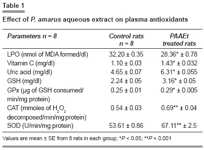
Plasma antioxidants
The levels of plasma LPO, vitamin C, uric acid and GSH are presented in [Table - 1]. A significant decrease (11.9%) in plasma LPO was observed in treated rats compared to control rats, whereas significant increase in plasma vitamin C (28.6%), uric acid (35.7%) and plasma GSH (41%) levels was observed in PAAEt treated rats compared to normal rats. The activities of plasma GPx, CAT and SOD of control and PAAEt treated rats are presented in [Table - 1]. A significant enhancement in the activities of plasma GPx (13.4%), CAT (28.4%) and SOD (25.18%) was observed in PAAEt treated animals compared to control animals.
DNA damage
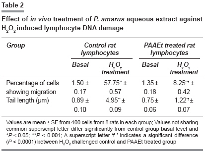
In vitro exposure of lymphocytes at different doses of PAAEt did not result in comet formation in SCGE indicating non-genotoxic nature of PAAEt. Results presented in [Table - 2] indicate the effect of supplementation of PAAEt 8 weeks on peripheral lymphocyte DNA damage and their ability to resist exogenous H 2 O 2 induced damage.
In vivo treatment of PAAEt has not resulted in lymphocyte DNA damage. A significant protection against H 2 O 2 induced DNA damage was observed in the lymphocytes of PAAEt treated rats compared to normal rats (85.3% decrease in number of damaged cells and 75.4% decrease in tail length of comets).
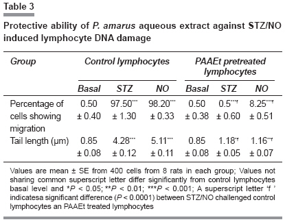
In vitro antigenotoxic effects of PAAEt against STZ and NO induced DNA damage are presented in [Table - 3]. PAAEt pretreated lymphocytes showed decrease in the number of damaged cells (89.2% and 91.1%) and length of comets (72.4% and 77.3%) against STZ and NO induced DNA damage respectively.
Discussion
Non-toxic nature of PAAEt was evident with the unaltered trend of body weight and plasma glucose levels in PAAEt treated rats compared to controls. The antioxidant potential of PAAEt was revealed by a significant decrease in the plasma LPO and a significant increase in plasma non-enzymatic antioxidants, vitamin C, uric acid and GSH in PAAEt treated rats compared to control.
Recently, free radical induced LPO has gained much importance because of its involvement in several pathologies such as aging, atherosclerosis, diabetes, wound healing, liver disorder, inflammation etc. Protection of the cell membrane from LPO could prevent, cure or delay the aforesaid pathologies. The enhanced plasma GSH, vitamin C, uric acid levels of PAAEt treated rats may be responsible for the observed decrease in the extent of plasma LPO.
Glutathione, SOD and CAT protect the cell constituents from oxidative damage. Despite these extensive defense systems, biomolecule damage may still occur and persist within the cell. The significant increase in the activities of SOD and CAT suggests a greater level of endogenous antioxidant associated with the PAAEt treatment resulting in an enhanced free radical scavenging activity. Plants are the sources for a wide variety of compounds like flavonoids and polyphenols. These compounds may be responsible for increasing antioxidant status.
The first screening of any compound, drug or potential nutraceutical starts with the genotoxicity test. [24] Endogenous levels of DNA damage remained unchanged under in vivo and in vitro treatment of PAAEt revealing that PAAEt is devoid of genotoxic and pro-oxidant property. The decreased levels of H 2 O 2 induced DNA damage in PAAEt treated rat lymphocytes is attributed to the increased scavenging of H 2 O 2 derived ROS by enhanced antioxidants.
Recent studies suggested that ROS, including superoxide (O 2° - ), H 2 O 2 , hydroxyl radical (°OH) and °NO play a central role in the mechanism of DNA damage and cytotoxicity of STZ. [25] Nitric oxide, generated from sodium nitroprusside in aqueous solution at physiological pH, interacts with oxygen to produce nitrite ions.
The decreased levels of STZ/NO induced DNA damage in PAAEt pretreated lymphocytes ( in vitro ) may be attributed to the possible direct antioxidant capacity of PAAEt. Phytochemical constituents of P. amarus may be responsible for scavenging ROS and protecting the DNA from ROS induced DNA damage. The concept of synergy is central to the holistic approach. The popular modern concept trend to isolate pure compounds may not achieve the desired results as observed in the natural version. Once an active principle is isolated from the natural product without its synergical colleagues to support and/or balance its action, it may lose its character as present in its natural form.
Conclusion Our present study indicates that PAAEt is devoid of genotoxicity and pro-oxidant property. The enhanced antioxidant status observed in PAAEt treated rats and its protective role against H 2 O 2 , STZ and nitric oxide generating system induced DNA damages might be due to the effect of different types of active principles acting individually or synergistically, each with a single or a diverse range of biological activities against oxidative stress, a widely recognized factor in many degenerative diseases.
References
| 1. | Anderson D, Phillips BJ, Tian-Wei YU, Edwards AJ, Ayesh R, Butterworth KR. Effects of vitamin C supplementation in human volunteers with a range of cholesterol levels on biomarkers of oxygen radical-generated damage. Pure Appl Chem 2000;72:973-83. Back to cited text no. 1 |
| 2. | Thyagarajan SP, Subramanian S, Thirunalasundari T, Venkateswaran PS, Blumberg BS. Effect of Phyllanthus amarus on chronic carriers of hepatitis B virus. Lancet 1988;2:764-6. Back to cited text no. 2 |
| 3. | Tran QL, Tezuka Y, Ueda J, Nguyen NT, Maruyama Y, Begum K, et al. In vitro antiplasmodial activity of antimalarial medicinal plants used in Vietnamese traditional medicine. J Ethnopharmacol 2003;86:249-52. Back to cited text no. 3 |
| 4. | Notka F, Meier GR, Wagner R. Concerted inhibitory activities of Phyllanthus amarus on HIV replication in vitro and ex vivo. Antiviral Res 2004;64:93-102. Back to cited text no. 4 |
| 5. | Mazumder A, Mahato A, Mazumder R. Antimicrobial potentiality of Phyllanthus amarus against drug resistant pathogens. Nat Prod 2006;20:323-6. Back to cited text no. 5 |
| 6. | Odetola AA, Akojenu SM. Antidiarrhoeal and gastro-intestinal potential of the aqueous extract of Phyllanthus amurus (Euphorbiaceae) . Afr J Med Sci 2000;29:119-22. Back to cited text no. 6 |
| 7. | Padma P, Setty OH. Protective effect of Phyllanthus against CCL4-induced mitochondrial dysfunction. Life Sci 1999;64:2411-7. Back to cited text no. 7 |
| 8. | Toyin YF, Stephen M, Michael A F, Udoka EO. Hepato protective potentials of Phyllanthus amarus against ethanol-induced oxidative stress in rats. Food Chem Toxicol 2008;46:2658-64. Back to cited text no. 8 |
| 9. | Raphael KR, Sabu MC, Kuttan R. Hypoglycemic effect of methanol extract of Phyllanthus amarus Schum and Thonn on alloxan induced diabetes mellitus in rats and its relation with antioxidant potential. Indian J Exp Biol 2002b;40:905-9. Back to cited text no. 9 |
| 10. | Kumar KB, Kuttan R. Chemoprotective activity of an extract of Phyllanthus amarus against cyclophosphamide induced toxicity in mice. Phytomed 2005;12:494-500. Back to cited text no. 10 |
| 11. | Rajeshkumar NV, Joy KL, Kuttan G, Ramsewak RS, Nair MG, Kuttan R. Antitumour and anticarcinogenic activity of Phyllanthus amarus e xtract. J Ethnoparmacol 2002;81:17-22. Back to cited text no. 11 |
| 12. | Kiemer AK, Hartung T, Huber C, Vollmar AM. Phyllanthus amarus has anti- inflammatory potential by inhibition of iNOS, COX-2, and cytokines via the NF-κB pathway. J Hepatol 2003;38:289-97. Back to cited text no. 12 |
| 13. | Das AK, Bhattacharjee AK. A systemic approach to phytochemical screening. Trop Sci 1970:12:54-8. Back to cited text no. 13 |
| 14. | Omaye ST, Turnbull JD, Sauberlich HE. Selected methods for the determination of ascorbic acid in animal cells, tissues and fluids. Methods Enzymol 1979;62: 3-11. Back to cited text no. 14 |
| 15. | Ellmans GL. Tissue sulfhydryl groups. Arch Biochem Biophys 1959;82:70-7. Back to cited text no. 15 |
| 16. | Utley HG, Bernheim F, Hochstein P. Effect of sulfhydryl reagents on peroxidation in microsomes. Arch Biochem Biophys 1967;118:29-32. Back to cited text no. 16 |
| 17. | Rotruck JT, Pope AL, Ganther HE, Swanson AB, Hafeman DG, Hoekstra WG. Selenium: Biochemical role as a component of glutathione peroxidase. Science 1973;179:588-90. Back to cited text no. 17 |
| 18. | Soon YY, Tan BKH. Evaluation of the hypoglycemic and antioxidant activities of Morinda officinalis in streptoztocin-induced diabetic rats. Singapore Med J 2000;43:77-85. Back to cited text no. 18 |
| 19. | Beers R, Jr Sizer JW. Spectrophotometric method for measuring breakdown of H 2 O 2 catalase. J Biol Chem 1952;195:133-40. Back to cited text no. 19 |
| 20. | Lowry OH, Rosebrough NJ, Farr AL, Randall RJ. Protein measurement with Folin- Phenol reagent. J Biol Chem 1951;193:265-70. Back to cited text no. 20 |
| 21. | Boyum A. Isolation of human blood monocytes with Nycodenz: A new non-ionic iodinated gradient medium. Scand J Immunol 1983;17:429-36. Back to cited text no. 21 |
| 22. | Singh NP, Mc Coy MT, Tice RR, Schneider EL. A simple technique of low levels of DNA damage in individual cells. Exp Cell Res 1988;175:184-91. Back to cited text no. 22 |
| 23. | Silvina BN, Laura MV, Daniel RC. A Silver staining method for single-cell gel assay. J Histochem Cytochem 2001;49:1183-6. Back to cited text no. 23 |
| 24. | Yen GC, Chen Y, Peng HH. Evaluation of the cytotoxicity, mutagenecity of emerging edible plants. Food Chem Toxicol 2001;39:1045-53. Back to cited text no. 24 |
| 25. | Bolzan AD, Bianchi MS. Genotoxicity of streptozotocin. Mutation Res 2002;512:121-34. Back to cited text no. 25 |
Copyright 2009 - Indian Journal of Pharmacology
The following images related to this document are available:
Photo images
[ph09019t2.jpg]
[ph09019t1.jpg]
[ph09019t3.jpg]
[ph09019f1.jpg]
|
