 |
| About Bioline | All Journals | Testimonials | Membership | News |
|
||||||
|
||||||
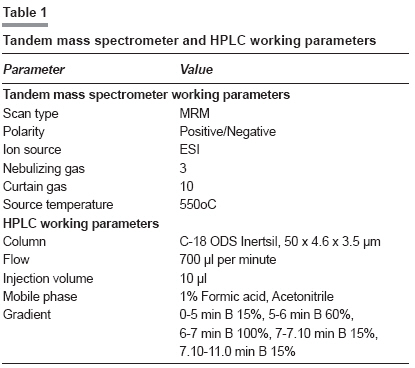
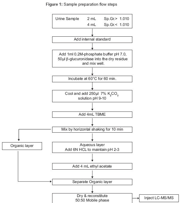
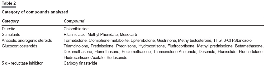
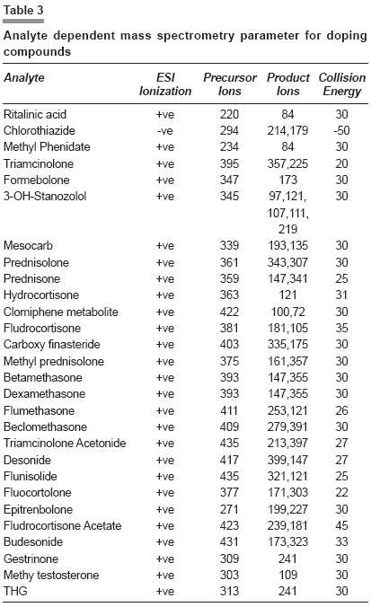
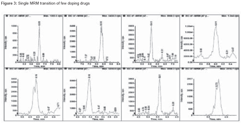
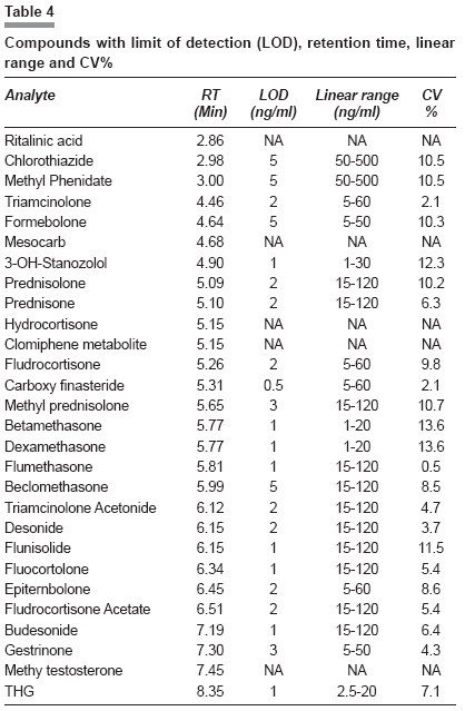
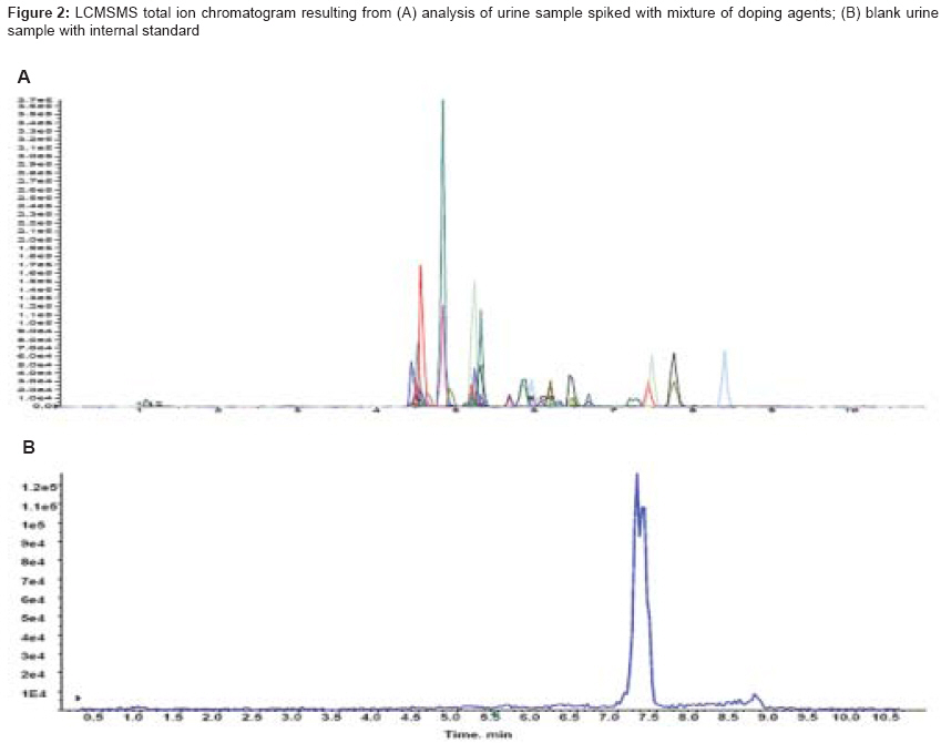
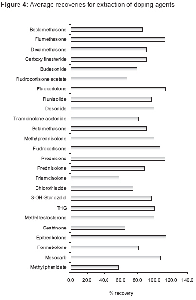
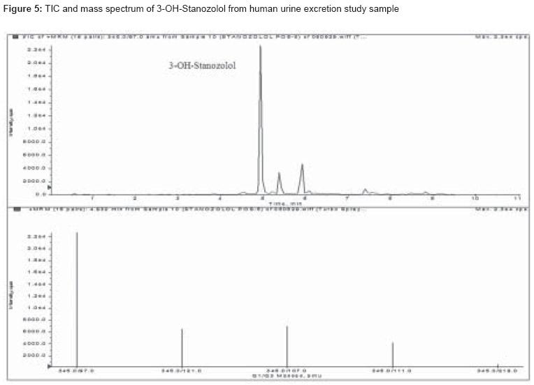
Indian Journal of Pharmacology, Vol. 41, No. 2, March-April, 2009, pp. 80-86 Research Article A simple and rapid ESI-LC-MS/MS method for simultaneous screening of doping agents in urine samples Reddy IMadhusudhana, Beotra Alka, Jain S, Ahi S National Dope Testing Laboratory, Ministry of Youth Affairs and Sports, J. N. Stadium, New Delhi Date of Submission: 06-Dec-2008 Code Number: ph09023 DOI: 10.4103/0253-7613.51347 Abstract Objective: The use of performance enhancing substances is banned in sports by the World Anti-Doping Agency (WADA). Though most prohibited substances can be detected by GC/MS, inclusion of corticosteroids and designer drugs has made it essential to detect these critical doping agents on LC/MS/MS due to their better separation and detection.Materials and Methods: A common extraction procedure for the isolation of acidic, basic and neutral drugs from urine samples was developed. A total of 28 doping drugs were analyzed on API 3200 Triple quadrupole mass spectrometer using C18 column in atmospheric pressure electrospray ionization. The mobile phase composition was a mixture of 1% formic acid and acetonitrile with gradient time period. Results: The method developed was very sensitive for detection of 28 doping agents. The linearity was performed for each drug and the total recovery percentage ranged from 57 to 114. Limit of detection is found to be 0.5 ng/ml for carboxy finasteride and 1-5 ng/ml for other drugs. The method was successfully used to detect positive urine samples of 3-OH-stanozolol, methyl phenidate, mesocarb, clomiphene metabolite and carboxy finasteride. Conclusion: The method developed based on controlled pH extraction method and HPLC-mass spectrometry analysis allowed better identification and confirmation of glucocorticosteroids and a few other drugs in different categories. The validated method has been used successfully for testing of 1000 In-competition samples. The method helped in detection of chemically and pharmacologically different banned drugs in urine in a single short run at a minimum required performance limit set by WADA. Keywords: Detection limit, glucocorticosteroids, LC/MS/MS, validation Introduction The use and abuse of performance-enhancing substances has been an issue in sports since ancient times. The availability of numerous synthetic steroids and peptide hormones has made testing an analytical challenge. [1] Acknowledging the facts of performance enhancing and deleterious side effects, the World Anti-doping Agency (WADA) banned these drugs in sports.[2] The techniques of dope testing have improved immensely from 1972 to 2008 using improved extraction methods [3] and sophisticated equipment. [4] Mass spectrometry was used for the first time in 1972 during the Munich Olympics. Since then various new chromatographic techniques viz. high-resolution mass spectrometry (Atlanta Olympics Games 1996), isotope ratio mass spectrometry (Special Olympic Winter Games, 1998) and liquid chromatography mass spectrometry (Athens Olympic Games, 2004) have come into dope testing. There are a total of 35 WADA accredited labs in the world. Each lab has their own set testing protocols utilizing various equipment viz. GC, GC/MS, HRMS, LC/MS/MS and IRMS. There are a number of methods available for individual groups of drugs viz. diuretics, [5] beta blockers, [6] stimulants and glucocorticosteroids [7] but during testing in our lab it was felt that apart from certain drugs like glucocorticosteroids for which it is mandatory to detect on LC/MS/MS, there are a few other drugs in various other categories of drugs viz. anabolic steroids (6), diuretics (1), stimulants (4) which are required to be included in the LC/MS/MS methods because of difficulty in detection by conventional GC and GC/MS. Therefore, the purpose of the present study was to explore the possibility of detecting by the glucocorticosteroids method a few other drugs which were difficult to detect at required lower concentration levels by GC and GC/MS methods.Materials and Methods Chemicals LC and MSMS Apparatus Sample Preparation Procedure Method development Electrospray ionization (ESI) and LC conditions LC conditions Results Internal standard Limit of detection (LOD) Calibration and quality control samples Calibration curve Precision and accuracy Specificity and matrix effect Total recovery Discussion The present method developed on HPLC-mass spectrometer could detect all the 28 doping agents in a single run with the limit of detection set by WADA. The total run time of the present method was short with better separation. In the last 3 to 4 years, a number of methods have been published on analysis of doping agents on LC/MS/MS. However, these methods are on specific group of drugs viz. diuretics, [5] beta-blockers, [6] glucocorticosteroids [7] and a few anabolic steroids. [8] The present method was developed for 14 glucocorticosteroids and 6 anabolic steroids and then extended to a few estrogenic drugs, stimulants and diuretics, thereby detecting a total of 28 drugs in a single screen. It helped to detect problematic drugs from diuretics (chlorothiazide), stimulants (methyl phenidate, mesocarb) and steroids (3-OH-stanozolol [Figure - 5]) thereby making one versatile method for various drugs. The ionization and fragmentation pattern differs for each category of drugs; the results suggest that glucocorticosteroids are neutral compounds to ionize [9] whereas steroids and diuretics are polar [10] and easily fragmentable. Also observed was no polarity switching between chlorothiazide (detected at a retention time of 2.98 min in negative mode) and methyl phenidate (detected at a retention time of 3.00 min in positive mode) though they are co-eluting substances. Furthermore, analysis shows stimulants and narcotics are highly sensitive and ESI appeared to be the best sensitive ionization for methyl phenidate. The ionization pattern and separation were earlier discussed in a few publications which suggest buffering agents are significant in ionization. [11] In the present study formic acid and acetonitrile are used as eluting mobile phases that gave good separation and sensitivity. The method was successfully used to detect positive urine samples of 3-OH-stanozolol, methylphenidate, mesocarb, clomiphene metabolite and carboxy finasteride. The positive urine samples were collected for 48-72 hrs after single dose administration of 3-OH-stanozolol, methyl phenidate, mesocarb, clomiphene and finasteride to healthy volunteers. The drugs and metabolites could be detected up to 48 hrs later. However, methyl phenidate and ritalinic acid were tested in a positive sample purchased from Australian Doping Lab. The total recovery percentage ranged from 57 to 114 where the lower end of the recovery may be due to the matrix associated with the samples at different pH conditions. Further, the recovery percentage for different categories of drugs done by this method is at concentrations of 10 ng/ml for steroids except 3-OH-Stanozolol at 2 ng/ml, 250 ng/ml for stimulants, 100 ng/ml for diuretics, 30 ng/ml for glucocorticosteroids and 100 ng/ml for carboxy finasteride. The extraction recovery percentage in the range of 80-120% is considered to be acceptable by the international validation protocols. [12] However, the protocol for bioanalytical method validation explains that the recovery of an analyte need not be 100%, but the extent of recovery of an analyte and of the internal standard should be consistent, precise and reproducible. [13] The validated method was used for testing of 1000 In-competition samples successfully. However, during this testing adverse analytical findings were reported for 3-OH-Stanozolol in ten samples. In conclusion, the method developed based on controlled pH extraction method and HPLC-mass spectrometry analysis allowed better identification and confirmation of chemically and pharmacologically different banned drugs in urine in a single short run. The method could detect target analytes at a concentration of minimum required performance limit set by WADA. The assay provides a useful alternative approach to individual methods for screening and confirmation of a few diuretics, stimulants, narcotics, steroids and glucocorticosteroids. The approach has great potential in doping and clinical testing and might simplify the analytical screening and confirmation cost effectively. Acknowledgments We thank Dr. Sangeeta Shukla, Professor, SOS of Zoology, Jiwaji University, Gwalior, who helped and provided constant support during this study.References
Copyright 2009 - Indian Journal of Pharmacology The following images related to this document are available:Photo images[ph09023t3.jpg] [ph09023t2.jpg] [ph09023f1.jpg] [ph09023t4.jpg] [ph09023t1.jpg] [ph09023f4.jpg] [ph09023f2.jpg] [ph09023f5.jpg] [ph09023f3.jpg] |
| |||||||||

