 |
| About Bioline | All Journals | Testimonials | Membership | News |
|
||||||
|
||||||
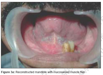
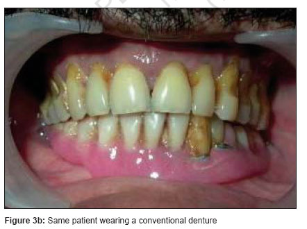
Indian Journal of Plastic Surgery, Vol. 40, No. Supp. 1, 2007, pp. 28-34 Review Article Reconstruction of the mandible Delacure Mark Institute of Plastic and Reconstructive Surgery, New York University School of Medicine, 530 1st Avenue. NY 10016 Correspondence Address: Dr Mark Delacure Associate Professor and Chief, Division of head and Neck Surgery, Institute of Reconstructive Plastic Surgery, New York University School of Medicine, New York 530 1S‘ Avenue, NY 10016 USA. E-mail: delacure@pol.net Code Number: pl07028 Abstract Segmental mandibulectomy leads to a great deficit in the form and function of the patient. Vascularised bone flaps have become the choice of the method of reconstruction of the mandible in an oncologic setting. Fibula osseous or oseteocutaneous flaps have become the favored method in centers across the world. This article reviews certain pertinent and practical points with regards to fibula flap reconstruction of the mandible. Methods of osteosynthesis, merits of inclusion of the FHL muscle in the flap and use of osteointegrated implants are discussed in detail. The reader will also be able at the end to understand the role of pre operative vascular imaging, steps taken when there is an irreversible flap failure and steps to maintain and rehabilitate occlusion as practiced in the author's service.Keywords: Fibula flap, mandibular reconstruction Effects of Mandibulectomy and Indications for Reconstruction While there is still a role for partial mandibulectomy in highly selected cases, these remain in the minority and are typified by alveolar lesions T1-2 and floor of mouth or tongue lesions, which approximate the mandibular bone. Unfortunately, it remains the case that segmental full-thickness mandibulectomy is required as the skeletal component of a composite resection all too often despite patient and physician and dentist education and the widespread use of advanced imaging techniques. Oromandibular function is generally impacted very little in marginal mandibulectomy, although commonly, in dentate patients, additional teeth must be removed to facilitate the creation of a keel or boat-shaped osteotomy so as not to compromise the biomechanical strength of the residual bone. The uneven removal of teeth will create an often-unbalanced masticatory force, which may even result in the pathologic fracture of the lower jaw. [1] In edentulous cases with significant vertical resorption and in those which have been previously irradiated, a load-sharing reconstruction plate may be advisable, spanning the weakened segment and affording it additional strength in function. [2] The creation of a continuity defect through the performance of segmental mandibulectomy mandates reconstruction in order to optimize both form and function. In selected cases, like previously edentulous or debilitated old patients who will be rendered with a posterior composite defect, no reconstruction being offered may be a reasonable and efficient approach. This causes drifting of the mandible to the resected side but results in minimal functional debility except for inability to wear dentures. More commonly than not, however, continuity must be restored to optimize the patient in function. Again, even in a dentate and younger mandible a posterior defect (i.e. posterior to the mid-body region) may not cause as much functional and aesthetic problems as with central mandibular defects. The dental malocclusion that follows the mandibular drift may be partially rectified by the use of prosthetic guiding flanges and the deformity that results may not be aesthetically and functionally as devastating as central mandibular defects. Hence the absolute indication for mandibular reconstruction remains a central symphyseal defect. [3],[4] Techniques for post-traumatic mandibular defects have a very restricted application in mandible reconstruction for malignancy due to "hostile" wound healing environments. These include compromised vascularity resulting from irradiation or previous surgery which involves ligation of vessels and dissection of tissue planes; orocervical communication, dimensions of the defect and age. As these nonvascularized grafts rely entirely upon the recipient bed for revascularization, healing and the maintenance of bone stock, they find very little application in contemporary reconstructive oncology. A bridging reconstruction plate (2.4 mm or greater profile) without actual replacement of bone, in combination with a regional myocutaneous flap or with collapse of the surrounding soft tissues has been used as a quick and effective measure in lateral defects to reestablish bicondylar joint function and optimize chewing function. [5],[6] But again this has a limited role in post-cancer reconstructions since they do not stand adjuvant treatments namely radiation. But they may have a limited role in the patients otherwise unsuitable for complex reconstructions. The microvascular transfer of bone and associated soft tissues from the leg (fibula) and hip (iliac crest - DCIA) has revolutionized the reconstruction of composite defects of the oral cavity since their widespread application in the mid-80s. All other methods might be worth mentioning in this era only for their contrast in terms of simplicity, lack of the requirement for elaborate equipment, technical capability and marathon operating room time and resources. Simplicity does not, however, always bring merit, particularly as mandibular defects become more centrally placed. Here, these conservative efforts bring more unpredictable results and often greater or more protracted complications. Free tissue transfer of vascularized bone has become the standard of care in present day reconstructive surgery in these situations, in the head and neck centres worldwide. Hence these methods will be discussed in detail and considerations in the use of these techniques alone will be addressed in this article. Choice of Vascularized Bone Flaps and Relative Merits Explorations of a variety of donor sites over the past several decades have resulted in the fibula as the flap of choice for the microvascular transfer of bone and associated soft tissues for the reconstruction of segmental defects of the mandible. [7],[8],[9],[10],[11],[12],[13],[14] The primary advantages of this donor site include: available length of bone, associated soft tissues available, near anatomic dimension, ability to accept bicortical implants and relatively low donor site morbidity. The ability to harvest the flap concurrent with the resection procedure optimizes case flow and minimizes length of procedure. Under nominal circumstances, the osteotomy site and interfaces with residual mandibular segments are well consolidated over the first three months. Lower extremity function has largely normalizes over the same period. The iliac crest or DCIA free flap rivalled the fibula in the defining days of mandible reconstruction, but has generally become a second-tier choice except in unusual circumstances. Disadvantages include shorter available bone and pedicle length, monocortical implant support and a significantly more morbid donor site. [9],[15],[16],[17],[18] The third-tier choice is represented by the scapula, whose requirement to intraoperatively reposition the patient for flap dissection (non-simultaneous), far outweighs the advantages of its additional degrees of freedom in the positioning of soft and hard tissue components. [19],[20],[21] Composite RAFF which had been proposed in the earlier years has become very much less used now due to the inadequate quality and quantity of the bone available as well as the need for prolonged protection of the forearm to prevent fracture of the residual radial bone. [22],[23],[24] Maintaining and Rehabilitating Occlusion In general, the body segment will require about 4 cm of bone, the symphysis 2 cm and each double closing wedge osteotomy, about 1 cm. These approximations serve well to allow simultaneous flap work during the resection and clearance of margins. In dentate patients, not only should the flap be inset in maxillary-mandibular fixation (MMF) [Figure - 1], but care must be taken to ensure seating of the condyles before MMF is secured and flap insetting must be absolutely neutral. Such intraoperative attention to detail is essential to prevent occlusal deformities as malocclusion problems after this procedure are complex and difficult to correct, often necessitating risky orthognathic procedures later on and threaten the entire construct and its healing. In general, multiple miniplates of the 2.0 mm locking screw variety have been adequate to maintain contour and occlusal relationships through the period of osteosynthesis with minimal hardware failure. The hardware failures are usually due to suboptimal technique. Refinements and Nuances - Fibula Microvascular Free Flap Osteosynthesis Unbalanced masticatory forces have occasionally resulted in the disruption of hardware due to the asymmetric requirements of most resections. Newer low-profile (2.0 mm) plating systems allow for less implant profile contribution to the aesthetic result while maintaining enough biomechanical strength to generate a stable construct. Decreased implant volume is also significant. Segment-to-segment shaping is preferred for defect bridging reconstruction plates to allow for subtleties in adjusting the shape of the neomandible for optimal contour. These should not be so numerous as to require extensive dissection for later plate removal when implant placement is planned. Locking screw plate designs are preferred to compensate for the latitude in contouring which may be somewhat less precise and also the need for less compressive clamping of the plate to the periosteal blood supply of the neomandible. [25],[26],[27] Plates should be placed transperiosteally and screw holes should be drilled monocortically into the flap. Screws on the native side i.e., on the residual original mandible should be placed bicortically and may be subperiosteal with limited dissection advisable. Unused screw holes may be used for myodesis of the suprahyoid musculature or for hyoid suspension. Insetting - contour Early concerns regarding the relative lack of vertical height of the fibula in comparison to the iliac crest free flap have given way to its general superiority in terms of bicortical architecture, overall contour and to advances in implantology that have made the lower neoalveolus height a non-issue. Usually, the flap should be inset so that the lower border of the mandible and fibula are aligned, assuring optimal aesthetic contouring in the submandibular region once oedema has receded and soft tissue contracture and atrophy have taken place. In certain cases e.g., as the construct advances posteriorly (body), the flap may be inset somewhat higher to facilitate implants and minimize unfavourable biomechanical lever forces that act more strongly upon a taller abutment. In general, the soft tissue component brought with the fibula flap undergoes very significant reduction in size over the first few years postoperatively. Bone-only flaps For massive oral cavity and full-thickness defects, the harvest of a skin paddle is usually required [Figure - 2]. Although helpful in monitoring the viability of the flap, the routine harvest of a skin paddle with the fibula flap should be individualized as it may be unnecessary in other cases. The use of a skin paddle often necessitates secondary procedures for those patients ultimately undergoing the placement of osseointegrated implants. When used as a bone-only flap the retention of an additional 1-2 mm of the peroneus longus muscle is advisable here to facilitate wound healing and to minimize potential complications. The mucosalized muscle cuff of a bone-only fibula free flap forms a superior neoalvelous for implant placement and obviates the need for skin graft vestibuloplasty and skin paddle excision in preparation for implants. In selected cases, where this bone-only technique is used, an ordinary tissue-borne denture prosthesis may be worn either transitionally (to implant abutment fitting) or even as a permanent prosthodontic measure [Figure - 3a, 3b]. When using the skin paddle, making it a sensate and neurotized fibula free flap have found little advantage and is not practiced routinely. Design of the incision I have found a serpigenous vertical incision to be aesthetically desirable to a straight linear one. The individualized use of skin paddle harvests will avoid the routine defect with the need for split-thickness skin grafts. Skin flap design using Doppler guidance to make it small and suitably placed with an intent to close by primary approximation often fails and will end with a STSG due to muscle and post-tourniquet oedema, mitigating the potential advantages of such designs. Such efforts have also resulted in wound healing complications such as tendon exposure which protract the postoperative course. [28],[29] Flexor hallucis longus (FHL) muscle In general, harvest of the entire FHL muscle is advisable as it has many uses during insetting. Generally, it is exploited to fill out the submandibular region and to bolster the intraoral closure and protect the anastomosis. As it atrophies significantly over the first six months, overcorrection of contour is not of concern. The muscle may also be myodesed over the lingual aspect of the construct to assist in hardware coverage, etc. There seems to be little advantage in retaining part of the muscle in the leg and the tenodesis effect of other foot flexors retains much of the great toe elevation that would seem to be lost in total muscle harvest. This loss of function seems to be of greatest importance to those for whom climbing, repetitive rise from crouched position and similar push-off motions are important. [28],[29] When trimming the muscle to fit the particular reconstructive needs of a particular defect, meticulous attention to detail in haemostasis is required due to bleeding from raw muscle edges and intramuscular blood supply, to avoid the development of haematoma, which may threaten the anastomosis via extrinsic compression. This is amplified where postoperative flap routines and protocols use heparin or aspirin. Airway considerations The inability to effectively myodese muscles formerly attaching to the anterior mandible (genial tubercle) may present airway difficulty in the early postoperative course as oedema peaks. This inability to anchor the muscles is due to the course of the peroneal vessels on the lingual aspect of the construct and the potential to compromise flap vascularity. Tracheotomy should be strongly considered for cases involving resection of the symphysis and parasymphyseal regions of the mandible. Ultimately, the retro positioning of these muscles does not seem to be an issue with regard to the ultimate decannulation. Acute perioperative peroral reintubation is to be avoided at all costs for reason obvious. Maxillary reconstruction Osseous reconstruction of the maxilla is a completely different and significantly more difficult endeavour than that of the mandible. This is largely due to the additional 3-5 cm of pedicle length that is required to perform anastomosis to the facial vessels in the neck. This is true even when the recipient vessels are dissected above the crossing of the marginal mandibular branch of the facial nerve. More commonly than not, short segment interposition vein grafts are required to complete the task, with attendant escalation of risk in additional operative times, additional donor sites and doubled numbers of anastomosis. When there are significant palatal defects, soft tissue deficiencies posterior to the new arch are difficult to reconstruct with this flap. However, in selected cases, this option should be seriously considered to maintain lip posture, afford osseointegrated implant restoration and to avoid the need for regularly removable and comparatively unstable and unwieldy prosthodontic options. The need to run the pedicle for an extended distance through a tunnel in the facial soft tissues mandates additional attention to detail in the haemostasis of small branches as haematoma within the tunnel (closed compartment), and the flow dynamics of the long pedicle, particularly on the low pressure venous side, may make this construct significantly more precarious than in the mandible. [30],[31],[32],[33],[34] In case of failure A detailed understanding of the microvascular architecture and physiology is necessary to understand the management of this flap and its components in the event of vascular compromise. In its normal state, the primary blood supply to the fibula i.e., two-thirds of cortical supply is via the medullary cavity in a centrifugal pattern of flow. About one-third of the cortical blood supply is via the periosteum in a centripetal direction. The nutrient artery enters the bone usually in its proximal third at its junction with the middle third. This supply is disrupted when the bone is osteotomized or when pedicle-lengthening procedures by the stripping of soft tissue from proximal bone are necessitated. When the bone is transferred, a reversal of the normal physiologic flow is effected through the musculoperiosteal attachments which are maintained through flap dissection and plating. That is, the flow becomes dependent on the centripetal flow through the peroneal vessels, into the muscular branches and on through the periosteal vessels, the cortex and into the medullary space. Although medullary flow is easily demonstrated after the flap is revascularized, it seems unlikely that the previously normal centrifugal flow is ever reconstituted. It is not logical, however, that more than a few millimetres′ cuff of the peroneus longus muscle be harvested, as an added margin of safety. This is because the peroneus longus does not have as its primary supply from the peroneal vessels and more muscle can often be a liability as it may be "parasitic" to the newly established pattern of flow. Furthermore, in the event of flap failure, the nonviable muscle cuff serves as a barrier to the potential neovascularization of the flap. It is the failure to understand this physiology that condemns efforts to retain the bone as a nonvascularized bone graft equivalent by "burying" or wrapping it with another muscle in the event of thrombosis of pedicle, exposure, etc. (e.g. by pectoralis major muscle flap). In the rare event that the bone remains covered, eventually, it will act as a sequestrum and ultimately extrude, masquerading initially as hardware failure as screws migrate from the bone which slowly undergoes resorption. [35] In general, in cases where the fibula free flap was the reconstruction of choice and complete failure has followed, the optimal solution remains this same flap - from the other leg - on a better day and under more favourable circumstances. Presurgical vascular imaging Conventional angiography or magnetic resonance angiography are recommended for selected case such as those over 50 years and those with co-morbidities like diabetes mellitus, hypertension, arteriopathy, histories of trauma and for anyone with an equivocal peripheral pulse exam. This being said, most younger patients with a normal clinical exam may do without such studies. [36],[37],[38] Osseointegrated implants Fibula flaps accept osseointegrated implants very well and these can be placed either in the primary sitting [Figure - 4] or after the lapse of few months. Logistic considerations, especially, the cost and loss of the implants if unfortunately the flap fails, make the choice of primary implantation less attractive. [39],[40],[41] Despite the ability to receive osseointegrated implants, only a minority of the patients actually makes it through the entire sequence due to expense, recurrence, emergence of distant metastases and the like. This ability cannot represent the main justification for the use of a microvascular bone transfer. Summary In the defects of the mandible, especially the anterior arch (mesial body, parasymphyseal and symphyseal) microvascular free tissue transfer has revolutionized the treatment, routinely returning to the patients unprecedented levels of form and function in comparison to similar patients just under three decades ago when these techniques began to be explored. Over the past decade in particular, persons and institutions worldwide have gained experience in the application of these techniques which had been previously prohibitive and unavailable. In nearly every region of the world, one can access units able to meet this relatively recently defined standard. Despite all of our more recent advances in allied fields, like, radiation oncology - its planning and technical advancement, in medical oncology now with its specific receptor blocking drugs of great promise and finally the explosion of technology in diagnostic imaging, it remains true that the microsurgical transfer of bone and related microvascular techniques have impacted the well-being of the head and neck cancer patient more than all of the former contributions combined. Free fibula flap remains the flap of choice in mandibular reconstruction. References
Copyright 2007 - Indian Journal of Plastic Surgery The following images related to this document are available:Photo images[pl07028f3b.jpg] [pl07028f4.jpg] [pl07028f3a.jpg] [pl07028f2.jpg] [pl07028f1.jpg] |
| |||||||||

![[Figure - 1]](/showimage?pl/photo/pl07028f1.jpg)
![[Figure - 2]](/showimage?pl/photo/pl07028f2.jpg)
![[Figure - 4]](/showimage?pl/photo/pl07028f4.jpg)