 |
| About Bioline | All Journals | Testimonials | Membership | News |
|
||||||
|
||||||
Tropical Journal of Pharmaceutical Research, Vol. 1, No. 1, June, 2002 pp. 3-14 Review Article
Experimental and modeling studies of mass transfer in microencapsulated cell systems Mattheus F. A. Goosen
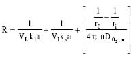
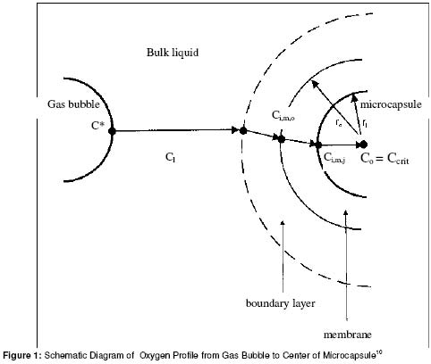
Department of Bioresource and Agricultural Engineering, College of Agriculture and Marine Sciences, Sultan Qaboos University, P.O. Box 34, Al-Khod 123, Sultanate of Oman. Fax: +968 515 418 Fax: +968 515 200 E-mail: theog@squ.edu.om (Business); mattheus@omantel.net.om (Home) Code Number: pr02002 ABSTRACTGaining a better understanding of mass transfer problems in encapsulated cell systems and in tissue engineering requires both experimental investigations and mathematical modelling. Specificmass transfer studies are reviewed including oxygen transfer in immobilised animal cell culture systems, modelling of electrostatic polymer droplet formation, and growth of plant somatic tissue encapsulated in alginate using electrostatics. INTRODUCTION Microencapsulation systems have found applications in encapsulated cell therapy/tissue engineering 1-3, bioseparations technology 4, immobilized biocatalysts 5, 6, and polymeric drug-delivery systems 7, 8. All areas, however, suffer from specific mass transfer problems. With drug-delivery systems, the release of the bioactive agent from the polymer matrix or capsule must be controlled so as to provide a constant steady release rate. In the case of immobilized cells, oxygen must be able to reach the viable cells at a sufficient rate to keep the cells alive, while the desired product, such as insulin in the case of diabetes treatment, must diffuse out of the capsule, along with low molecular weight waste products. With biocatalysts, whether they be enzymes or cells, the substrate must be able to reach the bead/capsule interior to allow the biochemical reaction to occur and the desired products must be able to diffuse out of the bead. This objective of this paper was to review specific mass transfer studies performed in our laboratory including oxygen transfer in immobilized animal cell culture bioreactors, modelling of electrostatic polymer droplet formation, and growth of somatic tissue encapsulated in alginate using electrostatics. Both experimental and modelling studies were combined so as to give the reader a better insight into common mass transfer problems. Modelling of the oxygen transfer process in immobilized cell systems It is not adequate simply to transfer sufficient oxygen to the bulk liquid culture medium in immobilized cell systems. Oxygen must also be transferred from the liquid to the cells. Consider for instance the transfer of oxygen from a gas bubble, through the culture medium to a microcapsule containing animal cells. The resistance to oxygen transfer from the gas phase to the inside edge of the microcapsule (i.e. gas to liquid, liquid to microcapsule, and trans-membrane resistance) (Figure 1) can be added together resulting in the following expression for the resistance to oxygen transfer, R 9, 10, 11:
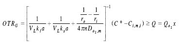
where VL is the volume of the liquid phase, n is the number of microcapsules, ro and ri are the outside and inside radius of the microcapsule, respectively, ksa is the volumetric mass transfer coefficient from a liquid to a solid, and DO2,m is the diffusivity of oxygen in the membrane. Employing Fick’s Law of Diffusion 12, it can be shown that the oxygen transfer rate from the gas phase to the inside edge of a microcapsule, OTRG, is given by:
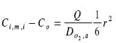
where C* is the oxygen concentration in equilibrium with the oxygen partial pressure in the gas phase, Ci,m,i is the oxygen concentration on the inside of the membrane, Q is the oxygen consumption rate of cells inside a microcapsule (mgO2/Lhr), QO2 is the oxygen consumption rate per cell, and x is the cell density. To enable the cells in the microcapsule to survive, the oxygen transfer rate, OTRG, must be greater than (or at least equal to) the oxygen consumption rate of cells inside the capsules, Q. Based on the work of Heath and Belfort13 the oxygen concentration profile within a microcapsule can be represented by:
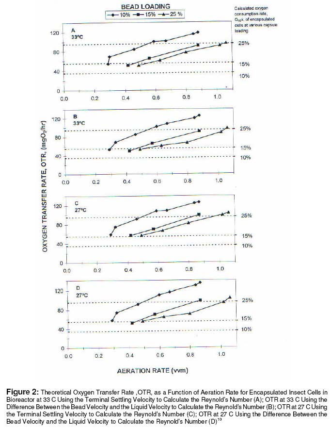
where r is the radius of the alginate core. If the concentration of oxygen in the center of the microcapsules, Co, is equal to a critical oxygen concentration, Ccrt, the concentration of oxygen at the inner surface of the membrane, Ci,m,i, can be determined from equation 3. The performance of a one liter external-loop air-liftbioreactor was investigated by studying the gas-liquid oxygen transfer at various aeration rates (0.1 vvm to 1.06 vvm). The influence of suspended alginate beads on the hydrodynamics and mass transfer of the system was examined over a range of microbead loadings (0 to 25% by volume). The intent was to investigate the effect of using various concentrations of cell immobilization matrices on the physical properties of the system. A mathematical correlation was developed for expressing the dependence of kla on aeration rate and microbead loading. A mathematical study of the mass transfer resistances from the gas phase to the interior of a microcapsule was also performed, using experimentally determined kla values, to enable the determination of the maximum bioreactor microcapsule loading. The oxygen probe time constant for our studies was 27 ± 7 sec and the maximum kla value found was 0.00861 sec-1. Since this is approximately equal to 1/5 ka, the oxygen probe response time was therefore not accounted for in the determination of the kla values in our investigation. According to Van't Riet 14, the response lag of an oxygen probe can be neglected if the oxygen probe time constant, t p , is less than 1/5 ka. To ensure that the kla studies would be measuring the gas-to-liquid mass transfer coefficient and not the mass transfer from the gas to the interior of the bead, the ka in the presence of alginate beads was compared to that in the presence of ion-exchange resin beads. The kla for a 10% loading of alginate beads it was determined to be 31.7 ± 0.7 hr-1 and for a 10% loading of ion-exchange resin beads it was 31.4 ± 1.5 hr-1, at an aeration rate of 0.67 vvm. The kla values are essentially equal. This suggested that the alginate beads were not acting as a "sink" for oxygen and that the desired quantity, gas-to-liquid mass transfer coefficient, was being measured. For immobilized cell systems, such as microcapsules, ensuring adequate oxygen transfer from the gas phase to the liquid medium does not necessarily ensure that adequate oxygen will reach the immobilized cells. It is possible that the kia may not be adequate for a certain microcapsule loading. A theoretical study was performed in our laboratory of oxygen transfer to cells immobilized in microcapsules in a bioreactor for various microcapsule loadings using equations 2 and 3 10. A schematic diagram of the oxygen concentration profile from a gas bubble to the center of a microencapsule is shown in Figure 1. Upon arbitrarily specifying the critical oxygen concentration in the center of the microcapsule, the rate of oxygen transfer from the gas phase to the inner surface of the microencapsule membrane (OTRG, mg/hr) for a certain microcapsule loading was compared to the oxygen demand of the cells (QO2,x) for the same microcapsule loading. The study was made for Spodoptera frugiperda (i.e. insect) cells cultivated in poly-1-lysine/alginatemicrocapsules at a maximum cell density of 8 x 10⁷ cells/mL capsules . An oxygendemand of 1.4 x 10⁻¹⁰ mmoleO2/cellhr was assumed. These insect cells are usually cultivated at 27 oC and 33 oC therefore our study was performed at both temperatures. It was necessary to estimate several of the parameters used to evaluate the oxygen transfer rate. The diffusivity of oxygen in sodium alginate (the immobilization agent inside the microcapsule) was estimated to be 86% of the diffusivity of oxygen in water (i.e. approximately the same as the diffusivity of oxygen in calcium alginate). According to King et al. 15, the membrane is 5 Rm thick and is composed of approximately 90% water. The diffusivity of oxygen through the membrane was therefore assumed to be equal to that of oxygen in water. A critical oxygen concentration in the center of the microcapsule was assumed to be 40% of air saturation. Figure 2 shows the results for the oxygen transfer rate, OTR attainable for microcapsule loadings of 10, 15 and 25% at 33 oC and 27 oC as a function of the aeration rate. The terminal settling velocity was used to calculate the Reynold's number. Comparing Figures 2A and 2B, the latter uses the difference between the bead and liquid velocities (determined experimentally) to calculate the Reynold's number, and indicates that there is not much difference (at most 8%) between the two methods. This suggests that the terminal velocity may be used as a good approximationof the relative velocity between the bead and the liquid if it is not feasible to determine the liquid and bead velocities experimentally. At 33 oC, for 10% bead loading, the oxygen demand of the cells was achieved at 0.29 vvm, which is the minimum vvm for suspension of the beads (Figure 2A). On the other hand, for 25% bead loadings a vvm of 1.06 is required to meet the oxygen demand of the cells. This is quite a high aeration rate thus it may not be feasible to operate at 25% bead loading. Decreasing the temperature to 27oC (Figures 2C and 2D) increased the oxygen transfer rate but only slightly (by approximately 8%). This was expected since a decrease in temperature increases the solubility of oxygen in the bulk liquid which increases the driving force for oxygen transfer. This, in turn, increases the oxygen transfer rate to the cells. The temperature did not, however, have a very significant effect on the oxygen transfer rate. These results suggest that for this bioreactor the cells will not be oxygen limited at microcapsule loadings of 10% and 15% (by volume). However, there is the potential for oxygen limitation at 25% microcapsule loadings if the reactor is not operated at a minimum aeration rate of 1.06 vvm. Mathematical modeling of electrostatic polymer droplet formation Droplet formation in the presence of an electric field has been analyzed previously 6. If gravity were the only force acting on the meniscus of a droplet attached to the end of a tube, large uniformly sized droplets would be produced. The gravitational force, Fg, pulling the droplet from the end of the tube is given by:
where r is the density of the polymer solution, r is the droplet radius and g is the acceleration due to gravity. The capillary surface force, Fg, holding the droplet to the end of the tube is given by:
where ro is the internal radius of the tube and g is the surface tension. Equating the gravitational force on the droplet to the capillary surface tension force holding the droplet to the tube (i.e. extrusion orifice) gives:
In the presence of an applied voltage, the electric force, Fe, acting along with the gravitational force, Fg, would reduce the critical volume for drop detachment resulting in a smaller droplet diameter. Equating the gravitational and electrical forces on the droplet to the capillary surface force, Fg, yields: Fg = Fg+ F (7) In the case of a charged needle, the stress produced by the external electric field at the needle tip is obtained by using a modified expression developed by De Shon and Carlson17:
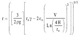
where H is the distance between the needle tip and collecting solution, V is the applied voltage, and eo is the permittivity of the air. The effect of applied potential on the droplet radius for a charged needle arrangement can be derived by substituting equations 4, 5 and 8 into equation 7:
Equation 6 was employed to calculate the microbead diameter in the absence of an applied voltage (i.e., 0 kV). The surface tension, g, of the alginate solution was g assumed to be 73 g/s2 which is the value eo for water against air 18. The density of the polymer solution was taken as 1 g/cm3. In the presence of an applied voltage, equation 9 was used to determine microbead size. The permittivity of air, , used in calculations was 1.0 g cm/s2 kV2. This value was estimated based on previous studies 16. Reasonably good agreement was obtained between calculated and experimental values of microbead diameter (Table 1) 19. For example, when the extrusion orifice diameter decreased from 1900 to 400 microns, the calculated bead diameters decreased from 4400 to 2600 microns, and the experimental valuesdecreased from 3700 to 2000 microns. When the extrusion orifice diameter was kept constant at 1900 microns and the applied voltage was increased from 0 to 10 kV, there was also a similar decrease in bead size from 4400 to 1690 microns for the calculated values and from 3700 to 1700 microns for the experimental values. Table 1: Comparison of experimental and calculated microbeaddiameter
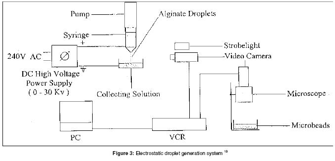
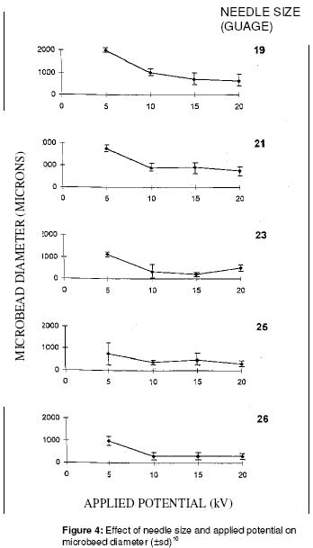
Encapsulation and growth of somatic tissue in alginate using electrostatics The mechanism of alginate droplet information using an electrostatic droplet generator has recently been investigated with a variable-voltage power supply 20-23. Animal cell suspensions were successfully extruded using the electrostatic droplet generator. The application of this technology to plant cell immobilization has only recently been 11, 19, 23. A major concern in cell and bioactive agent immobilization has been the production of very small microbeads so as to minimize the mass-transfer resistance problem associated with large-diameter beads (>1000 µm). Klein et al. 24 reported production of alginate beads with diameters from 100 to 400 µm using compressed air to quickly pass the cell/gel solution through a nozzle. Few attempts have been made in the application of electric fields to the production of micron size polymer beads for cell immobilization 25. Somatic embryogenesis is a new plant tissue culturetechnology in which somatic cells (i.e., any cell except a germ or seed cell) are used toproduce an embryo (i.e., plant in early state of development) 26. The technique of somatic embryogenesis in liquid culture, which is believed to be an economical way for future productionof artificial seeds, may benefit from cell immobilization technology. Encapsulation may aid in the germination of somatic embryos by allowing for higher cell densities, protecting cells from shear damage in suspension culture, allowing for surface attachment in the case of anchorage dependent cells, and being very suitable for scale-up in bioreactors. The long-term objective of the project reported in this section is the development of an economical method for the mass production of artificial seeds using somatic embryogenesis and cell immobilization technology. The short-term aim was to investigate the production of small alginate microbeads using an electrostatic droplet generator. Callus tissue from Carnation leaves was also immobilized and cultured. The section starts with a detailed experimental description of electrostatic droplet generation for those not familiar with the technique 22 (Figure 3). Production of Alginate Beads using Electrostatics Attach a syringe pump to a vertical stand. Usea 10 mL plastic syringe and 22- or 26gage stainless steel needles. A variable high voltage power supply (0-30 kV) with low current (less than 0.4 mA) is required. Wehave used a commercial power supply model 230-30R from Bertan (Hicksville, NY). Prepare 1.5% (w/v) CaCl2 in saline (0.85 g NaCl in 100 mL distilled water). Saline can be replaced with distilled water if an alginate solution without cells is being extruded. Place the CaCl2 solution in a petri dish on top of an adjustable stand. The stand allows for fine tuning of the distance between the needle tip and collecting solution. Prepare 1 to 4 % (w/v) low viscosity sodium alginate by dissolving alginate powder with stirring in a warm waterbath. Slowly add the 1 to 4 g sodium alginate to 100 mL warm saline solution (or distilled water), stirring continuously. It may take several hours to dissolve all of the alginate. Add about 8 mL of the alginate solution to a 10-mL plastic syringe, put back the plunger, and attach the syringe to the upright syringe pump. Make sure that the stainless steel needle, 22gage, is firmly attached and the syringe plunger is in firm contact with the moveable bar on the pump. Position the petri dish (or beaker) containing CaCl2 so that the needle tip is about 3 cm from the top of the CaCl2 hardening solution. This is the primary reason for using an adjustable stand. Attach the positive electrode wire to the stainless steel needle and the ground wire to the collecting solution. The wires may need some additional support to prevent them from bending the needle. Switch on the syringe pump and wait for the first few drops to come out of the end of the needle. This could take a minute or two. Doing it this way also ensures that the needle that the needle is not plugged. After the first drop or two has been produced, switch on the voltage supply. Make sure that the voltage is set low, less than 5 kV. If this is the first time that you have tried electrostatic droplet generation, raise the voltage slowly and observe what happens to the droplets. The rate at which they are removed from the needle tip increases until only a fine stream of droplets can be seen. The changeover from individual droplets to a fine stream can be quite dramatic. The most effective electrode and charge arrangement for producing small droplets is a positively charged needle and a grounded plate. Two other arrangements are also possible; positively charged plate attached to needle, and a positively charged collecting solution. Make sure that the positive charge is always on the needle. This ensures that the smallest microbead size is produced at the lowest applied potential. With a22-gage needle and an electrode spacing of 2.5 -4.8 cm there will be a sharp drop in microbead size at about 6 kV. This can be noticed visually by observing the droplets coming from the needle tip. Standard commerciallyavailable stainless steel needles can be employed. However, when going from a 22- to 26-gage (or higher) needle, needle oscillation may be observed. This needle vibration will produce a bimodal bead size distribution with one peak around 50 mm diameter beads and another around 200 mm. If a syringe pump is not available, remove the syringe plunger and attach an air line with a regulator to the end of the syringe. Varying the air pressure on the regulator can control the alginate extrusion rate. Lumps of sodium alginate often form if the powder is added all at once to the warm saline. Sprinkle the alginate powder into the saline a small amount at a time with gentle mixing. Once it has dissolved (up to 1-2 h), allow the viscous solution to cool and then transfer it to several plastic test tubes, cap and store in the refrigerator until needed. This prevents bacterial growth. If the alginate solution is very viscous, air bubbles will be trapped during the stirring. These bubbles will disappear if the viscous solution is left to stand overnight. If the needle is plugged, place it in dilute citrate solution for a few minutes. Passing a fine wire through the needle also helps. Resuspending cells in 1 % (w/v) sodium alginate solution will dilute the alginate and could give tear-drop shaped beads when the solution is extruded. To solve this problem, increasethe concentration of sodium alginate solution to 3 or 4 %. Extrusion of alginate droplets using a 5.7 KV fixed-voltagepower supply showed that there is a direct relationship between the electrode distanceand the bead diameter. For example, at 10-cm electrode distance, the bead diameter was 1500 µm while at 2 cm it decreased to 800 µm. The greatest effect on bead diameter was observed between 2 and 6 cm electrode distance. While there was overlapin bead sizes between 6, 8 and 10 cm electrode distance, there was a significant difference (i.e., no overlap in SD) between bead sizes at 2 and 6-cm electrode distance. An inverse relationship between needle size and microbead diameter was observed. Aside from the 23 G needle there was a significant difference between bead sizes produced by all needles (i.e., no SD overlap). As the needle size decreased from 19 to 26 G, the bead size decreased from 1400 to 400 µm, respectively. These results support previous work reported by Bugarski et al. 21. The present investigation showed that the alginate concentration does not appear to be important due to overlapping SD intervals for all data points. The bead diameter was found to be 800 µm at both 1% and 3 % alginate concentration. Lookingmore closely at the effect of needle size on bead diameter, as a function of applied potential (Figure 4) we see that the decrease in microbead size was greatest between 5 and 10 KV for all needle gages tested. For example, when the applied potential was increased from 5 to 10 KV, the microbead diameter decreased from 2000 to 1000 µm and from 1000 to 250 µm for the 19 and 26 G needles, respectively. The smallest bead, 200 µm, was produced with a 26 G needle at 20 KV. Immobilised Callus Tissue Growth Immobilized callus cells from carnationleaves retained viability as observed by cell growth and plantelet formation 11, 19, 23. In a related study, Shigeta 27 was able to germinate and grow encapsulated somatic embryos of carrot using a 1% sodium alginate solution, as compared to a 2% alginate solution used in the present investigation. The main findings of our experiment, though, indicated that s o m a tic tissue could be electrostatically extruded and aseptically cultured while retaining viability. Plantlets obtained from 4% alginate b eads on aga r , o rigina ll y immobilized at 10 kV, 6 cm distance, were transferred to sterilized potting mixture at two months culture. The plantlets grew well and showed complete leaf and root development by four months19. Suspension culture of encapsulated somatic tissue was less successful. Piccioni28 in a recent study investigated the growth of plantlets from alginate encapsulated micropropagated buds of M.26 apple rootstocks. He showed that the addition of growth regulators (e.g., indolebutyric acid) to the somatic tissue culture several days prior to the encapsulated, as well as the addition of the same regulators to the encapsulation matrix, improved the production of plantlets in suspension culture from 10% to more than 60%. We can speculate that culturing the Carnation leaf callus tissue in the presence of growth regulatorsprior and during encapsulation may enhance the production of plantlets from suspension culture. Electrostatic droplet generation does not appear to have a negative impact on somatic tissue viability since cell growth and plantlet formation was observed. This is in agreement with similar studies reported for insect cells 29 and mammalian cells 25, where it was shown that high electrostatic potentials did not affect cell viability. In closing, the technique also has great potential in medicine for encapsulating genetically engineered cells 30 and in environmental engineering for removal of heavy metals from water using gel beads 31. CONCLUDING REMARKSIn the coming decade, the development of commercially-successful microencapsulated cell systems will necessitate close collaboration between scientists with different areas of expertise such as engineering, microbiology, biochemistry, pharmacology and medicine. As our knowledge of the physicochemical and mass transfer characteristics of such systems increases, we can expect to see many new areas of application. This is an opportunity that should not be missed. ACKNOWLEDGEMENTSThe financial support of the Natural Science and Engineering Research Council of Canada, and Sultan Qaboos University, Collegeof Agriculture (Grant AGBIOR 9505 to Mattheus Goosen) is gratefully acknowledged. REFERENCES
Full text of this journal is also available online at http://www.tjpr.freehosting.net © 2003 - Pharmacotherapy Group, Faculty of Pharmacy, University of Benin, Benin City, Nigeria. The following images related to this document are available:Photo images[pr02002f4.jpg] [pr02002f1.jpg] [pr02002f3.jpg] [pr02002f2.jpg] | ||||||||||||||||||||||||||||||
| |||||||||

 (1)
(1) (2)
(2) (3)
(3) (6)
(6) (8)
(8) (9)
(9)