 |
| About Bioline | All Journals | Testimonials | Membership | News |
|
||||||
|
||||||
Tropical Journal of Pharmaceutical Research, Vol. 2, No. 2, Dec 2003, pp. 215-221 Research Article An alternative colorimetric method for the determination of chloramphenicol Chukwuenweniwe J Eboka1, Johnson Smart and Sunday A Adelusi Faculty of Pharmacy, University of Benin, Benin City, Nigeria Code Number: pr03013 ABSTRACTPurpose: To develop a simple, cheap, fast, accurate, sensitive and precise colorimetric method that can be used for the determination of chloramphenicol. Key words: Chloramphenicol, colorimetric assay, microbiological assay, p-dimethyl-aminobenzaldehyde. INTRODUCTION Chloramphenicol is one of the first widely used antibiotics. It was discovered in 1947 by Ehrlich et al1 amongst the metabolic products of Streptomyces venezuelae. The ability of chloramphenicol to inhibit rickettsiae in-vitro as well as a wide range of gram-positive and gram-negative bacteria gave rise to its immediate interest. This interest was further enhanced when the antibiotic was found to be well absorbed from the alimentary tracts of mice and dog1. It is bacteriostatic for most organisms but bacteriacidal to Haemophilus influencae. Chloramphenicol acts by interfering with protein synthesis specifically by inhibiting the enzyme that transfers the peptide chain to the amino acid (puromycin) on the ribosome2. Recently, it has been shown that the drug causes single–strand breaks in Escherichia coli deoxyribonucleic acid3, 4. Chloramphenicol is commercially available in different dosage forms and can be administered as capsule, injection, eye drops and ear drops. Various methods are available for the assay of the compound in bulk drug, in dosage forms and in biological fluids5-20. These include colorimetric5-8, microbiological9-11, radio-immunoassay12,13, high performance liquid chromatography14-17, gas liquid chromatography18, spectrophotometric19,20 , and polarographic21 methods. Although various colorimetric methods are available for the assay of the compound, they often lack speed and the experimental conditions are cumbersome and are thus prone to errors. The objective of the present work is to develop an alternative colorimetric method for the routine assay of chloramphenicol in bulk drug and dosage forms. This method is simple, fast, cheap, accurate and reproducible. EXPERIMENTAL MaterialsAll the reagents were of analytical grade and were used as received. Para-dimethylamino benzaldehyde and methanol were obtained from BDH Chemical Limited (Poole, United Kingdom). Titanium (III) chloride (14.5 – 15.5%), glacial acetic acid and dimethyl sulphoxide were obtained from May and Baker (Dagenham, United Kingdom). Nutrient agar was obtained from Oxoid Limited (Bassingstoke, United Kingdom). Pure sample of chloramphenicol powder was obtained from Parke Davis Medical (East Leigh Hants, United Kingdom). Five different brands of chloramphenicol capsules, five brands of chloramphenicol injections, four brands of chloramphenicol eye drops and four brands of chloramphenicol ear drops were purchased from local pharmacies in Benin City, Nigeria. The equipments used include a Spectronic D21 ultra – violet/visible spectrophotometer (Milton Roy Limited, USA), steam bath, shaker bath, hot box oven, autoclave and incubator (Gallenkamp, Birmingham, United Kingdom). MethodsColorimetric method Pure chloramphenicol powder (0.1 g) was accurately weighed into a 20 ml volumetric flask containing a mixture of 3 ml each of glacial acetic acid and distilled water. The contents of the flask were shaken gently to dissolve the powder. Titanium (III) chloride (3 ml) was then added to the flask and the contents mixed gently. The mixture was left to stand at room temperature for 10 min to achieve complete reduction of the nitro group of chloramphenicol to a primary aromatic amino group. The resulting solution (1 ml) was then pipetted into a 25 ml volumetric flask and made up to volume with methanol. From this solution, 0.2, 0.4, 0.6, 0.8 and 1.0 ml was each pipetted into different 25 ml volumetric flasks. The content of each flask was made up to 1.0 ml by adding 0.8, 0.6, 0.4, 0.2 and 0.0 ml of methanol, respectively. Para–dimethyl-aminobenzaldehyde (10 ml of 0.1% w/v in methanol) was then added to each flask. The contents were mixed and heated on a steam bath for 20 min. The flasks were brought out, allowed to cool to room temperature and each made up to volume with methanol. Blank was obtained by repeating the procedure but omitting the drug in the preparations. Time for the completion of each of the two reactions i.e. the reduction and the reaction with para–dimethylaminobenzaldehyde were noted. The wavelength of maximum absorption (λmax ) of the product of reaction was also noted. Stability of the final product was investigated by measuring the absorbance of the final product at λmax at 0.5, 1.0, 2.0, 3.0 and 4.0 hr after its formation. To test the sensitivity, reproducibility and limit of detection of the method developed, various standards of chloramphenicol were prepared. The concentrations of chloramphenicol in the final solutions of these standards ranged from 3.6 to 17.8 μg/ml. The limit of detection was taken as twice the minimum concentration that could be determined reproducibly and falling in the linear portion of a regression line of a range of concentrations of the compound. To investigate the effects of starch and lactose, 0.2 g of each was separately mixed with 0.1 g of chloramphenicol. Each mixture was used in the place of the drug in the procedure above. For capsule and injection dosage forms, the weights of granules and powders respectively equivalent to 0.1 g of chloramphenicol were quickly weighed and used in the place of pure chloramphenicol in the procedure above. For chloramphenicol eye and ear drops, 2.0 ml of each eye and ear drop was added to a mixture of 3.0 ml glacial acetic acid and 1.0 ml of distilled water. The procedure was then continued as for the pure drug above. A Beer’s plot was made at the wavelength of maximum absorption from where the concentrations of unknowns were interpolated. Microbiological Method The method outlined in the British Pharmacopoeia19 was adopted. Chloramphenicol solution (5 mg/ml) was made in dimethyl sulphoxide and diluted with distilled water to produce a 1 mg/ml solution. This solution was diluted serially with distilled water to produce different standard solutions containing 30, 60, 90 and 120 µg/ml of chloramphenicol. An overnight broth culture of Sarcina lutea (ATOC 9341) obtained from the Department of Pharmaceutical Microbiology, University of Benin, Benin City was used in inoculating the surface of five nutrient agar plates. A sterile 4 mm cork borer was used to make wells in the agar plates. The standard chloramphenicol solutions (0.4 ml each) were placed into the wells. Dimethyl sulphoxide (without the drug) diluted serially as above was used as control and 0.4 ml was also placed inside the well. The plates were maintained at room temperature for 30 min to allow diffusion of the drug into the medium and incubated at 37° C for 24 hr. The diameter of the zones of inhibition was then measured. Readings were done in duplicate and their averages taken. For capsule and injection dosage forms, their granules and powders respectively were used to prepare solutions equivalent to 5 mg/ml chloramphenicol in dimethyl sulphoxide. Thereafter, the procedure for the pure drug was followed. For the eye and ear drops, 1.0 ml was taken and added to 9.0 ml of dimethyl sulphoxide. This was mixed and diluted further with dimethyl sulphoxide to produce a solution equivalent to 5 mg/ml chloramphenicol. Each was then diluted serially with dimethyl sulphoxide to make different standard solutions and the procedure for the pure-drugs followed. A semi–logarithmic plot of logarithm of concentration versus average diameter of zone of inhibition was made from where concentrations of unknowns were interpolated. Analysis of data A statistical package called Instat (GraphPad Inc, USA) was used to compare the results obtained from the method developed and the microbiological assay of chloramphenicol for various dosage forms using 2-tailed paired Student t-test. At 95% confidence interval, 2- tailed p – values less than 0.05 were considered to be statistically significant. RESULTS The procedure used in obtaining the final product involved two separate reactions. These are the initial reduction of the aromatic nitro group in chloramphenicol to a primary aromatic amine which in turn was reacted with p–dimethylaminobenzaldehyde. By withdrawing samples after different times of reduction, we established that the reduction of the nitro group in chloramphenicol using titanium (III) chloride under the experimental condition adopted here, reached completion in less than 10 min. We also established that 10 ml of 0.1 % w/v p–dimethylaminobenzaldehyde in methanol added to the reduction product and heated for 20 min was the optimum condition for the quantitative yield of the final product. The final product was greenish-yellow in colour and had a wavelength of maximum absorption (λmax) of 440 nm. At this wavelength, and under the experimental conditions followed here, Beer’s law was obeyed for chloramphenicol reaction product. The regression equation of absorbance as a function of the concentration of chloramphenicol in the final product is A = 0.0074 + 0.054C (r2 = 0.9995) where A is absorbance, C is concentration in μg/ml and r is correlation coefficient. Limit of detection was found to be 1.05 μg/ml. Coefficients of variation ranged from 0.61 to 2.17% at a concentration range of 1.05 μg/ml to 17.8 μg/ml. The excipients (lactose and starch) in the capsules did not interfere with the assay of chloramphenicol using this method developed. Recoveries of chloramphenicol from lactose and starch were 98.8 ± 0.6% and 97.9 ± 0.1 %, respectively. Results of the assay of chloramphenicol in the different dosage forms using both the method developed and microbiological method are shown in the Table. We found no statistical difference between the method developed and the microbiological assay (p < 0.05). Table: Percentage of label claim of chloramphenicol in
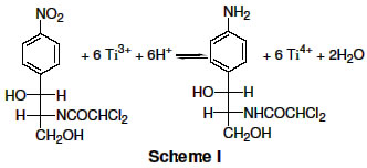
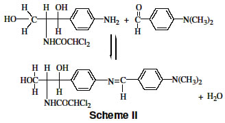
DISCUSSION We have developed an alternative colorimetric method in this study for the determination of chloramphenicol. This method is a modification of the method of Glazko and Wolf5 which involves the reduction of the nitro group in chloramphenicol with potassium borohydride in alkaline medium containing palladium and reacting the primary aromatic amine produced with p–dimethylamino benzaldehyde to give a coloured product. The method of Glazko and Wolf5 is associated with problems including (1) the reducing agent is not very soluble in the medium used necessitating filtration to be carried out after the reduction process, and (2) the reaction is not quantitative. Titanium (III) chloride in glacial acetic acid chosen as the reducing agent of the nitro group of chloramphenicol in this study has been known to reduce aromatic and heter–aromatic nitro groups easily and quantitatively at room temperature22 . This reduction process is shown in Scheme I. To ensure that the reaction proceeded favourably to the right, the amount of water must be minimised. The initial medium of equal volumes of glacial acetic acid : water (1:1 v/v) has been established to be the optimum condition for aromatic nitro group reduction22. Thus by simply mixing chloramphenicol and titanium (III) chloride in the solvent system, the reduction took place easily at room temperature within 10 min. Prolonged reaction time at high temperature was avoided for the reduction. Colour development was achieved with pdimethylaminobenzaldehyde as it reacted with the reduced chloramphenicol to form a coloured compound as shown in Scheme II. The reaction product contained an extensive conjugated double bond (chromophore) that absorbed in the visible region of the electromagnetic spectrum (λmax = 440 nm). Water is also a product of this reaction and should be minimised for product formation to proceed quantitatively. Use of methanol at all stages of the reaction minimised the amount of available water and also ensured that the products of the reactions dissolved easily. Under the experimental conditions adopted, Beer’s law was obeyed for different concentrations of chloramphenicol; thus each stage of the reaction was quantitative. At the wavelength of maximum absorption of the reaction product, 1.05 µg/ml of chloramphenicol gave an absorbance value of 0.05. Thus, the method is very sensitive. Also, the final product was stable for over 4 hr. The reactions took place homogeneously, and can be easily carried out. Of particular note is the reduction reaction which took place quickly at room temperature. When applied to assay chloramphenicol in different dosage forms, the method developed was found to be very precise. Assay results of chloramphenicol in all the different dosage forms investigated (except one brand of chloramphenicol capsule) met the requirements of chloramphenicol in the different dosage forms in respect of the percentage label claim19. Both the method developed and microbiological method proved that one of the products investigated was substandard with content of chloramphenicol being 11.4 and 11.8%, respectively. At 95% confidence interval, no significant difference was found between the data obtained using the method developed and the microbiological method. Chloramphenicol has been determined colorimetrically using vanillin for colour development of the reduced chloramphenocol23. A rapid and simple enzymatic assay of chloramphenicol has been developed which combined the specificity of the enzyme chloramphenicol acetyltransferase with the convenience of colorimetric detection24. The assay of chloramphenicol using this method was linear over the concentration range of 1.5 –65 μg/ml. The linear range of the method developed in this study falls within this range. Colorimetric detection after high pressure liquid chromatography has also been used to study the pharmacokinetics of chloramphenicol in cows after intramuscular application and to monitor the levels of chloramphenicol in the blood and milk of goats following oral administration25, 26. The method developed here can thus facilitate the colorimetric detection of chloramphenicol in the various applications. CONCLUSIONAn alternative colorimetric method has been developed that is very fast, sensitive, reproducible, simple and cheap to carry out yet gave accurate results. The developed method can be used for the routine assay of chloramphenicol in bulk drug and in pharmaceutical dosage forms. The colour development in the final product involved the reaction of p-dimethyl-aminobenzaldehyde and the primary aromatic amine of reduced chloramphenicol. Any trace of primary aromatic amine in the reaction system will thus interfere in the colour development and hence affect the final result. In order to make this method specific for chloramphenicol, it is recommended that it should be extracted first or that any product that will yield any primary aromatic amine in the reaction system is removed. REFERENCES
Copyright @2002-2004. TJPR Faculty of Pharmacy, University of Benin, Benin City, Nigeria The following images related to this document are available:Photo images[pr03013s1.jpg] [pr03013s2.jpg] |
| |||||||||
