 |
| About Bioline | All Journals | Testimonials | Membership | News |
|
||||||
|
||||||
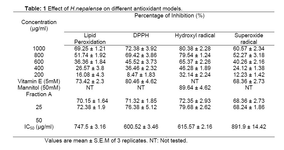
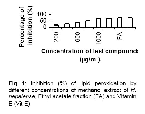
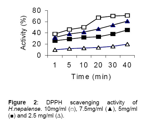
Tropical Journal of Pharmaceutical Research, Vol. 4, No. 1, June 2005, pp. 341-347 Research Article Antioxidant and antimicrobial activities of Heracleum nepalense D Don root Suvakanta Dash1*, Lila Kanta Nath1, Satish Bhise2 and Nihar Bhuyan1 1Himalayan Pharmacy Institute, Majhitar, Rangpo, East Sikkim-737132, India. 2Govt College of Pharmacy, Vidyanagar, Satara, Karad- 415124, India. Code Number: pr05003 Abstract Purpose: The aim of the present study was to
investigate antioxidant and antimicrobial effects of the methanol extract
of Heracleum nepalense D.Don roots. Keywords: Heracleum nepalense, Lipid peroxidation, Superoxide scavenging, DPPH assay, Antimicrobial effect, Flavonoid. Introduction Reactive oxygen species (ROS) such as superoxides, peroxides and hydroxyl radicals are known to play an important role and have been identified as major contributors to all cell and tissue damage in many disease conditions1. In living organisms, various ROS can form in different ways. Normal aerobic respiration stimulates polymorphonuclear leukocytes and peroxisomes appear to be the main endogenous sources of most of the oxidants produced by cells. Exogenous sources of ROS include tobacco smoke, alcohol, pesticides, certain pollutants and microbial infections 2-3. In recent years, there has been increased incidence of antibiotic resistance in pathogenic organisms and the persistence of pathogens in immune compromised individuals is of great concern4. Therefore, actions must be taken to reduce this problem such as controlling the use of antibiotics, carrying out research to better understand the genetic mechanisms of resistance and continuing investigations aimed at the development of drugs from natural sources. The recent years have witnessed resurgence of interest in herbal drugs globally as more people are turning to the use of herbal medicinal products in health care. About 80% of individuals from developing countries use traditional medicine, which involves compounds derived from medicinal plants. It is high time the hidden wonders of plant molecules were revived with the modern tools of target-based screening to develop newer advanced generation antioxidants and antimicrobials with novel modes of action. Antioxidants play an important role in inhibiting and scavenging free radicals, thus providing protection to human body against infection and degenerative diseases. Heracleum nepalense D.Don (Apiaceae) is a small shrub occurring in Nepal and Sikkim 5. The plant is used in veterinary medicine. It exhibits stimulant property and increases blood pressure in goats 6. The roots of the plant are used in folk medicine as digestive, carminative and antidiarrhoeal (Authors personal experience). The roots of the plant are reported to have coumarins 7 and steroids 8. The present study was aimed at evaluating antioxidant and antimicrobial properties of Heracleum nepalense. Experimental Plant materials and phytochemical screening The fresh dried roots of Heracleum nepalense (HNSE) were collected from the southern district of Sikkim. The plant was authenticated by Botanical Survey of India, Gangtok, Sikkim. The voucher specimen was preserved in our laboratory for future reference. The dried roots were powdered and stored in a well-closed container. 1 kg of powder (40 mesh size) was extracted by cold percolation with 3 liters of 70% v/v methanol in a percolator for 72 h at room temperature 9 .The residue was removed by filtration. The extract was then evaporated to dryness under reduced pressure in a rotary evaporator at 42-45°C. The concentrated extract of leaves was kept in a dessicator for further use. The preliminary phytochemical tests of the root extract were done by Pollock and Stevense method 10. Concentrated methanol extract was suspended in hot distilled water, cooled and the blast precipitate was filtered off. The filtrate (aqueous solution) was fractioned by extracting it successively with petroleum ether, ethyl acetate and acetone. The ethyl acetate fraction of the crude extract on purification yielded one major fraction A (flavonoid) with some fatty substances. ChemicalsThiobarbituric acid was obtained from Loba Chemie, India. 1,1-Diphynyl-2-picryl hydrazyl (DPPH), NADH and nitroblue tetrazolium (NBT) were obtained from Sigma chemicals, St. Louis, USA. Deoxy ribose was obtained from Merck India. Dimethyl sulphoxide, ethylene diamine tetra acetic acid, ferrous sulphate, trichloroacetic acid, hydrogen peroxide, ascorbic acid, mannitol, potassium dihydrogen phosphate, potassium hydroxide, deoxy ribose, phenazine methosulphate were of analytical grade and were obtained from Ranbaxy fine chemicals. Determination of antioxidant activity Assay of lipid peroxidation The extent of lipid peroxidation in goat liver homogenate was measured in vitro in terms of formation of thiobarbituric acid reactive substances (TBARS) by using standard method 11 with minor modifications 12. Goat liver was purchased from local slutter house. Its lobes were dried between blotting papers (to remove excess blood) and were cut into small pieces with a heavy-duty blade. They were then homogenized in glass-Teflon homogenizing tube in cold phosphate buffer saline (pH 7.4). It was centrifuged at 2000 rpm for 10 min and supernatant was diluted with phosphate buffer saline up to final concentration of protein 0.8-1.5 mg/0.1 ml. Protein concentration was measured by using standard method of Lowery et.al 13. To study the comparative response, the experiment was divided into nine groups. Liver homogenate (5%, 3ml) was aliquoted to nine different 35mm glass petri dishes. The first two groups were treated as control and standard where buffer and vitamin E were added. In the third to seventh group, different concentrations of methanol extract (200-1000 µg.ml-1) and fraction A (25, 50 µg.ml-1) were added. Lipid peroxidation was initiated by adding 100µl of 15mM ferrous sulphate solution to 3ml of liver homogenate 14. After 30 minutes, 100 µl of this reaction mixture was taken in a tube containing 1.5 ml of 10% Trichloro acetic acid. After 10 minutes, tubes were centrifuged and supernatant was separated and mixed with 1.5ml of 0.67% thiobarbituric acid. The mixture was heated in a water bath at 85ºC for 30 min and in a boiling water bath to complete the reaction. The intensity of pink colored complex formed was measured at 535nm in a spectrophotometer (Shimadzu model 1601). The percentage of inhibition of lipid peroxidation was calculated by comparing the results of the test with those of controls as per the following formula: Inhibition (%) = [(control − test) ×100] / control------- Eqn 1. DPPH radical scavenging activity DPPH scavenging activity was measured by spectrophotometric method14. To a methanolic solution of DPPH (100 µM, 2.95ml), 0.05 ml of methanol extract as well as fraction A (25, 50 µg.ml-1) and Standard compound (vitamin E) were added at different concentrations. Equal amount (0.05 ml) of methanol was added to a control. Absorbance was recorded at 517nm at regular intervals of 1 to 5 min. The percentage of scavenging was calculated by comparing the control and test samples with the Eqn 1. Hydroxyl radical scavenging activity Hydroxyl radical scavenging activity was measured by studying the competition between deoxy ribose and test compounds for hydroxyl radical generated by the Fe3+– ascorbate – EDTA –H2O2 system (Fenton reaction) according to the method of Kunchandy & Rao 15. The reaction mixture containing, a final volume of 1.0 ml, 100 µl 2-deoxy-ribose, 500 µl of the various concentrations of the methanol extract as well as fraction A (25, 50 µg.ml-1) and standard compound (Mannitol 50 mM) in KH2PO4- KOH buffer (20mM, pH 7.4), 200 µl 1.04 mM H 2O2 and 100 µl 1.0mM ascorbic acid was incubated at 37ºC for 1 hour. One milliliter 1% trichloroacetic acid was added to each test tube and incubated at 100ºC for 20 min. After cooling to room temperature, absorbance was measured at 532nm against a control preparation containing deoxyribose and buffer. Percent inhibition was determined by comparing the results of the test and control samples with the above mentioned Eqn 1. Superoxide scavenging activity The superoxide scavenging activity of methanol extract was determined by the method described by Nishimik et al 16, with slight modification. About 1.0 ml NBT solution containing 156 µM NBT dissolved in 1.0 ml 100 mM phosphate buffer, pH 7.4, 1.0 ml NADH solution containing 468 µM NADH dissolved in 1.0 ml 100mM phosphate buffer, pH 7.4, and 0.1 ml of various concentration of the methanol extract as well as fraction A (25, 50 µg.ml-1) and standard compound (vitamin E) were mixed and the reaction was started by adding 100 µl of phenazine methosulfate solution containing 60µM phenazine methosulphate in 100 mM phosphate buffer, pH 7.4. The reaction mixture was incubated at 25 ºC for 5 min and absorbance at 560 nm was measured against control sample. Percent inhibition was determined by comparing the results of the test and control samples with the above mentioned Eqn 1. Antimicrobial activity Bacteria A total of 257 bacterial strains belonging to different genera were tested in this study. The test organisms were obtained from Department of Bacteriology, Calcutta School of Tropical Medicine and Institute of Microbial Technology (IMTECH), Chandigarh, India. Three multiresistant Staphylococcus strains (Staphylococcus aureus ML 275, Staphylococcus aureus NCTC 8530 and Staphylococcus epidermidis 865) were kindly provided by Prof. (Mrs.) Sujata Ghosh Dastidar, Department of Pharmacy, Jadavpur University, and Kolkata, India. They were aseptically isolated and identified by the Barrow and Feltham’s method and preserved in the freeze-dried state 17. Gram-positive strains were grown in nutrient broth (NB, Oxoid brand) and Gram-negative bacteria were grown in peptone water (PW, Oxoid brand, bacteriological peptone plus Nacl 0.5%) for 18 h before use. Determination of antimicrobial activity Sensitivity tests were performed by disc diffusion method18. The nutrient agar plates (Oxoid brand), containing an inoculum size of 105-106 cfu.ml-1 of bacteria were used. Previously prepared crude methanol extract (Concentration 128-2000 µg.ml-1) and fraction A (Concentration 128-2000 µg.ml-1) discs were placed aseptically on sensitivity plates. The discs containing methanol and known antibiotics (Amoxycillin and Gentamicin) served as negative and positive controls. All the plates were then incubated at 37°C±2°C for 18 h. The sensitivity was recorded by measuring the clear zone of inhibition on agar plate around the discs. The MICs were determined by the standard agar dilution method 19. All the test compounds were dissolved in methanol. These were then individually added at each final concentrations of 0-2000 µg.ml-1, to molten agar (Oxoid brand), mixed thoroughly, pH adjusted to 7.2 to 7.4 and poured into sterile Petri dishes. Bacterial cell suspensions were spot inoculated on the plates using a bacterial planter (10 µl). The final number of cfu inoculated onto the agar plates was 104 for all strains. The inoculated plates were then incubated at 37°C±2°C for 18 h. The lowest concentration of the plate, which did not show any visible growth after incubation, was considered as MIC. The agar plate containing only methanol and Amoxycillin was served as negative and positive control. Statistical analysis Data are reported as the mean ± SD of three measurements. Statistical analysis was performed by the student t-test and by ANOVA. IC50 values for all the above experiments were determined by linear regression method. A p-value less than 0.05 was considered as indicative of significance. Results and Discussion Antioxidant activity Assay of lipid peroxidation The results presented in Table-1 showed that the methanol extract of the HNSE inhibited FeSO4 induced lipid peroxidation in a dose dependent manner. The extract at 1000 µg.ml-1 exhibited maximum inhibition (69.25 ± 1.21%) of lipid peroxidation, on the other hand fraction A at 50 µg.ml-1 concentrations showed (72.38 ±1.9%) inhibition, nearly equal to the inhibition produced by vitamin E ( Fig. 1). The IC50 value was found to be 747.5 ±3.16 µg.ml-1. The inhibition could be caused by the absence of ferryl-perferryl complex or by changing the ratio of Fe3+ / Fe2+ or by reducing the rate of conversion of ferrous to ferric or by chelating the iron itself or combination thereof 20. DPPH scavenging activity DPPH is a stable free radical that can accept an electron or hydrogen radical to become a stable dimagnetic molecule. Due to its odd electron, the methanolic solution of DPPH shows a strong absorption at 517 nm. DPPH radical reacts with suitable reducing agents and then electrons become paired off and the solution loses colour stoichometrically with the number of electrons taken up 21. Such reactivity has been widely used to test the ability of compound/plant extracts to act as free radical scavengers. Reduction of the DPPH radicals can be observed by the decrease in absorbance at 517 nm. The DPPH scavenging capacity of the extract was found to be 72.38 ± 3.92 % at 1000 µg.ml-1. The fraction A at 50 µg.ml-1 doses, on the other hand, exhibited 76.38 ± 5.12 % inhibition compared with 80.46 ± 4.62 % for the standard drug vitamin E at 5mM ( Table-1). The IC50 value was found to be 6.5 mg.ml-1. The activity was also dependent on time ( Fig. 2). Hydroxyl radical scavenging activity Hydroxyl radicals are the major active oxygen species causing lipid peroxidation and enormous biological damage 22. Ferric-EDTA was incubated with H2O2 and ascorbic acid at pH 7.4. Hydroxyl radicals were formed in free solution and were detected by their ability to degrade 2-deoxy-2-ribose into fragments that formed a pink chromogen upon heating with TBA at low pH 23. When the test compounds were added to the reaction mixture they removed hydroxyl radicals from the sugar and prevented their degradation. The extract of HNSE significantly inhibited (80.38 ± 2.28 %) degradation of deoxy-ribose mediated by hydroxyl radicals at the dose of 1000 µg.ml-1 ( Table-1), compared to that of a known scavenger mannitol (50mM). The concentration of the methanol extract needed for 50% inhibition was 615.57 µg.ml-1. The fraction A at 50 µg.ml-1 on the other hand, exhibited (78.68 ± 2.62 %) inhibition compared with (89.64 ± 2.84 %) for the standard mannitol. Superoxide scavenging activity Superoxide radical O2¯ is a highly toxic specie, which is generated by numerous biological and photochemical reactions. Both aerobic and anaerobic organisms possess superoxide dismutase enzymes, which catalyse the breakdown of superoxide radical 24. Reduced phenazine methosulfate assay was used to measure the superoxide dismutase activity of HNSE. The results presented in Table-1 showed that the scavenging capacity of the extract was 60.57 ± 2.34% at 1000 µg.ml-1. The fraction at 25 µg.ml-1 exhibited 68.36 ± 2.73 % inhibition of superoxide radicals. IC50 was found to be 8.9 mg.ml-1. Inhibition was proportional to the amount of the extract added. Antimicrobial activity The methanol extract of HNSE roots exhibited a significant in vitro antimicrobial activity against 257 Gram-positive and Gram-negative bacteria including multiresistant Staphylococcus (MRSC) strains. All the three reference strains of bacteria were found to be sensitive within 1000 µg.ml-1. The preliminary biological screening of the fractions showed that the ethyl acetate part was more active than other fractions (Data not shown). The results of the antimicrobial spectrum of the roots extract presented in Table 2 showed that out of 257 bacteria, the growth of 197 isolates were inhibited at a concentration of 128 – 512 µg.ml-1. 57 isolates were resistant up to 1000µg.ml-1, while the remaining 03 isolates where resistant up to >2000 µg.ml-1, the highest concentration tested. The MICs tests revealed that 63 out of 75 Gram-positive bacteria were sensitive between 128 and 256 µg/ml (zone diameter 10–16 mm); while out of 179 Gram-negative isolates, 120 were sensitive between 256-512 µg.ml-1 (zone diameter 10-14 mm). Hence, it appears that the antimicrobial activity of the methanol extracts was directed both against Gram-positive and Gram-negative bacteria. The ethyl acetate fraction (fraction A) was also tested for antimicrobial activity. The result revealed that all the isolates were sensitive at 128-256 µg.ml-1 (Table 3). It was interesting to note that all the MRSC were susceptible to fraction A at a concentration of 128 µg.ml-1, while they were resistant to the two test antibiotics. Thus, ethyl acetate fraction may become clinically relevant, particularly for antibiotic-resistant strains. However, the activity has to be studied using isolated pure compounds from the fraction to which the more resistant strains were susceptible in order to confirm these findings. AcknowledgementThe authors are thankful to the All India Council for Technical Education, Government of India, New Delhi and Prof (Dr) S.G.Dastidar, Division of Microbiology, Jadavpur University, Kolkata, India for providing the research facility. References
Copyright @2002-2006. TJPR Faculty of Pharmacy, University of Benin, Benin City, Nigeria The following images related to this document are available:Photo images[pr05003f1.jpg] [pr05003f2.jpg] [pr05003t1.jpg] |
| |||||||||
