 |
| About Bioline | All Journals | Testimonials | Membership | News |
|
||||||
|
||||||
Tropical Journal of Pharmaceutical Research, Vol. 5, No. 1, June 2006, pp. 539-544 Research Article Evaluation of Moringa oleifera Leaf Extract on Alcohol-induced Hepatotoxicity Nadro, M.S., Arungbemi, R.M. and *Dahiru, D. Department of Biochemistry Federal University of Technology, Yola P.M.B.
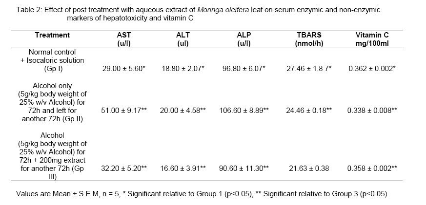
2076, Yola, Adamawa State Code Number: pr06003 Abstract A study on the protective effect of Moringa oleifera leaf extract in acute alcohol-induced hepatotoxicity in rats was evaluated. Rats fed alcohol only produced significant increase in the levels of enzyme markers of tissues damage (ALT, AST and ALP), lipid peroxidation (TBARS) and decreased serum vitamin C levels compared to normal control rats. Pretreatment with 100 and 200mg/kg body weight of extract significantly decreased the levels of enzyme markers, lipid peroxidation and markedly increased serum vitamin C level in a dose-dependent manner. Post-treatment with 200mg/kg body weight of extract significantly enhanced the recovery of animals from hepatic damage compared to untreated control. Lipid peroxidation and depletion of vitamin C due to oxidative stress could be the possible mechanisms of alcohol induced toxicity and the protective effect of the extract could be as a result of its ability to inhibit lipid peroxidation and prevent the depletion of vitamins C. Key words: Moringa oleifera, hepatotoxicity, lipid peroxidation, vitamin C INTRODUCTION Alcohol abuse is a major health problem worldwide. Alcoholic liver disease is responsible for 15-30% of all admissions in many general hospitals in South Wales1. Although important progress has been made in understanding the pathogenesis of alcoholic liver disease, current therapies for this disease are not effective. Novel therapeutic approaches such as utilizing agents that successively correct the fundamental cellular disturbances resulting from excessive alcohol consumption are attractive2. Alcohol administration has been found to cause accumulation of reactive oxygen species, which in turn causes lipid peroxidation of cellular membranes and proteins and DNA oxidation resulting in hepatocyte injury2. Based on the hypothesis that oxidative stress occurs only when the antioxidant capacity is insufficient to cope with the generation of prooxidants many studies have focused on the alcohol-associated changes in the liver antioxidants. In spite of the tremendous advances made in allopathic medicine, no effective hepatoprotective medicine is available. Plant drugs are known to play a vital role in the management of liver diseases. At the same time, surprisingly, we do not have readily available plant drugs/formulations to treat severe liver disease3. Focus on plant research has increased all over the world and a large body of evidence has been collected to show immense potential of medicinal plants used in various traditional systems4. Such scientific studies have led to isolation of chemical substances with therapeutic properties. And many of the isolates have found use as modern drugs while others have served as substrates for the synthesis of drugs. Unfortunately, a greater proportion of plants known traditionally to possess medicinal properties and which are used in herbal medicine have not been subjected to scientific evaluation which is necessary not only because of the need to discover new drugs but also the need to assess the toxicity risks. Beside, it is important that traditionally claimed therapeutic properties of such plants are confirmed even if the active principles are not ultimately discovered5. As a consequence of an increasing demand for the biodiversity in screening programs, seeking hepatoprotective therapeutic drugs from natural products, there is the need to develop interest at locally screening medicinal plants used in the treatment of liver problems. Moringa oleifera commonly known as Zogale in Hausa (Nigeria) is used both as a source of food and medicine. The leaves have been implicated in the treatment of liver diseases in traditional medicine. However there is no scientific evidence to support that the leaves can be used in the prevention or treatment of alcohol-induced hepatotoxicity. This research work was aimed at evaluating the effect of the leaf extract of Moringa oleifera Lam on alcohol-induced hepatotoxicity and the possible mechanism of action of the plant material. The University Ethics Committee for Animal Experiments approved the study. Procedures for the Animal care conformed to the international laws and Guidelines for the Use of Animals in Biomedical Research6. MATERIALS AND METHODS Animals Male albino rats, weighing between 130-150g were obtained from the Veterinary Research Institute Vom, Jos, Plateau State, and were housed in a well-ventilated room under 12h light/dark cycle. The animals were fed with a commercial diet (Vital Feed: Grand Cereals and Oil Mill Ltd, Jos) and water ad libitum. Plant material The plant material, Moringa oleifera leaves (fresh) were collected from within Yola metropolis and dried under shade at 28 ± 2oC. The dried material was made into powder using a mortar and pestle, and sieved with sieve of 0.3mm aperture size (Endicott Ltd, London). Hepatotoxin Absolute ethanol Analar (99.8%), the alcohol used to prepare alcohol solution was purchased from Mallinckrodt Chemical Works St. Louis, USA. A 5g/Kg bw of 25% w/v alcohol solution was administered as the hepatotoxin. Preparation of aqueous extract One hundred grams of dry fine powder of Moringa oleifera leaf was suspended in 600ml of water and stirred magnetically overnight (12hrs) at 37oC. This was repeated three consecutive times. The residue was removed by filtration and the extract evaporated at <40oC under reduced pressure using rotary evaporator7 to yield a semi-solid extract weighing 19.66g (196.60g/kg). Animal treatment Forty rats were divided into eight groups of 5 animals each. Two protocols were employed to study pretreatment and post-treatment effects of the extract. Protocol 1 consisted of five groups. Group I normal control; group II extract control (extract plus isocaloric solution); group III experimental control (alcohol only); group IV and V, pretreated with extract 100 and 200mg/kg bw. Protocol 2 consisted of 3 groups. Group I normal control; group II experimental control (alcohol only for 3 days) and group III (alcohol for 3 days followed by treatment for another 3 days). In both protocols 5g/kg bw of 25%w/v alcohol solution, isocaloric solution and extract were administered by oral gavage (intragastrically). Collection of blood sample At the end of the experimental period, the animals were sacrificed after ether anesthesia and blood collected without the use of anti-coagulant for serum preparation. The blood samples were collected by direct cardiac puncture and allowed to stand for 10 min before being centrifuged at 2,000 rpm for another 10 min and the serum were collected using rubber micropipette and used for the analysis. The levels of alkaline phosphatase (ALP) was by analyzed by the method of Wright et al.8, alanine aminotransferase (ALT) and aspartate aminotransferase (AST) were analysed according to Reitman and Frankel9, serum lipid peroxidation (thiobarbituric acid reactive substances, TBARS) was as described10, while serum vitamin C was determined as described earlier11. Statistical analysis Results were presented as mean ± standard deviation (Mean ± S.D) for all values. Student‘t’ test was used for test of significance between two values. SPSS statistical software (Chicago, USA) was used for statistical analysis. RESULTS The levesl of ALT, AST, ALP, lipid peroxidation as assayed by TBARS and vitamin C levels of pretreatment with or without Moringa oleifera extract are as shown in table 1. Group III rats (alcohol only) developed hepatic damage compared to group I (normal control). This was evidenced by a marked elevation (p< 0.05) in the levels of hepatic enzyme markers studied. However, the levels of the enzyme markers were significantly decreased (p< 0.01) in groups pretreated with 100 and 200mg/kg bw of the aqueous extract of Moringa oleifera. The levels of the enzymes (ALT, AST, and ALP) in 200mg/kg bw extract pretreated group were also significantly reduced compared to the group pretreated with 100mg/kg bw extract. The level of lipid peroxidation as assayed by TBARS significantly increased in alcohol only administered group compared to normal control. Lipid peroxidation was found to be significantly reduced in groups pretreated with the 100 and 200mg/kg bw of the extract and the decrease in lipid peroxidation was dose dependent between the pretreated groups. Administration of alcohol caused a significant decrease in the levels of serum vitamin C compared to normal control rats. The extract pretreated groups (100 and 200mg/kg bw) before alcohol administration showed significant (p< 0.05) increase in vitamin C levels compared to group administered alcohol only. Table 2 shows the results of the effect of post treatment of Moringa oleifera extract on acute alcohol induced hepatotoxicity. Alcohol was observed to cause marked increase (p< 0.05) in serum enzyme markers studied in the group administered alcohol for 3 days and left for another 3 days without treatment compared to normal control. Treatment with 200mg/kg extract for 3 days after administration of alcohol was found to enhance recovery from tissue damage as seen by the significant (p< 0.01) decrease in levels of serum enzyme markers studied, a significant decrease in lipid peroxidation (p< 0.05) and increased serum vitamin C concentration. DISCUSSION Alcohol is a commonly abused drug and acute ingestion of it may cause liver damage leading to other liver problems. Alcohol treatment of rats is known to cause the translocation of fat from the peripheral adipose tissue to liver, kidney and brain for accumulation12. The animals treated with alcohol only (group 1) had a significant hepatic damage as indicated by the elevated levels of serum enzyme markers of tissue damage studied. The rise in the ALT level is usually accompanied by an elevation in the levels of AST, which plays a role in the conversion of amino acids to keto acids13. Pretreatment with the extract of Moringa oleifera decreased levels of serum enzyme markers, thus suggesting that the extract possessed compounds that protected the hepatocytes from alcohol-induced liver injury and subsequent leakage of enzymes in to the circulation. Decreased levels of the enzyme markers in the post-treated group compared to control were an indication that the extract also possessed a curative effect. Acute alcohol ingestion is known to promote oxidative stress in animals and humans14. In the present study, significantly increased levels of lipid peroxidation in blood serum of rats treated with alcohol were observed, indicating the activation of the lipid peroxidation system. Lipid peroxidation is initiated by the abstraction of a hydrogen atom from the side chain of polyunsaturated fatty acids in the membrane lipids. The high vulnerability of neutral tissues to oxidative damage is partly due to its high lipid content15. Pretreatment with extract before alcohol administration significantly decreased the levels of lipid peroxidation in the blood. The antiperoxidative effect may be due to the presence of antioxidants in the leaves such as β-carotene, α-tocopherol and vitamin C as earlier reported16. Post treatment with the extract also resulted in marked decrease in the level of lipid peroxidation compared to the group left without post treatment. Thus, the extract also has the ability to not only protect the animals from the effect of lipid peroxidation induced by alcohol but also to scavenge the already produced radicals and reverse the effect of the observed lipid peroxidation. Increased lipid peroxidation under ethanol treated conditions of rats left without pre or post treatment was due to increased oxidative stress as a result of depletion of the antioxidant scavenger system which might have resulted in changes in the cellular metabolism of the liver14. The observed decreased in the level of serum vitamin C (an antioxidant) in alcohol treated group only could be as a result of increased utilization of this antioxidant in scavenging the free radicals generated or produced during acute alcohol induction17. Pretreatment of rats with the extract at the different doses administered resulted in significant increase in the levels of vitamin C. This indicates that the extract was able to raise the vitamin C levels of the pretreated animals, and increased the ability to combat produced free radicals. Raised level of serum vitamin C after post-treatment of extract enhance the recovery of the animals from alcohol-induced damage compared to untreated group. In conclusion, the study has further supported the fact that acute alcohol intake induces hepatotoxicity. The probable mechanisms of alcohol induced tissue damage were found to be due to increased lipid peroxidation and depletion of antioxidant reserves as assayed by serum vitamin C level. Pretreatment with the extract was also found to protect the liver from acute alcohol induced damage, while post-treatment with the extract exhibited a therapeutic effect. Both protective and curative effects of the extract have been attributed to the rich antioxidant nature of the leaf extract, which exhibited its effects in a dose dependent manner. References
Copyright 2006. TJPR Faculty of Pharmacy, University of Benin, Benin City, Nigeria The following images related to this document are available:Photo images[pr06003t2.jpg] [pr06003t1.jpg] |
| |||||||||
