 |
| About Bioline | All Journals | Testimonials | Membership | News |
|
||||||
|
||||||
Tropical Journal of Pharmaceutical Research, Vol. 5, No. 1, June 2006, pp. 557-560 Research Article In Vitro Antimicrobial Activity of Stevia Rebaudiana Bertoni Leaves Manish B. Tadhani and Rema Subhash
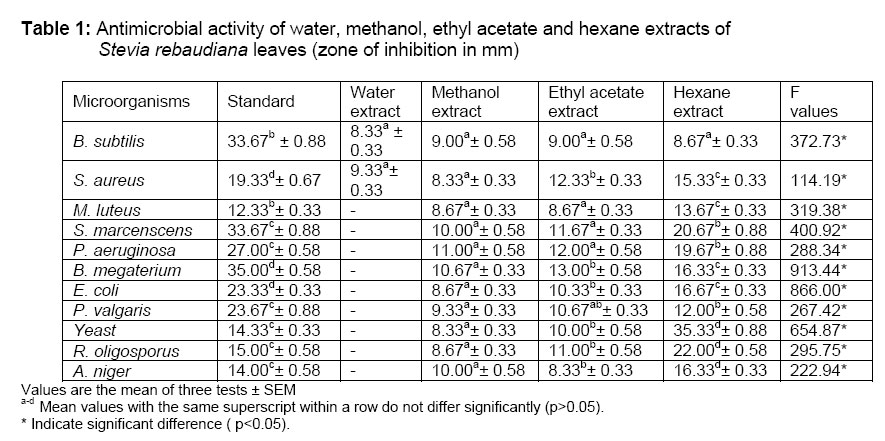
Food Biotechnology Laboratory, Post Graduate Department of Home Science, Sardar Patel University, Vallabh Vidyanagar-388 120 Gujarat, INDIA Code Number: pr06006 Abstract Purpose: The aim of the present study was to investigate antimicrobial activity of the various extracts of Stevia rebaudiana leaves. Keywords: Stevia rebaudiana, antimicrobial activity, leaf extracts INTRODUCTION The use of natural products with therapeutic properties has a long history, plant, animal, and mineral products were the main source of medicines1. Many efforts have been made to discover new antimicrobial compounds from various kinds of sources such as micro-organisms, animals, and plants. Systematic screening of them may result in the discovery of novel effective antimicrobial compounds2. Plants can possess antimicrobial natural products to protect themselves from microbial infection and deterioration3. In the developing countries, synthetic drugs are not only expensive and inadequate for the treatment of diseases but are also often with adulterations and side effects4. In recent years, concern over pathogenic and spoilage microorganisms in foods has increased due to the increase in outbreaks of food borne disease5. There are growing interests in using natural antimicrobial compounds, especially extracted from plants, for the preservation of foods. In addition, there are more consumers who tend to question the safety of synthetic additives and would prefer natural foodstuffs6-7. There is therefore the need to search for plants of medicinal value. The plant used in the present study was Stevia rebaudiana (Asteraceae), which is used traditionally for the source of natural sweetener. The dry extract from the leaves of stevia (Stevia rebaudiana Bertoni) contains sweet diterpene glycosides, flavonoids, alkaloids, water-soluble chlorophylls and xanthophylls, hydroxycynnamic acids (caffeic, chlorogenic, etc.), neutral water-soluble oligosaccharides, free sugars, amino acids, lipids, essential oils, and trace elements8. To the best of our knowledge, nobody has investigated the potential antimicrobial effect of Stevia rebaudiana leaves in India until now. Therefore, the presents study was planned to study the antimicrobial activity of water, methanolic, ethyl acetate and hexane extracts of Stevia rebaudiana leaves against the selected microorganisms. MATERIALS AND METHODS Plant Material and Microorganisms: Dried stevia leaves were supplied by Growmore Biotech Ltd., Hosur, Tamilnadu, India. Leaves were packed in Polyethylene bags and stored at -18º C until used. The bacterial strains used were obtained from the stock culture of the Department of Microbiology, V.P. Science College, Vallabh Vidyanagar, India. The organisms included in the present study were B. subtilis, S. aureus, M. luteus, S. marcenscens, P. aeruginosa, B. megaterium, E. coli, P. vulgaris, Yeast, A. niger and R. oligoporus. All the bacterial strains used for the experimental purpose were grown and maintained on nutrient agar medium. Yeast was isolated from curd sample on Sabouraud agar medium in the laboratory and maintained on the same medium. R. oligosporus and A. niger were grown and maintained on potato dextrose agar medium. Preparation of extracts: Dry powdered stevia leaves were extracted separately with water, methanol, ethyl acetate and hexane (all solvents were of HPLC grade) using environmental shaker (New Brunswick, USA) at 250 rpm for 24 hours. Then, the extracts were filtered using Whatman No. 1 filter paper and the filtrates were then evaporated to dryness under reduced pressure. Residues were stored in labeled sterile screw-capped bottles at -20°C. Antimicrobial Assay: The yield of water, methanol, ethyl acetate and hexane extracts of the leaf were found to be 22.44, 30.20, 10.31 and 1.74g %, respectively, on dry weight basis. Respective solvents were used to prepare a final concentration of 50 mg ml-1 and sterilized by filtration through a 0.45 µm nylon membrane filter. Various extracts of Stevia leaves were subjected to antimicrobial assay using the cup-plate method9. Nutrient agar plates were prepared by pouring 20 ml of nutrient agar in sterile Petri dishes for antibacterial assay. Similarly, potato dextrose agar plates were prepared for antifungal assay. These were allowed to solidify. The bacterial cultures used for assay were 24 hours old whereas fungus cultures were 4 to 5 days old. Concentration of these organisms was prepared to contain approximately 1× 10⁶ cfu/ml. Sugar tubes containing molten agar (10 ml) were sterilized and cooled to about 40-42o C. The tubes were then inoculated with 0.1 ml of the appropriate culture suspension of bacterium or fungus, mixed gently and poured onto previously solidified nutrient agar or potato dextrose agar plates, respectively. After setting, a cup borer (6 mm diameter) was properly sterilized by flaming and used to make four uniform cups in each Petri dish. The cups were then filled with different Stevia leaf extracts and allowed to diffuse for 45 minutes. The solvents used for extraction were analyzed similarly as control. Ciprofloxacin (10 µg/disc) and amphotericin-B (100 units/disc) were used as standards for bacteria and fungi, respectively. The plates were incubated at 37o C for 24 hours. At the end of the period, inhibition zones formed on the medium were evaluated in mm using a scale. The experiment was carried out in triplicates. Statistical analysis: Analysis of variance (ANOVA) and Duncan’s test were carried out using SPSS package for statistical analysis. RESULTS The in vitro antimicrobial activity of water, methanol, ethyl acetate and hexane extracts of dried Stevia leaves are shown in Table 1. The solvents used to prepare extracts showed no activity. Water extract showed activity against B. subtilis (8.33 mm) and S. aureus (9.33 mm) only whereas other three extracts showed activity against all tested microorganisms. All the four extracts did not show significantly different activity against B. subtilis. Ethyl acetate (12.33 mm) and hexane extracts (15.33 mm) showed significantly (p<0.05) higher activity compared to the water (9.33 mm) and methanol (8.33 mm) extracts against S. aureus. Activity of methanol, ethyl acetate and hexane extracts against M. luteus, S. marcenscens and P. aeruginosa were found to be 8.67, 8.67 and 13.67; 10.00, 11.67 and 20.67; 11.00, 12.00 and 19.67 mm, respectively. In the case of M. luteus, S. marcenscens and P. aeruginosa, methanol and ethyl acetate extracts activity did not differ significantly but these were found to be significantly lower (p<0.05) compared to activity of hexane extract. Methanol, ethyl acetate and hexane extracts zones of inhibition for B. megaterium and E. coli were 10.67, 13.00 and 16.33; 8.67,10.33 and 16.67 mm, respectively, which were significantly (p<0.05) different for each other. P. valgaris, ethyl acetate extract (10.67 mm) activity was not significantly different compared to methanol extract (9.33 mm) and hexane extract (12.00 mm) but hexane extract gave significantly higher activity compared to methanol extract. In the case of bacterial species, standard ciprofloxacin (10 µl/disc) showed significantly higher (p<0.05) activity compared to all the extracts except hexane extract against M. luteus. Zones of inhibition was found to be 8.33, 8.67 and 10.00 mm for methanol extract, 10.00, 11.00 and 8.33 mm for ethyl acetate extract and 35.33, 22.00 and 16.33 mm for hexane extract against yeast, R. oligosporus and A. niger, respectively. Hexane extract showed a significantly higher (p<0.05) activity compared to methanol and ethyl acetate extract as well as standard amphotericin B (100 units per disc) against yeast and fungi. DISCUSSION Antimicrobial activities of various herbs and spices in plant leaves, flowers, stems, roots, or fruits have been reported by many workers3-6-10. It is worthwhile to note that there are no data in the literature to indicate previous investigation of the antimicrobial activity of Stevia rebaudiana leaves for comparison. Water extract showed activity against only two microorganisms among the selected microorganisms. Methanol and ethyl acetate extracts have more or less similar activities but hexane extract showed significantly higher activity compared to other extracts against most microorganisms tested. The results of present study indicate that the stevia leaf extracts have inhibitory activities against microorganisms, although their antibacterial activities are lower than that of the standard (ciprofloxacin). However, in the case of fungi, hexane extract had significantly higher activity than the standard (amphotericin-B). The results of the present work indicate that stevia leaf extracts may be an ideal candidate for further research into their uses for food preservation as well as pharmaceutical and natural plant-based products. Acknowledgement The authors are indebted to Growmore Biotech Ltd., Hosur, Tamilnadu for supplying dried leaves of Stevia rebaudiana. References
Copyright 2006. TJPR Faculty of Pharmacy, University of Benin, Benin City, Nigeria The following images related to this document are available:Photo images[pr06006t1.jpg] |
| |||||||||
