 |
| About Bioline | All Journals | Testimonials | Membership | News |
|
||||||
|
||||||
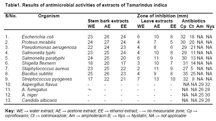
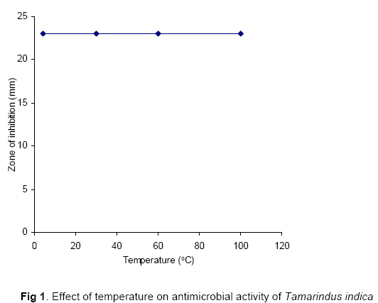
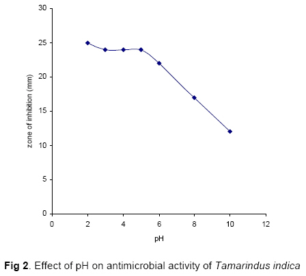
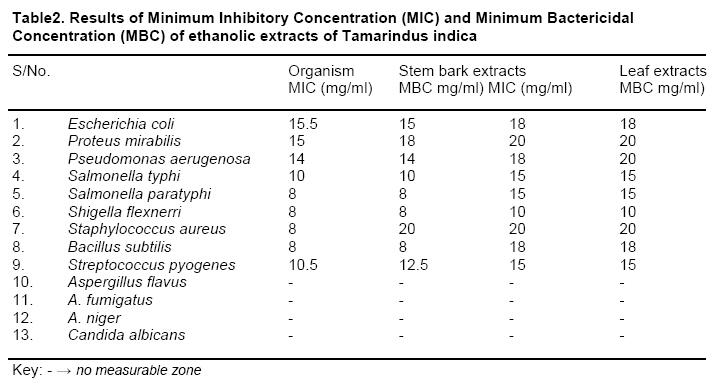
Tropical Journal of Pharmaceutical Research, Vol. 5, No. 2, December 2006, pp. 597-603 Research Article Antimicrobial Activity of Tamarindus indica Linn JH Doughari Department of Microbiology, Federal University of Technology P.M.B 2076, Yola; Adamawa State. Nigeria Corresponding: E-mail: jameshamuel@yahoo.com Code Number: pr06011 AbstractPurpose: Tamarindus indica is a plant that is used in traditional medicine for the treatment of cold, fever, stomach disorder, diarrhea and jaundice and as skin cleanser. To evaluate the scientific basis for the use of the plant, the antimicrobial activities of extracts of the stem bark and leaves were evaluated against some common gram negative and gram positive bacteria and fungi. The study also investigated the chemical constituents of the plant and the effect of temperature and pH on its antimicrobial activity. Key words: Tamarindus indica, Antimicrobial activity, minimum inhibitory concentration, minimum bactericidal concentration, chemotherapy, infectious disease. INTRODUCTION Plants remain the most common source of antimicrobial agents. Their usage as traditional health remedies is the most popular for 80% of world population in Asia, Latin America and Africa and is reported to have minimal side effects1,2. In recent years, pharmaceutical companies have spent a lot of time and money in developing natural products extracted from plants, to produce more cost effective remedies that are affordable to the population. The rising incidence in multidrug resistance amongst pathogenic microbes has further necessitated the need to search for newer antibiotic sources. Tamarindus indica Linn. (commonly called Tamarind), family Fabaceae, subfamily caesalpiniaceae is a tropical evergreen tree native to fertile areas throughout Africa and Southern Asia. It is widely cultivated as an ornamental tree and for its acidic fruits used in making drinks and a popular component of many decoctions used as health remedies. In Northern Nigeria, the fresh stem bark and fresh leaves are used as decoction mixed with potash for the treatment of stomach disorder, general body pain, jaundice, yellow fever and as blood tonic and skin cleanser. Because of its wide usage and availability, this study was set out to investigate the antimicrobial activity of the plant and to determine the effect of temperature and pH on the efficacy of the plant as an antimicrobial agent. MATERIALS AND METHODSPlant materials were collected from the wild in Yola North local government area of Adamawa state, Nigeria and were identified and authenticated at the Biological Sciences Department, Federal University of Technology, Yola. Adamawa State, Nigeria. Preparation of ExtractsThis was carried out as earlier described with slight modifications3. The freshly collected stem bark and fresh mature leaves were chopped into pieces and shade dried at room temperature (32-35oC) to constant weight for 5 days. 50g of each of the plant parts were coarsely powdered using a mortar and pestle and were further reduced to powder using an electric blender. The powder was transferred into closed containers. Each of the powdered air-dried plant material was extracted with water, acetone and ethanol. 25g of each powdered sample was mixed in a conical flask with 100ml of deionised distilled water or organic solvent, plugged, then shaken at 120 rpm for 30 minutes and kept for 24 h. After 24 h, each of the extracts was filtered rapidly through four layers of gauge and then by a more delicate filtration through Whatman no1 filter paper. The resulting filtrates were then concentrated in a rotary evaporator and subsequently lyophilized to dryness. The yield of powder was 51% from water extracts, 32% from acetone and 17% from ethanol extracts for the stem bark while the respective values of 49%, 32% and 19% (w/w) were obtained for the leaves. Test OrganismsBacteria and fungal isolates used for this work. They included Escherichia coli, Proteus mirabilis, Pseudomonas aerugenosa, Salmonella typhi, Salmonella paratyphi, Shigella flexnerri for gram negative bacteria and Staphylococcus aureus, Bacillus subtilis and Streptococcus pyogenes for gram positive bacteria, all clinical isolates obtained from the Microbiology Laboratory of the Specialist Hospital Yola, Adamawa State, Nigeria. Fungal isolates used included Aspergillus flavus, A. fumigatus, A. niger and the yeast Candida albicans and were laboratory isolates obtained from the Microbiology Laboratory of Federal University of Technology Yola, Adamawa State, Nigeria. All the bacterial strains were suspended in nutrient broth and incubated at 37oC for 48 h. Nutrient agar (NA) and Potato dextrose agar (PDA) were used for testing the antibacterial and antifungal activity respectively. Phytochemical analysisThe freshly prepared extracts were subjected to standard phytochemical analyses to test for the presence of the phytoconstituents tannins, saponins, sesquiterpenes, alkaloids and phlobatamins 4. Determination of antimicrobial activityAntimicrobial activity of the aqueous and organic extracts of the plant sample was evaluated by the paper disc diffusion method4. For determination of antibacterial activity, bacterial cultures were adjusted to 0.5 McFarland turbidity standard and inoculated onto Nutrient agar (oxoid) plates (diameter: 15cm). For the determination of antimycotic activity, all the fungal isolates and Candida albicans were first adjusted to the concentration of 106 cfu/ml. Cultures of Candida albicans were suspended in sterile solution of 0.9% normal saline and the spores of the other filamentous fungi were suspended in Tanquay buffer and all the cultures were inoculated onto Sabroud Dextrose Agar plates. Sterile filter paper discs (diameter 6mm for bacteria and 13mm for fungi) impregnated with 100µl of extract dilutions reconstituted in minimum amount of solvent at concentrations of 50 and 100mg/ml were applied over each of the culture plates previously seeded with the 0.5 McFarland and 106 cfu/ml cultures of bacteria and fungi respectively. Bacterial cultures and those of Candida albicans were then incubated at 37oC for 18 h while the other fungal cultures were incubated at room temperature (30 – 32oC) for 48 h. Paper discs impregnated with 20µl of a solution of 10mg/ml of ciprofloxacin and cotrimoxazole (for bacteria) and nystatin and amphoteracin B (for fungi) as standard antimicrobials were used for comparison. Antimicrobial activity was determined by measurement of zone of inhibition around each paper disc. For each extract three replicate trials were conducted against each organism. Determination of MIC and MBCThe minimum inhibitory concentration (MIC) of the exrtracts was estimated for each of the test organisms in triplicates. To 0.5ml of varying concentrations of the extracts (20.0, 18.0, 15.0, 10.0, 8.0, 5.0, 1.0 0.5, 0.05 and 0.005mg/ml), 2ml of nutrient broth was added and then a loopful of the test organism previously diluted to 0.5 McFarland turbidity standard for (bacterial isolates) and 106 cfu/ml (for fungal isolates) was introduced to the tubes. The procedure was repeated on the test organisms using the standard antibiotics (ciprofloxacin and cotrimoxazole for bacteria and nystatin and amphoteracin B for fungal isolates). A tube containing nutrient broth only was seeded with the test organisms as described above to serve as control. Tubes containing bacterial cultures were then incubated at 37oC for 24 h while tubes containing fungal spore cultures were incubated for 48 h at room temperature (30 – 32oC). After incubation the tubes were then examined for microbial growth by observing for turbidity. To determine the MBC, for each set of test tubes in the MIC determination, a loopful of broth was collected from those tubes which did not show any growth and inoculated on sterile nutrient agar (for bacteria) and saboraud dextrose agar (for fungi) by streaking. Nutrient agar and saboraud agar only were streaked with the test organisms respectively to serve as control. Plates inoculated with bacteria were then incubated at 37oC for 24 hours while those inoculated with fungi were incubated at room temperature (30 – 32oC) for 48 h. After incubation the concentration at which no visible growth was seen was noted as the minimum bactericidal concentration. Effect of Temperature and pH on antimicrobial activity of extractsFive milliliters of 100mg/ml of acetone extracts were constituted in test tubes and treated at 4, 30, 60 and 100oC in a water bath for 30minutes and tested for antimicrobial activity. To determine the effect of pH, acetone extracts were treated at pH ranges of 2.5 to 10 using 1N HCl and 1N NaOH solutions respectively in series of test tubes for 30minutes. After 30 minutes of treatment, each of the treated extracts were neutralized (pH 7) using 1N HCl and 1N NaOH as the case may be, and then tested for antimicrobial activity. RESULTSPhytochemical constituents present in the plant extract included tannins, saponins, sesquiterpenes, alkaloids, and phlobatamins. Results of the antimicrobial activity of the plant extracts are shown in Table 1. The result shows that the plant extracts were effective against organisms. The highest activity (diameter of zone of inhibition 27mm) was demonstrated by the acetone extracts of stem bark against Proteus mirabilis while the lowest activity (diameter of zone of inhibition 2mm) was demonstrated by the water extract against Staphylococcus aureus. The leaf extracts generally showed lower activity against the test organisms compared to the stem bark extracts. Result of the effect of temperature and pH on the plant extracts showed that various temperature ranges of 4, 30, 60 and 100oC had no effect on the antimicrobial activity of the extracts (Fig 1), but the activity slightly increased at acidic pH (2 to 6). While at alkaline pH the activity of the plant extracts reduced (Fig 2). Results of minimum inhibitory concentration (MIC) and minimum bactericidal concentration (MBC) are shown in Table 2. The result showed that Staphylococcus aureus had the highest MIC (18 mg/ml) and MBC (17.5 mg/ml), while the lowest MIC of 8 mg/ml was shown by Salmonella paratyphi and Bacillus subtilis respectively. Salmonella typhi had MIC and MBC values of 10 mg/ml for the stem bark extracts. The MIC and MBC values were generally lower for the leaf extracts against the test organisms compared to those of the stem bark extracts. DISCUSSIONPhytochemical constituents such as tannins, flavonoids, alkaloids and several other aromatic compounds are secondary metabolites of plants that serve as defense mechanisms against predation by many microorganisms, insects and herbivores5, 6. This may therefore explain the demonstration of antimicrobial activity by the stem bark and leaf extracts of Tamarindus indica. The demonstration of antibacterial activity against both gram positive and gram negative bacteria may be indicative of the presence of broad spectrum antibiotic compounds7. This will be of immense advantage in fighting the menace of antibiotic refractive pathogens that are so prevalent in recent times. The result also showed that the stem bark extracts are more effective than the leaf extracts. This may be due to the fact that the stem bark was more developed and mature than the leaves and may contain fewer pigments and other phenolics which have been reported to interfere with the antimicrobial activity of the extracts. Out of the three solvents used for extraction, the acetone extracts showed the highest activity against the test organisms, followed by the ethanol extracts and water extracts. Different solvents have been reported to have the capacity to extract different phytoconstituents depending on their solubility or polarity in the solvent6. Acetone extracts in this study might have had higher solubility for more phytoconstituents, consequently the highest antibacterial activity. The demonstration of antimicrobial activity by water extracts provides the scientific basis for the use of these plants in the traditional treatment of diseases, since most traditional medicine men use water as their solvent in which the decoctions are prepared. Although the plant is used as a decoction with other plants as skin cleanser, all the plant extracts tested did not show any antimycotic activity against any of the fungi at the tested concentrations. Their cleansing activity may be as a result of their synergy with components from other plants and some other metabolites. The temperature resistance may be an indication that the phytoconstituents can withstand higher temperatures. This also explains the traditional usage of these plant parts where a very high temperature is used to boil them and for a longer period of time. The antibacterial activity of the extracts slightly increased at acidic pH. Increase in activity of phyotoconstituents in the presence of acidic medium has earlier been reported8. The local application of these plants involves the addition of high doses of potash which is a strong basic salt, and for the fact that the activity of the extracts reduced at alkaline pH in this study, it may explain why the plant concoction is taken for longer period of time before any curative effect is noticed. The highest MIC and MBC values of Staphylococcus aureus is an indication that either the plant extracts are less effective on some gram positive bacteria or that the organism has the potential of developing antibiotic resistance, while the low MIC and MBC values for other bacteria is an indication of the efficacy of the plant extracts. CONCLUSIONThe demonstration of broad spectrum of antibacterial activity by Tamarindus indica may help to discover new chemical classes of antibiotic substances that could serve as selective agents for infectious disease chemotherapy and control. This investigation has opened up the possibility of the use of this plant in drug development for human consumption possibly for the treatment of gastrointestinal, urinary tract and wound infections and typhoid fever. The effect of this plant on more pathogenic organisms and toxicological investigations and further purification however, needs to be carried out. REFERENCES
Copyright 2006. Pharmacotherapy Group, Faculty of Pharmacy, University of Benin, Benin City, Nigeria. The following images related to this document are available:Photo images[pr06011f1.jpg] [pr06011t2.jpg] [pr06011t1.jpg] [pr06011f2.jpg] |
| |||||||||
