 |
| About Bioline | All Journals | Testimonials | Membership | News |
|
||||||
|
||||||
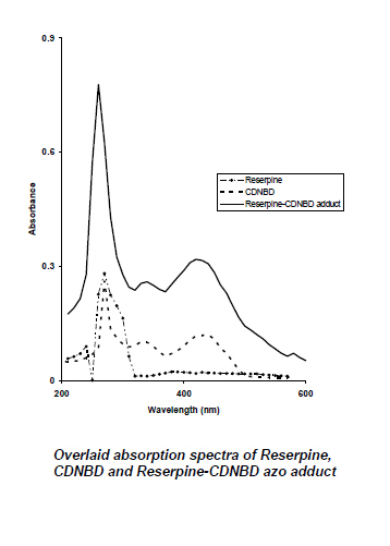
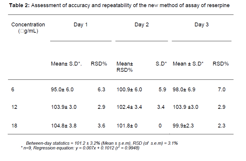
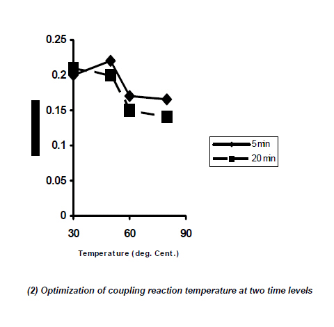
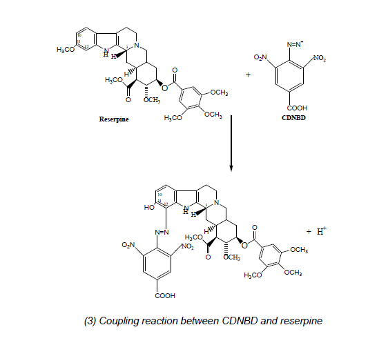
Tropical Journal of Pharmaceutical Research, Vol. 6, No. 2, June 2007, pp. 695-703 Research Article Improved Colorimetric Determination of Reserpine in Tablets Using 4-Caboxyl-2,6-dintrobenzene diazonium ion (CDNBD) Olajire A Adegoke, Sunday O Idowu* and Ajibola A Olaniyi Department of Pharmaceutical Chemistry, Faculty of Pharmacy, University of Ibadan, Nigeria *Correspondence: +234 805 8427072 olakunleid@yahoo.com Code Number: pr07009 AbstractPurpose: To develop a simple, rapid and improved colorimetric method for the assay of reserpine in tablets KEYWORDS: Reserpine, colorimetry, 4-Caboxyl-2,6-dintrobenzene diazonium ion (CDNBD), diazo coupling INTRODUCTION Reserpine is an alkaloid obtained from the roots of certain species of Rauwolfia (Apocyanaceae) mainly R. serpentina and R. vomitoria or by synthesis. The material obtained from natural sources may contain closely related alkaloids 1. Descriptions of the medicinal use of the root of Rauwolfia serpentina (Benth.), a climbing shrub indigenous to India, are present in ancient Hindu Ayurvedic writings2. Reserpine has been used in the management of hypertension and in chronic psychoses such as schizophrenia. It has also been tried in the management of Raynaud's syndrome Chemically, reserpine is (3β, 16β, 17α, 18β, 20α)-11, 17-dimethoxy-18 [(3, 4, 5trimethoxybenzoyl)-oxyl] yohimban-16-carboxylic acid methyl ester or 3,4,5-trimethoxybenzoyl methyl reserpate3. Only the active pharmaceutical ingredient is official in the BP 2002 4 and determined by a UV procedure after nitrosation. The USP 24/NF 195, uses a UV method for all Rauwolfia preparations after exhaustive and extensive solvent extraction. Other multi-ingredient preparations containing reserpine are assayed by HPLC methods in the USP 24/ NF 19. The determination of reserpine and other indole alkaloids from Rauwolfia serpentina and R. vomitoria by HPLC and HPTLC has been reported. Best separation on HPLC was achieved with 10% CH3CN and 0.1% trifloroacetic acid in water6. Qualitative and quantitative analyses of a reserpinechlorothiazide mixture in two steps by HPLC have also been described7 . Other chromatographic procedures are also described8 . Many flourimetric procedures have been described for reserpine either in bulk, dosage forms or in biological fluids. Agents that have been used include hydrogen peroxide, selenious acid, p-toluenesulphonic acid and vanadium pentoxide9 , hexa-amine cobalt (III) tricarbanato cobaltate10 and 2-iodoxybenzoate in aqueous acetic acid11 . A flow-injection assembly has also been adopted following fluorescence derivatization11 . A chemiluminometric determination of reserpine and two other Rauwolfia alkaloids, rescinnamine and yohimbine based on reaction with KMnO4/polyphosphoric acid has been described12 . Extraction of reserpine into chloroform from pH 4.0 phosphate buffer and then ion-pair formation with bromocresol purple or methyl orange have also been reported9. The most commonly used colorimetric procedure for reserpine involved oxidation of the compound to 3,4-didehydroreserpine with sodium nitrite and measurement of the absorbance of oxidation product at about 390nm. Oxidation of reserpine with nitrite in acetic acid followed by extraction into chloroform has also been done. Reserpine has also been analysed colorimetrically by reaction with vanillin, aminopyrimidine, xanthydrol (50 -500µg), phenylisocyanate, iodine and sodium glyoxalate9. Many of the above mentioned procedures suffer from the disadvantages of complexity of method, extensive solvent extraction and are time consuming. In continuation of our work on the development of relatively simple colorimetric methods for the assessment of organic compounds of pharmaceutical importance13-17, we report a new colorimetric method for the determination of reserpine in bulk and tablet dosage form using the newly developed 4carboxyl-2, 6-dinitrobenzene diazonium ion (CDNBD) 18-20. EXPERIMENTALChemicals and Reagents Brinerdine (Novartis Pharma SPA Torre Annunziata Italy for Novartis Pharma AG Basle, Switzerland), Regroton (Norvatis Pharma AG Levent-Istanbul), reserpine (CRS number 92808, Sandoz Pharma), ethanol, ethylacetate, glacial acetic acid, concentrated sulphuric acid, sodium nitrite (all analytical reagents from BDH, Poole, England), pre-coated thin layer chromatographic plates GF254, 0.2mm (Merck, Germany), 4amino-3,5-dinitrobenzoic acid (ADBA) synthesized in our laboratory. Equipment: UV/VIS spectrophotometer (Unicam Aurora, Helios Scan Software v 1.1, Pye Unicam, England), analytical balance H80 (Mettler, UK), ultrasonic bath (Langford Electronics, UK), vortex mixer (Griffins and George Ltd, UK), digital colorimeter, model 6051 (Jenway, U.K.) METHODPreparation of stock solutions An optimized process was used for the preparation of the CDNBD reagent solution using ADBA in concentrated sulfuric acid as previously reported 20. Reserpine stock solution was made by dissolving 6mg in 10ml of glacial acetic acid. This gave 0.6mg/mL solution. Optimization studies Temperature and reaction time were optimized using the method of steepest ascent21. Aliquot of the reserpine stock solution (100 µL) was added to the reagent solution (500 µL) in a test tube and the reaction mixture was mixed in a vortex mixer for 10 sec followed by incubation in turn at 30, 50, 60 and 80oC for 5 and 20 mins. Each determination was carried out in duplicate. The reaction was terminated by addition of ice-cold water (5mL) to the reaction mixture kept in icebath. The aqueous solution was extracted with ethylacetate (5 mL) as previously optimized13 and kept in a vial wrapped with aluminum foil. A blank reagent solution was similarly prepared but replacing reserpine stock solution with glacial acetic acid. The absorption spectrum of the reaction mixture extract was determined against the absorption of the blank reagent extract, using the UV/VIS spectrophotometer. The absorption wavelength, 470 nm, where the difference in absorptivity between the adduct and the reagent was optimal was selected for sample determination on the colorimeter. The optimal reaction time was determined by adding aliquot of reserpine stock solution (100 µL) in turn to the reagent solution (500 µL) in eight test tubes. Coupling reaction was carried out by incubation at 60oC for 0, 2, 5, 10, 20, 25 and 30 minutes. Ethylacetate extracts of the reaction mixture was prepared as usual after each reaction time and the absorbance was measured at 470 nm using the colorimeter. An optimum reaction time was then determined as the time corresponding to the maximal absorption of the samples. All determinations were done in duplicates. Stoichiometric ratio of drug-reagent adduct formation: Equimolar solutions (0.918 mM) of the reagent and the drug stock solution were prepared using the procedure described above. Into seven different test tubes, 0, 0.25, 0.33, 0.50, 0.67, 0.75 and 1.0 mL of the reagent solution was added respectively. Each tube was then made up to 1.0 mL with drug stock solution. Blank determinations were similarly carried out using glacial acetic acid in place of the drug stock solution. The mixtures were mixed on a vortex mixer for 10 sec and kept at 30oC for 10 minutes and extracted into 5 mL ethylacetate. The absorbance was measured at 470 nm against the blank and the absorbance values obtained were plotted against the mole fraction of the reagent solution. Each determination was carried out in duplicate. Assay of dosage forms Weight uniformity test was carried out on the two brands of the tablet obtained. For Brinerdine(R) tablets, an amount of powdered tablet equivalent to 0.4mg reserpine was weighed out and dissolved in 20 mL chloroform. After dissolution, the solution was clarified and 2mL aliquots were dried in test tubes. The sample was reconstituted in 0.25mL glacial acetic acid and then diazotized with 0.5mL of CDNBD reagent solution. The sample was treated as before. For Regroton ®, an amount equivalent to 0.5mg was dissolved in 25mL chloroform and treated as Brinerdine®. The USP 2000 spectrophotometric procedure for reserpine and hydrochlorothiazide was adopted as reference procedure. Validation of methods Calibration lines using standard solutions of 0, 3, 6, 12, 18 and 24 µg/mL reserpine were carried out using the optimal analytical conditions as described above. Linear regression analysis was used to calculate the slope, intercept and the coefficient of determination (r2) of each calibration line. The assay precision and accuracy were determined as documented by USP 22. The limit of detection was computed as previously described as the analyte concentration giving a signal equal to the blank signal plus three standard deviations of the blank23. Assessment of method selectivity Two approaches were adopted for the assessment of the selectivity of this new procedure for the assay of reserpine. In the first procedure, standard solutions of reserpine (12µg/mL) were spiked into each of starch, gelatin, lactose, magnesium stearate and a mixture of these excipients. Sample analysis was done through the optimized procedure described for reserpine above. The accuracy was compared with that of reserpine reference standard solution. In the second approach, powdered samples of the two brands of tablets investigated and the reserpine reference sample were kept at 100oC in the oven for 5 hours. Afterwards, methanolic solution of the tablets and the reference were analysed for possible degradation products. Three separate chromatographic systems were adopted [EtOAc: MeOH (9:1); EtOAc: MeOH (6:4) and Hex: EtOAc (5:5)] in order to reveal the presence of any degradation products. Stability of the azo adduct under diffuse light Standard test solutions containing 12µg/mL of reserpine were prepared in four sample vials. Two of the vials were wrapped with aluminum foil, while the other two were left unwrapped. Both sets were kept on the laboratory bench. The absorbance reading of the extracts at 470nm were taken at 30 minutes interval for a period of three hours. RESULTSReserpine was found to couple instantly with CDNBD and the colour remained stable at room temperature for days. The adduct, however, lacked fluorescence often inherent in reserpine when the TLC plate (containing the adduct and pure sample of reserpine) was examined under the UV lamp at 365nm. This indicates the formation of a new compound. The absorption of reserpine, the blank reagent and the azo adduct between them is presented in Figure 1. Reserpine was found to exhibit a major peak at 290nm while there is no significant light absorption in the visible region. The spectrum of the adduct however revealed a new λmax in the visible region at 420nm. The maximum difference in the absorptivity between the adduct and the reagent was found at 470nm. One peculiar feature of the absorption spectrum of the adduct is that it appears more of a hyperchromic shift when compared with that of the reagent (λmax 255, 340 and 430 nm). However, the colour of the adduct (reddishbrown to bright orange) is distinct from that of the yellow colour of the reagent. Absorbance of the adduct was highest at 30oC and 10 min reaction time. Maximum absorbance of the adduct was obtained at a mole fraction of 0.5 for the reagent solution and the absorbance was found to decrease at both lower and higher mole fractions. The azo adduct formed by the coupling reaction was stable when wrapped for a period of 3 hours. There was no significant difference between the content of reserpine in the tablets assayed by the new method and the USP method as shown in Table 1 (p>0.05). The linear regression equation for the calibration line under the optimal conditions is y = 0.007x + 0.1012 with a correlation coefficient of 0.9974 (r2 = 0.9948). The limit of detection for the assay of reserpine using CDNBD was found to be 2.245µg/mL. The 95% confidence limit for the slope and intercept are 0.007±0.001 and 0.1012±0.0091 respectively. Recovery studies for the assay of reserpine using the CDNBD method were done at concentration levels of 6, 12, and 18µg/ml. The three-day assessment of the accuracy and reproducibility is presented in Table 2. The overall accuracy was found to be 101.17±3.16 with a coefficient of variation of 3.12%. The photostability of the reserpine azo adduct to diffuse light in ethylacetate done over three-hour period revealed that for samples exposed to the laboratory atmosphere, the absorbance was found to drop gradually to a constant value of 0.16 from 90 to 180 minutes. The absorbance of the wrapped samples was however constant over the three-hour period. In the assessment of method selectivity, the following recoveries were obtained in the presence of the excipients; starch (102.39 ± 2.51), lactose (103.40±3.1), magnesium stearate (103.49 ± 2.90), gelatin (102.69 ± 2.49) and a mixture of these excipients (102.39 ±2.51) when compared with the reserpine alone (103.9 ± 3.0). In the second approach, the tablet samples gave a single spot in the three mobile phases investigated. It appears that reserpine is thermally stable. DISCUSSIONThe colorimetric determination of reserpine via diazo coupling reaction, using CDNBD, is the first of such methods to be carried out. While most previously reported methods are based on measurement of the fluorescence of reserpine, a cheaper and readily adaptable colorimetric method would be desirable. Optimization of coupling reaction temperature was investigated at 30, 50, 60 and 80oC at both 5 and 20 minutes. The optimization of reaction temperature is depicted in Figure 2. One prominent observation is that at 5 and 20 minutes, the absorbance of the adduct gradually falls after 50oC. At 5 minutes, a very slight increase (0.02 absorbance units) was observed when the temperature was increased by 20oC (i.e. 30oC to 50oC). The absorbance of the azo adduct thereafter dropped. Similarly, the absorbance at 30oC, 20 minutes is only 0.01 higher than that at 30oC, 5 minutes. Since a gain in time of analysis will be achieved on using the most convenient temperature, the room temperature of 30oC was selected as the temperature for the reaction of reserpine with CDNBD. The optimization of coupling time gave 10 minutes as the optimum time. Maximum absorbance for the reserpine azo adduct was observed at the mole fraction of reagent being 0.5. This implies that the reserpine molecule required just a molecule of CDNBD for the dye formation. The absorbance gradually increased from lower mole fractions of the reagent and peaked at 0.5; the value decreased thereafter. The 1H NMR of reserpine reveals that the proton on the indole ortho (C-12) to the methoxyl group is the most shielded24. Thus electrophilic attack by CDNBD will be on this position. Judging from the spectra observed for indomethacin16 which shares the indole nucleus with reserpine, the structure of reserpine-CDNBD adduct is proposed as shown in Figure 3. This is further corroborated by the 1:1 mole ratio obtained between reserpine and CDNBD. The recovery studies showed results that represent a great improvement over other previously reported spectrophotometric methods that adopted non-specific reactions for isolation and determination of reserpine. This high recovery obtained here is comparable with those obtained with some flourimetric methods especially that described by Walash et al 10 where the recovery of 100.8±1.4% was obtained. The lower concentration value adopted (6 µg/mL) was found to give the least precise results. This can be attributed to the fact that it is close to the limit of quantitation (LOQ = 3 x LOD = 6.7335 µg/mL). However, better recoveries were observed for 12 and 18 µg/ml analyte sizes. Since 12 µg/mL represents the midpoint of the calibration line and since it also gave precision results close to 18 µg/mL concentration levels, it was selected as working concentration for subsequent analysis. The result of the photostability study revealed that the new method (CDNBD) has a common property with all methods (including the BP and USP methods) used for the analysis of reserpine and that, all sample solutions be protected from light. However, if the analysis could be carried out within thirty minutes in the CDNBD method only 0.03 absorbance loss will be observed (from 0.21 at 0 minute to 0.18 at 30 minutes). It can therefore be concluded that the azo adduct of reserpine is stable up to three hours when sample solutions in ethylacetate are wrapped in aluminum foil. This new procedure described for reserpine for the first time offers a great advantage of simplicity. It is also the first colorimetric procedure for reserpine based on azo dye formation. We have previously demonstrated the reactivity of CDNBD as a useful agent for the detection and assay of phenol ethers 16, 17, 25. Essentially, the ability of the method to determine reserpine in multicomponent formulations without recourse to extensive extraction procedure are of great advantage. Table 1: Assay of reserpine in Brinerdin® and Regroton®
USP requirement: content of reserpine = 90-110% of labeled amount a; n =5 b; n =4 This new method via azo dye formation gave higher accuracy than the chemiluminometric determination of Pinotsis et al 12. When compared with the use of vanillin and xanthydrol as derivatizing reagents, CDNBD method is more sensitive. Another advantage, when compared with the reference USP method, is that this method offers some degree of selectivity. The USP method uses nitrous acid generated in situ for nitrosation and any residual or contaminant aromatic skeleton will also react. However, CDNBD will only react with an activated skeleton. CONCLUSIONThis new method for reserpine represents an improved approach for its determination. It offers the advantages of speed, simplicity and selectivity. Moreover, colorimeters are readily available and affordable. It is of equivalent accuracy with the official procedure and it could find application as an in-process analytical method for reserpine. The utilization of CDNBD as a pre-column derivatization reagent for HPLC analysis of reserpine is under investigation in our laboratory. REFERENCES
Copyright 2007. Pharmacotherapy Group, Faculty of Pharmacy, University of Benin, Benin City, Nigeria. The following images related to this document are available:Photo images[pr07009t2.jpg] [pr07009f2.jpg] [pr07009f1.jpg] [pr07009f3.jpg] [pr07009t1.jpg] | ||||||||||||
| |||||||||
