 |
| About Bioline | All Journals | Testimonials | Membership | News |
|
||||||
|
||||||
Tropical Journal of Pharmaceutical Research, Vol. 6, No. 2, June 2007, pp. 725-730 Research Article Evaluation of Phytochemical Constituents, Antibacterial Activities and Effect of Exudate of Pycanthus Angolensis Weld Warb (Myristicaceae) on Corneal Ulcers in Rabbits DN Onwukaeme1 TB Ikuegbvweha1 and CC Asonye21Department of Pharmacognosy, Faculty of Pharmacy, University of Benin, Benin City, 2Department of Optometry, Faculty of Life Sciences, University of Benin, Benin City, Nigeria *Correspondence: Tel: +234-8033714849 E-mail: nkemmkparu@yahoo.com Code Number: pr07013 AbstractPurpose: A 1: 1: 2 mixture of exudates of Anchomonas difformis, Cyrtrospherma senegalense and Pycanthus angolensis is claimed to be used for the treatment of corneal ulcers. The purpose of the study is to evaluate the phytochemical constituents, antibacterial activities and the effectiveness of a mixture of these exudates in the treatment of corneal ulcers as claimed in Traditional medicine practice. Key words: Pycanthus angolensis, exudates, corneal ulcers, rabbits. INTRODUCTION Pycanthus angolenses Welw Warb (Myristicaceae) is also known as “African nutmeg or false nutmeg'1 The Nigerian Local names of the plant are, Abakang (Ibibio) Akwa mili (Ibo) Abora (Itsekiri), Akamo, Akujaadi (Yoruba), Nupe (Kpokgi), and Abaororo (Urhobo). Others are: Etena (Cameroon), and Loioka (Zaire)1 It is a forest tree of about 30.5 m high 2.45 m in girth. The bark is grey, longitudinally fissured, flaking in patches in old trees and exudes reddish coloured juice. It is widely used for ethnomedical purposes2 An infusion of the bark is reported to be effective in the treatment of leprosy3 and for purification of breast milk in Guinea. A terpenoid quinone with potential use in treatment of Type 2 diabetes was isolated from the plant4 Herbal medicine practioners in some parts of Delta State, Nigeria, claim that a 1: 1: 2 mixture of exudates of Anchomones difformis: Cyrtosperma senegalense: and Pycanthus angolensis respectively is used for treatment of corneal ulcers. (Personal communication with Tega Ikuegbvweha, the second-named author). This work was done to verify this claim. EXPERIMENTALPlants exudates. Parts of Anchomonas difformis, and Pycanthus angolensis plants were collected from Ikpoba Hill, Benin City and Crytosperna senegalense was from Urhobo by the second named-author. They were identified by Alhaji A. Abubakar a Taxonomist formally with the Department of Pharmacognosy, Faculty of Pharmacy, University of Benin. Exudates from each plant was collected into a separate bottle, reduced to dryness on the boiling water bath and the residue subjected to the tests. Phytochemical tests: Phytochemical tests for bioactive constituents were carried out on portions of the residual material using standard phytochemical procedures5- 8. Fehling's test for reducing sugars (in glycosides): The residue was re-dissolved in water on the water bath. To 2ml of the solution, in the test tube was added, 1ml each of Fehling's solutions A and B. The mixture was shaken and heated in a water bath for 10min. The colour obtained was recorded. A brick-red precipitate indicates reducing sugar. Frothing test for saponins: Water extract was obtained by boiling on the water bath . The extract was transferred into a test tube and shaken vigorously then was left to stand for 10 mins and the result noted. A thick persistent froth indicates saponins. Borntrager's test for anthraquinone derivatives: Chloroform extract of the material was obtained by boiling on the water bath. To 2 mls of this extract, 1ml of dilute (10 %) ammonia was added and the mixture was shaken. Any colour change was recorded. A pink-red colour in the ammoniacal (lower) layer shows anthracene derivatives. Test for Flavonoids Water extract of the sample was reduced to dryness on the boiling water bath. The residue was treated with dil. NaOH, followed by addition of dilute HCl, solubility and colour were noted. A yellow solution with NaOH, which turns colourless with dil HCl confirms flavonoids. Ferric chloride solution test for tannins: Water extract was treated with 15 % ferric chloride test solution. The resultant colour was noted. A blue colour indicates condensed tannins, a green colour indicated hydrolysable tannins. Colour tests for alkaloids: 500 mg of plant material was extracted with 500 mls of methanol for 20 minutes, on a water bath The extract was then filtered off and allowed to cool. This extract was dispensed in 2 ml of portions into four different test tubes. Either the Dragendorff's or Hager's or Mayer's or Wagner's alkaloidal reagent was added to each tube and the presence or absence of colours of any precipitates were noted in each test tube. Keller- Kiliani test (for de-oxy sugars in cardiac glycosides): Methanol extract was obtained and the extract reduced to dryness. 50 mg of this was dissolved in 2 ml chloroform. H2SO4 was added to form a layer and the colour at interphase recorded. Brown ring at interphase is characteristic of deoxysugars in cardenolides. The results showed the presence of only reducing sugars in A. difformis and C. senegalense but showed that P. angolensis contained reducing sugars, as well as tannins and flavonoid glycosides. These are constituents that could have biological activity, hence the biological tests were carried out on the P. angolensis exudates only. Table 1: Phytochemical constituents of exudates of Pycanthus angolensis
Table 3: Effects of exudates of P. angolensis on chemical-induced corneal ulcers in rabbits
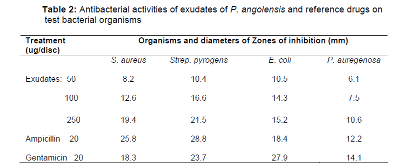
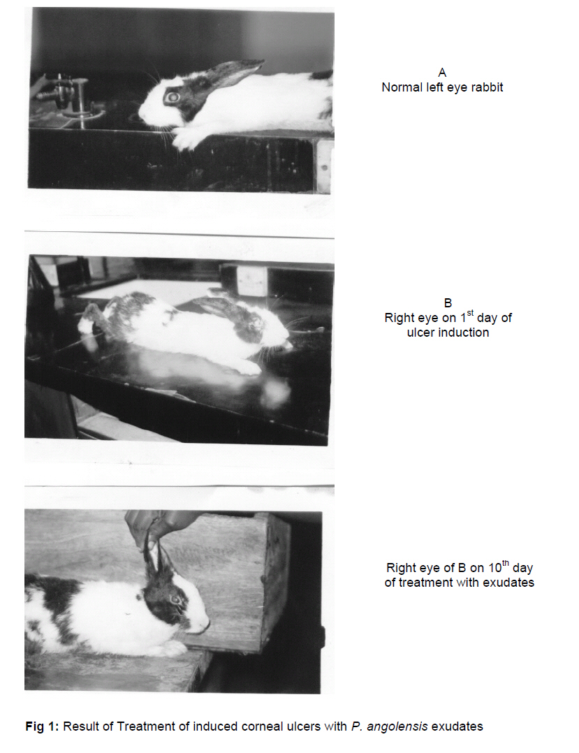
Test Organisms: Standard Laboratory strains of the organisms were obtained from the University of Benin Teaching Hospital and sub cultured in the Laboratory of Pharmaceutical Microbiology, University of Benin. The organisms used were Escherichia coli (Gram-negative), Pseudomonas aeruginosa (Gram-negative) Staphylococcus aureus (Gram-positive) and Streptococcus pyrogens (Gram-positive). The bacterial cultures were maintained on nutrient agar (Oxoid Ltd). Prior to use, they were sub-cultured into nutrient broth and incubated overnight at 37 0C. Antibacterial tests The activities of exudates on the organisms were evaluated using the paper disc diffusion method (9). 100 mg of dried residue was redissolved in 10 ml normal saline and from this stock solution volumes equivalent to 50, 100 and 250 µg/disc were spotted on antibiotic sterile assay paper discs (Whatman No. 1 (0.6cm diameter). A blank containing normal saline and another containing 20 µg/disc of a particular reference drug (Ampicillin or gentamicin) were also set up. The discs were allowed to air-dry and then placed on pre-sterilized agar plates (10 cm diameter) containing 10 ml of Agar Muller Hinton 3.5% medium, and spread with 10 µl of a standardized inoculum (5 x 10⁵ cells/ml) of the test bacterium. Plates were incubated for 24 hr. at 37oC and zones of inhibition measured (mm). Experiments were set up in four replicates. The results are presented in Table 2. Effect of exudates on corneal ulcers in rabbits. Animals: Rabbits, 300-450 g body weight were bought from Aduwawa in Benin City. They were maintained on potato leaves, elephant grass and mature unripe pawpaw fruits and water for a period of 8 weeks for them to acclimatize. Animal Care: They were de-wormed by 0.1ml invome SuperR subcutaneously (sc) administered. Also to improve their immunity and check infection and stress, keproceryl and vitalite were given orally. Induction of ulcer. Animals were anaesthetized with ketamin injection (sc) 2 drops of 4% Novesea tropical anesthetic agent instilled into the cornea of right eyes of 2 rabbits, (A) followed 20 mins later with 2 drops of 1M solution of NaOH instilled into the eyes. The 2 left eyes (B) were left as control. The 2 animals showed ulcerated cornea (A) and so were used for the test. The dried residue was re-dissolved in normal saline to make a stock of 2% w/v from this, 2 drops were daily instilled into the right ulcerated eyes (A). The extract was administered 3 times daily for nine consecutive days following induction. Animals were allowed free food and water. During this period, observations were made for any signs of toxicity, infection and discharge, status of the ulcerated cornea, any healing. Photograph of the ulcerated, non-ulcerated and treated eyes were taken on the 1st day of treatment and on the and 10th day of experimentation. The photographs are enclosed. (Fig. 1) RESULTS AND DISCUSSIONExudates of A. difformis and C. senegalense, contained only reducing sugars. Exudate of P. angolensis contained reducing sugars, flavonoids and tannins. In the antimicrobial test, (Table 2) 50 µg/ml exudate had least activity against all the organisms. It was most active against Strep pyrogens. Though 250 µg/ml showed significant activity on the organisms, these were less than those of the reference drugs. The induced ulcers were healed within nine days (Fig. 1). There was no sign of toxicity in the animals that received the exudates. Phenolic compounds are generally noted for their antimicrobial activities.5,8 Specifically, antimicrobial activities of tannins and flavonoids of Allium cepa 10 and Thymus serphylum11 have been reported. These phenolic constituents present in the exudates could then be responsible for its antibacterial effect as noted in this work. In addition, tannins act externally as water proof to the external layers of the exposed tissue since they precipitate proteins, thus protecting the underlying layers. They also have vasoconstriction effect on small superficial or exposed vessels. By limiting fluid losses and by preventing external aggressions, tannins enhance tissue regeneration in case of superficial wounds or burns8,10. This further strengthens the healing activity of the exudates. Thus the healing effect of exudates could also be attributed to the above actions of Flavonoids and tannins. CONCLUSIONThis work has identified the phytochemical constituents in exudates of A. difformis, C. senegalense and P. angolensis. A the antibacterial activities of exudates of P. angolensis, though less than the activities of the reference drugs, ampicillin and gentamicin, has been demonstrated. The P. angolensis exudates alone was active , it healed corneal ulcers within nine days of treatment, in contrast to the claim that the mixture of exudates of the three plant materials exerts the effect. REFERENCES
Copyright 2007. Pharmacotherapy Group, Faculty of Pharmacy, University of Benin, Benin City, Nigeria. The following images related to this document are available:Photo images[pr07013t3.jpg] [pr07013t2.jpg] [pr07013f1.jpg] [pr07013t1.jpg] | |||||||||||||||||||||||||||||||||||||||||||||||||||||||||||||||||||||||||||||||||||||||||||||||
| |||||||||
