 |
| About Bioline | All Journals | Testimonials | Membership | News |
|
||||||
|
||||||
Tropical Journal of Pharmaceutical Research, Vol. 7, No. 1, March 2008, pp. 867-877 Research Article A Mathematical Analysis of Intravitreal Drug Transport R Avtar, D Tandon* Department of Mathematics, Harcourt Butler Technological Institute, Kanpur 208002, India Code Number: pr08002 Abstract
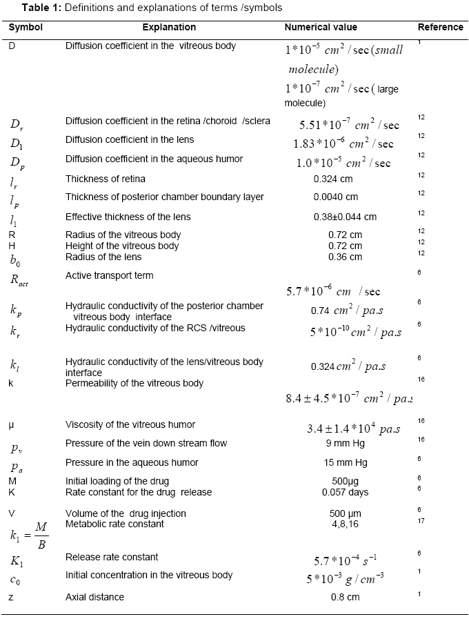
Purpose: The aim of our present work is the development of a quasi steady-state model for the distribution of intravitreally injected drugs and investigation of the effects of various model parameters on the drug distribution in normal and diseased eyes. Keywords: Convective-diffusive transport, intravitreal injection, line-Jacobi iterative technique, release rate. INTRODUCTION Several vitroretinal diseases such as cytomegalovirus retinitis, age- related macular degeneration (AMD), retinitis pigmentosa (R.P), diabetic retinopathy and a combination of similar retinal diseases are currently being treated by using drug intravitreal injection or control release implant of drugs1. The diffusion of drug, convection of vitreous outflow, enzymatic reaction (metabolism), drug binding and efficacy of delivery system mainly control the bioavailability of drug after its intravitreal injection and controlled release implant. Many drugs used to treat vitreoretinal diseases have a narrow concentration range in which they are effective and may be toxic at higher concentrations 2, 3. Therefore, it is critical to know the drug distribution within the vitreous following delivery by intravitreal injection or controlled release implant. The ability to predict drug distribution can maximize the therapeutic benefits and minimize potential adverse effect such as possible tissue damage caused by excessively high concentrations of drug. A mathematical analysis of the drug concentration and theoretical investigation of the effects of physiological parameters on the concentration may elucidate the mechanism of drug transport in the vitreous and may contribute to the improvement of present understanding of the bioavailability of drugs required for the treatment of vitroretinal diseases. Several studies4, 5, 6 ,7 have analyzed intravitreal drug distribution and the elimination of drug from the vitreous of the eye. Previous studies6, 8 have assumed that the vitreous humor was stagnant, ignoring convective drug transport within the vitreous body. It is well known that during the pathogenesis of glaucoma, intraocular pressure (IOP) is elevated (40-80 mm Hg) due to the obstruction of the aqueous outflow system which may increase the flow of aqueous humor through the vitreous. Under extreme pathophysiologic conditions, e.g rhegmatogenous retinal detachment, the integrity of the retina is broken, allowing vitreous outflow to pass into the subretinal space. The rate of fluid movement through a retinal tear was estimated to be 1.2 µL/min9, signifying a ten-fold increase and a maximum condition for elevated flow, when compared to normal vitreous outflow of 0.1 µL/min10.This increase in the vitreous outflow may be caused by the enhanced hydraulic conductivity of the retina. Thus, the vitreous outflow can play a significant role in drug distribution in glaucomatous or rhegmatogenous retinal detached eyes. Stay MS. et al 6 developed a mathematical model for prediction of the biodistribution of drug in the vitreous body released from injectable biodegradable polymer microspheres. They considered the diffusive and convective mass transport of drug within the vitreous and neglected the metabolic consumption and degradation of drug in vitreous body. Kakuji 11 has also presented a pharmacokinetic model for ocular drug delivery in the spherical modified cylindrical eye based on Fick’s law of diffusion but neglected the convective transport. The present work is concerned with the development of a simple mathematical model for the quasi-steady state concentration distribution of drug in the viteous body. The objective of the present work was to investigate the effects of the parameters metabolic rate and intraocular pressure on the drug concentration distribution of the intravitreally injected drugs and on change in drug concentration with time at the centre of retina. Besides the some other physiological parameters on the drug concentration also has been observed. MATHEMATICAL FORMULATION
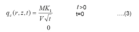
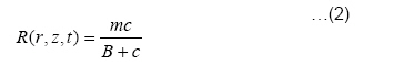
where m, B are the reaction rate and Michaelis Menten rate constants, respectively, for the metabolic process in the vitreous body. In the present study, the metabolic and degradation of drug is approximated by a first order metabolic rate constant in the vitreous body for intravitreal injection. The delivery of drug at the injected site is described by the following form 10
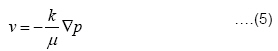
where M is the initial loading of the drug (kg), K1 (sec−1) the release rate constant of the drug, and V the volume of the drug injected (m3) .The release term is defined only at the injection site andis zero for all other positions within the vitreous body. Now, Eq. (1) can be written in the form: The aqueous flow in the porous vitreous body is described by Darcy’s Law:
µ where k is the permeability of the vitreous body, µ , the viscosity of the permeating aqueous humor, and p, the fluid pressure in the vitreous body. A ratio Using continuity equation ∇.v= 0 ,we get: ∇2 p = 0 ….(6) where, ∇2 is the Laplacian operator in a cylindrical co-ordinate system. Boundary Conditions for Pressure: The physiologically relevant and mathematically consistent boundary conditions required for the determination of velocity components of vitreous body are described below:
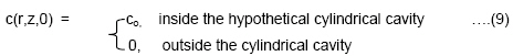
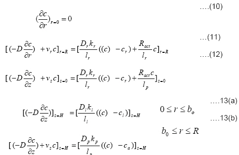
where, kr the hydraulic conductivity of the retina , kp the hydraulic conductivity of posterior chamber –vitreous body interface , p the fluid pressure in the vitreous body, pv ,the pressure of the vein downstream flow, and pa ,the intraocular pressure. Initial Condition for Concentration: For the intravitreal injection of drug, the initial condition is described as follows:
Boundary Conditions for Concentration: The boundary conditions for the drug concentration are given as follows:
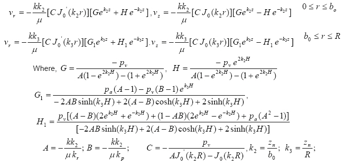
Where c is the drug concentration in the vitreous body,bo the effective radius of lens.The subscripts p,l,r refer to as the posterior chamber membrane, posterior lens surface and the RCS membrane, respectively. Solution to the model. The analytical solution to Eq. 6 subject to the boundary conditions 8(a,b)-10(a,b) is given by : p(r, z) =[CJ0(k2 r)][Gek2 z + Hek2 z ] 0 ≤ r ≤ b0 …..(14) p(r, z) = [CJ0(k3r)][G1ek3 z + H1 ek3 z ] b0 ≤ r ≤ R ......(15) And, the radial and axial velocity components of vitreous outflow are obtained as given below:
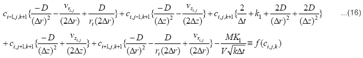
where zn are the possible zeros of the Bessel function. The approximate steady-state distributions of radial and axial velocities were computed and use in numerical solution of the mathematical model. Numerical Solution to the Model: A numerical solution of the partial differential equation governing the quasisteady state distribution of drug in the vitreous body is obtained by using d in numerical solution of the mathematical model. the implicit Crank-Nicolson scheme16. The finite difference analogue of Eq.4, obtained by using the implicit Crank-Nicolson’s scheme16, is given by:
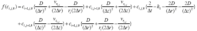
where f(ci, j,k ) denotes the term corresponding to the equation for location (i,j) in the grid and is given by:
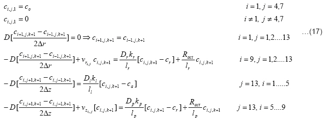
Finite difference analogues of the initial condition (9) and boundary conditions (10-13(b)) are given below
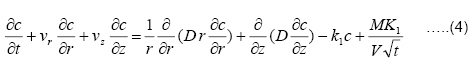
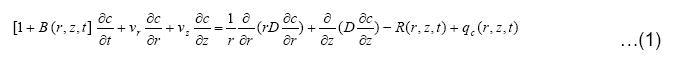
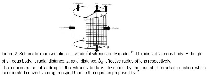
The concentration of a drug in the vitreous body is described by the partial differential equation which incorporated convective drug transport term in the equation proposed by 10:
where c is the concentration of drug in the vitreous body, D the diffusion coefficient of drug, R(r,z,t) the metabolism and degradation rate, B(r,z,t ) drug binding term and qc(r, z,t) release rate of injected drug. Effect of drug binding in the drug concentration is negligible compared to that of convection, diffusion and metabolism of drug. The metabolism and degradation of drug in the vitreous body is assumed to follow Michaelis Menten kinetics described by11,13.
where m, B are the reaction rate and Michaelis Menten rate constants, respectively, for the metabolic process in the vitreous body. In the present study, the metabolic and degradation of drug is approximated by a first order metabolic rate constant in the vitreous body for intravitreal injection. The delivery of drug at the injected site is described by the following form Kakuji11
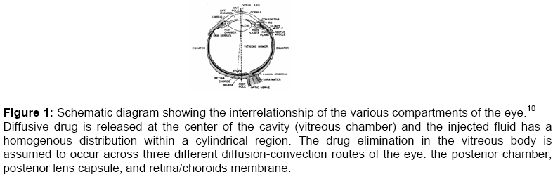
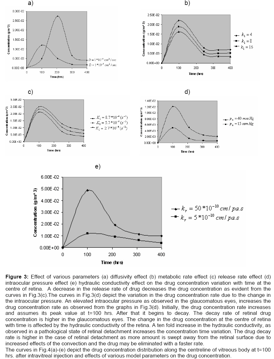
The almost spherical vitreous body Fig.(1) is represented as a porous fluid filled cylinder in contacts with the retina/choroids membrane, the lens posterior capsule (chamber), and the posterior aqueous humor Fig.(2). A hypothetical cylindrical cavity is assumed placed on the axis of symmetry behind the lens in the vitreous body. We assume that the diffusion of drug and moving liquid will originate in this cavity. The implicit iterative scheme given by Eq.16 is simplified in the light of finite difference analogues of the initial and boundary conditions 10-13(b) and the resulting system of algebraic equations written in the pentadiagonal matrix form Aci, j,k +1 = f (ci, j,k ) was solved by the line-Jacobi iterative technique. Computations were conducted and post processed on a COMPAQ-PC, Intel Pentium IV 2.40 GHz processor, with 1.0 GB of RAM, and 80 GB hard disk. The results were plotted in Excel. RESULTS The computational results of the present model have been obtained by solving the system of algebraic equations obtained from Eq.16 along with Eq.17 and using estimated values of the physiological parameters listed in Table 1. The concentration plot of the drug at the centre of retinal surface with respect to time and effects of various model parameters on the concentration change with time have been displayed in Fig.(3)(a)-(e). It is observed from curves that the drug concentration increases with time in initial hours after intravitreal injection. The concentration of high diffusivity drugs is lower than that of low diffusivity drugs in initial hours. After 100 hours the concentration of high diffusivity drugs becomes higher than that of low diffusivity drugs. As is evident from the graphs in Fig.(3)(b) an increase in the metabolic reaction rate decreases the drug concentration at the centre of retina. This is true because when the value of metabolic reaction rate is increased, more amount of drug will be consumed and degraded leading to a decrease in the drug concentration at the centre of retina. The drug concentration change with time is influenced by the rate of drug delivery.
DISCUSSION The concentration distribution of intravitreally injected drug and the change in drug concentration with time at the centre of retina are relevant for treatment of some vitro-retinal diseases such as, age-related macular degeneration (AMD), retinitis pigmentosa, glaucoma, rhegmatogenous retinal detachment etc. The present analysis has been primarily concerned with the theoretical investigations of independent effects of the intraocular pressure and metabolic consumption of drug on the concentration distribution in the vitreous body and timevariation in the concentration of low and high diffusivity drugs at the centre of retina. As has been observed a rise in the intraocular pressure and a retarded metabolic consumption causes an increase in the drug concentration and concentration change at the retinal surface. Some future experimental studies must be directed towards the investigations of such effects and focus on the similar analysis. The present analysis has been concerned with the investigation of independent effects of various model parameters on the drug concentration. But in practical situations these parameters will interact and there should be a consideration for the interactive effects. For the purpose, future studies in this field should try to propose and establish relationships among/ between model parameters. However the interactive effects of the parameters can be observed by using different sets of values of model parameters of interest. The mathematical model proposed in this study will be a useful approach for predicting the drug availability and its elimination in normal and pathological states. The model results may be useful in the design of therapeutic procedures required for the safe and effective therapeutic use of a drug. CONCLUSION The concentration of intravitreally injected drug at the centre of retina an along centreline of vitreous body is reduced as the metabolic reaction(consumption) rate and drug release rate constant increase in normal and diseased eyes. It is seen from the various curves in graphs that drug concentration decay rate in the diseased eye is higher than that in the normal eye. ACKNOWLEDGMENT The authors gratefully acknowledge the constructive and fruitful comments of the reviewers of original manuscript of the paper. REFERENCES
© Pharmacotherapy Group, Faculty of Pharmacy, University of Benin, Benin City, Nigeria. The following images related to this document are available:Photo images[pr08002f3.jpg] [pr08002f2.jpg] [pr08002t1.jpg] [pr08002f4.jpg] [pr08002f1.jpg] |
| |||||||||
