 |
| About Bioline | All Journals | Testimonials | Membership | News |
|
||||||
|
||||||
Tropical Journal of Pharmaceutical Research, Vol. 7, No. 1, March 2008, pp. 879-885 Research Article Characterization Of Glipizide-Loaded Polymethacrylate Microspheres Prepared By An Emulsion Solvent Evaporation Method BC Behera*a, SK Sahooa, S Dhala, BB Barika, BK Guptab aUniversity Department of Pharmaceutical Sciences, Utkal University, Vani-Vihar, Bhubaneswar, Orissa. Pin- 751004. bDept. of Pharmaceutical Technology, Jadavpur University Kolkata, India Code Number: pr08003 Abstract
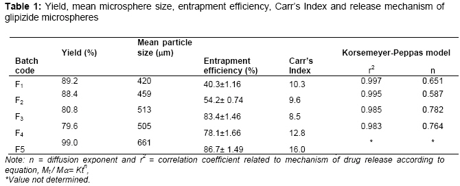
Purpose: The purpose of the present investigation was to formulate and evaluate microencapsulated glipizide produced by the emulsion – solvent evaporation method, Keywords: glipizide, Eudragit®, microspheres, controlled release, polymethacrylate. INTRODUCTION Microspheres have been widely accepted as a means to achieve oral1 and parenteral controlled release2 drug delivery system. The microsphere requires a polymeric substance as a carrier and a core material. Among the various methods developed for formulation of microspheres, the solvent evaporation method has gained much attention due to its ease of fabrication without compromising the activity of drug. In the present investigation, Eudragit® RS 100 and Eudragit® RS 100 in combination with RL 100 microsphers were used as encapsulation materials. Eudragit ® RS 100 and Eudragit® RL 100 are referred to as ammoniomethacrylate copolymers, with the former having 5% functional quaternary ammonium groups and the latter having 10% functional quaternary ammonium groups. Eudragit® RS 100 is a water-insoluble polymer that is widely used as a wall material for sustained release microcapsules 3. This is due to its biocompatibility, good stability, easy fabrication and low cost. The drug of choice, glipizide, is an effective antidiabetic drug particularly in Type II diabetes (Non-insulin dependent diabetes mellitus). It is a secondgeneration sulfonylurea that actually lowers the blood glucose level in human by stimulating the pancreatic cell and thereby releasing the insulin. It has a short biological half-life of 3.4 ± 0.7 h2, 4 which make it more suitable to be designed as a controlled release formulation. The main purpose of the present research was to develop a controlled drug delivery system of glipizide for per-oral administration using biocompatible Eudragit® polymers in order to increase its biological half and to determine the influence of formulation and preparation variables on microparticle characteristics, such as drug incorporation and in vitro drug release rate. MATERIALS AND METHODS Materials Glipizide was received as a gift from M/s Stadmed Private Ltd, Kolkata, India. Eudragit® RS 100 and RL 100 were obtained from Röhm Pharma, GmbH, Darmstadt, Germany. All other reagents and solvents used were of pharmaceutical or analytical grade. Method Glipizide microspheres were prepared by solvent evaporation techniques5-7. Different amounts of Eudragit® RS or Eudragit® RS / RL combination were dissolved in 8.5 ml acetone separately by using a magnetic stirrer (Remi Equipments, Mumbai, India, model 2MIH). The core material, glipizide, was added to the polymer solution and mixed for 15 minutes, followed by magnesium stearate (100mg) and then mixed thoroughly. The resulting dispersion was added in a thin stream to a mixture of 90 ml light liquid paraffin and 10 ml n-hexane contained in a 250 ml beaker, while stirring at 700 rpm using a mechanical stirrer (Remi Motors, Model No.RO-123R, Mumbai, India.). Stirring was continued for 3 h until the acetone evaporated completely. The microspheres formed were filtered using Whatman no.1 filter paper. The residue was washed 4-5 times with 50 ml portions of nhexane. The product was then dried at room temperature for 24 hours. Formulations containing drug: polymer ratio 1:1.5,1:2 and 1:3 were coded F1, F2 and F3, respectively, while the formulation with a Eudragit® RS: RL combinations of 0.9:0.1 (drug: polymer ratio1: 3) was codeed F4. A Eudragit® RS formulation in a drug: polymer ratio of 1:3 and containing 50 mg magnesium stearate coded F5 .All batches were prepared in triplicate. Micromeritic properties of beads The flow properties of the microspheres were investigated by measuring the bulk density, tapped density and Carr’s index1, 3. The bulk and tapped densities were measured in a 10 ml graduated measuring cylinder. The sample contained in the measuring cylinder was tapped mechanically by means of constant velocity rotating cam. The initial bulk volume and final tapped volume were noted from which, their respective densities were calculated. Carr’s index was determined by using the following formula: Carr’s index =[(Tapped density-bulk density) / Tapped density] X 100 Mean particle size The mean particle sizes of the beads were determined by sieving method1,3 Microspheres were separated into different size fractions by sieving for 10 minutes using mechanical sieve shaker (Cuprit Electrical Co. India) containing standard sieves having apertures of 1000, 710, 500, 355, 250 & 180 µm (Indian Pharmacopoeia, 1996). The particle size distribution of the microspheres for all the formulations was determined and mean particle size of microspheres was calculated by using the following formula.
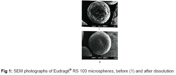
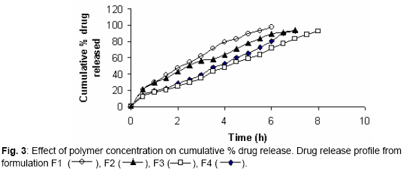
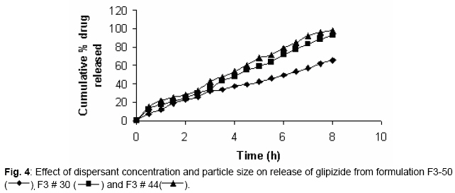
Determination of glipizide About 50 mg microspheres were accurately weighed and added to 50 ml of phosphate buffer (pH 7.4). The resulting mixture was agitated on a mechanical shaker for 24 h. The solution was then filtered and the drug content was measured at 275.2 nm spectrophotometrically (Systronic 2101 UV-Visible double beam spectrophotometer, India) after suitable dilution. FTIR Study FTIR spectra of glipizide, blank microspheres and drug-loaded microspheres were obtained in KBr pellets using a Perkin-Elmer model 883 Spectroscope in the ranges, 500 to 4000 cm-1. Scanning electron microscopy (SEM) A scanning electron microscope (JEOL, JSM6360) was used to characterize the surface topography of the microspheres after gold coating. Drug release behavior 8 The in vitro dissolution studies were carried out in 500 ml of phosphate buffer, pH 7.4, maintained at 37 ± 0.5° and 100 rpm by using United States Pharmacopoeia basket type dissolution test apparatus (LABINDIA, Disso-2000, Mumbai, India) under sink conditions. Accurately weighed samples of the microspheres were added to the dissolution medium and at preset time intervals, 2 ml aliquots were withdrawn and replaced with an equal volume of fresh dissolution medium. After suitable dilution, the samples were analyzed spectophotometrically at 275.2 nm. The concentration of glipizide in test samples was corrected and calculated using a regression equation of the calibration curve. The dissolution studies were carried out in triplicate and the mean values were plotted as percentage cumulative release versus time. Kinetics of drug release In order to understand the mechanism drug release, the first 60 % of drug release was fitted to the Korsmeyer-Peppas model 9. Mt / Mα =kt n Where M t / Mα is the fraction of the drug released at time, t, k is the rate constant and ‘n’ is the release exponent. The n value is used to characterize different release mechanisms and is calculated from the slope of the plot of log of fraction of drug released vs log of time. RESULTS The resulting microspheres formulated by solvent evaporation method was found to be spherical and free flowing in nature. The mean particle size of microspheres ranged from 420 -660 µm (see Table 1). It was noticed that mean particle size increased with increase in polymer concentration and decrease in magnesium stearate concentration. The encapsulation efficiencies ranged from 40.27 - 86.67 %. The encapsulation efficiency was also found to be dependant on nature of polymer used in the formulation. From the in vitro drug dissolution studies it was found that the sustaining effect of microspheres depended on the polymer concentration, amount of dispersant used and the type of polymer used in the formulation. DISCUSSION Increase of mean particle size with increase in polymer concentration may have occurred due to the fact that as polymer concentration increases it produces a significant increase in the viscosity in a fixed volume of solvent, thus leading to an increase of the emulsion droplet size and finally a higher microsphere size. 10-11 The flow properties of the microspheres are expressed in terms of Carr’s Index (Table 1). The Carr’s index for all formulations was less than 16, which indicate excellent flow properties and suggests that the microspheres can be easily handled during processing The encapsulation efficiency of the drug depended on the solubility of the drug in the solvent and continuous phase. An increase in the concentration of polymer in a fixed volume of organic solvent resulted in an increase in encapsulation efficiency12. Generally, the encapsulation efficiencies of the microspheres prepared with Eudragit® RS 100 were higher than those of microspheres prepared with the Eudragit® RS/RL combination. This can be attributed to the high content of the ammonium group in Eudragit® RL, which might have facilitated the diffusion of some of the entrapped drug to the surrounding medium during formation of the microspheres. Higher entrapment of drug was noticed when a lower concentration of magnesium stearate was used. This may be due to the formation of large-size microspheres at lower magnesium stearate levels, which provided less surface area for drug escape to the external processing medium. Scanning electron microscopic photographs of microspheres are shown in Fig. 1. The surface of the microspheres appeared spherical but rough. The microsphere were not agregated. Glipizide showed prominent peaks at 1651, 2943, 3354, 1529 and 1689 due to the presence of C = N aliphatic group, C-H2 aliphatic, N-H stretching of NH2 , C-H aliphatic and C=O stretching (Fig. 2). The same peaks were also observed in the formulation indicating the stable nature of the drug during encapsulation. Fig. 3 shows that the release of glipizide from the microspheres and it illustrates that the rate of drug release from the microspheres depended on the polymer concentration and the type of polymer used. The decrease in release rate with increasing content of the polymer can be explained by a decreased amount of drug present close to the surface and also by the fact that the amount of uncoated drug decreases with increase in polymer concentration13. When Eudragit® RL was used in combination with Eudragit RS, the drug released at a faster rate compared to Eudragit® RS alone. This is due to the fact that the amount of quaternary ammonium groups of Eudragit® RS is lower than that of Eudragit RL, which renders Eudragit® RS is less permeable14. The effect of magnesium stearate on drug release rate is shown in Fig. 4 .The faster drug release dispersing agent concentration rose may be due to decrease in particle size, which provided a larger surface for drug release. The effect of particle size on the drug release was also studied using microspheres of different size fractions (355 and 500 µm, respectively) as shown in Fig. 4. The release profile was in line with the theory of the effect of the particle size on dissolution rate 1. As microsphere size decreased, the drug release increased as result of higher surface area. In order to determine the mechanism of drug release the data obtained were fitted to the Korsemeyer-Peppas model in order to determine the ‘n’ value, which describes the drug release mechanism9. The ‘n’ value of all the formulations was between 0.5 and 1 indicating that the mechanism of drug release was non-Fickian type diffusion. CONCLUSION Eudragit® microspheres containing glipizide can be prepared successfully by using an emulsion solvent evaporation technique. The surface structure of the microspheres was spherical and rough. The encapsulation efficiencies were over 40 % and the mean size was in the range of 420 - 660 µm. The release rate of Eudragit® RS 100 microspheres was much slower than that than those prepared with the combination of Eudragit® RS/RL 100. The release pattern of the microspheres was found to be of the non-Fickian. ACKNOWLEDGEMENT The authors greatly acknowledge M/s Stadmed Private Ltd, Kolkata, India, for the supply of glipizide free of charge. The authors are also grateful to the Indian Institute of Technology (IIT), Kharagpur, India, for help in performing characterization studies. REFERENCES
© Pharmacotherapy Group, Faculty of Pharmacy, University of Benin, Benin City, Nigeria. The following images related to this document are available:Photo images[pr08003t1.jpg] [pr08003f4.jpg] [pr08003f1.jpg] [pr08003f2.jpg] [pr08003f3.jpg] |
| |||||||||

