 |
| About Bioline | All Journals | Testimonials | Membership | News |
|
||||||
|
||||||
Tropical Journal of Pharmaceutical Research, Vol. 7, No. 1, March 2008, pp. 913-919 Research Article Studies on Wound Healing Properties of Quercus infectoria SP Umachigia, KN Jayaveerab, CK Ashok Kumarc, GS Kumard, BM Vrushabendra swamye and DV Kishore Kumarf aDepartment of Pharmaceutics, dDepartment of Pharmacognosy, eDepartment of Pharmacology, fDepartment of Code Number: pr08007 Abstract
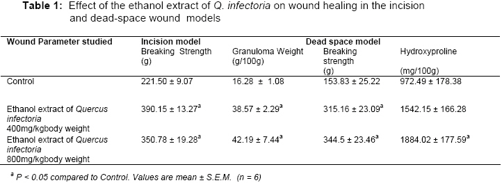
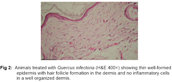
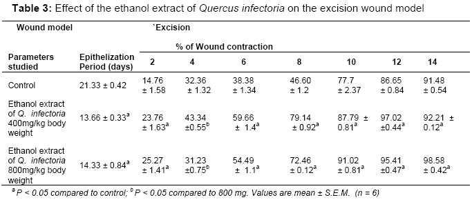
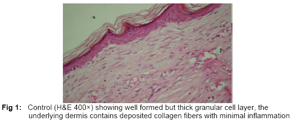
Purpose: The aim of the present study was to investigate the wound healing activity of the selected Indian medicinal plant Quercus infectoria. Key words: Quercus infectoria, wound healing, superoxide, catalase INTRODUCTION Quercus infectoria Olivier (Fagaceae) is a small tree found in Greece, Asia Minor and Iran. The galls arise on young branches of this tree as a result of attack by the gall-wasp Adleria gallae-tinctoria1 In Asian countries, the galls of Q. infectoria have been used for centuries in oriental traditional medicines for treating inflammatory diseases.2 Majuphal, a widely known plant in Indian traditional medicine has been used as dental powder and in the treatment of toothache and gingivitis3,4 The galls of Q. infectoria have also been pharmacologically documented to possess astringent, antidiabetic,5 antitremorine, local anaesthetic,6 antiviral,7 antibacterial,8 antifungal,9 larvicidal10 and antiinflammatory11 activities. The main constituents found in the galls of Q. infectoria are tannin (50-70%) and small amounts of free gallic acid and ellagic acid. 12-14 We are unable to find any information on the woundhealing properties of this plant. The present study is therefore an attempt to assess the efficacy of the galls using different parameters of wound healing in rats, and to study the influence of antioxidant enzymes on this property. MATERIALS AND METHODS Plant materials The galls of Q. infectoria used in this study were obtained from the local market and were identified based on its physical characteristics. The galls were crushed to small pieces using pestle and mortar and powdered in an electric grinder. Phytochemical Screening The powder of the galls of Quercus infectoria (50g) was subjected to successive extraction with different solvents in increasing order of polarity from petroleum ether (60°–80°), to benzene, chloroform, acetone, and alcohol, and finally to chloroform: water15. The dry extracts were subjected to various chemical tests in order to detect the presence of different phytoconstituents 16. Preparation of Ethanol Extract The shade-dried, powdered galls (1 kg) were extracted exhaustively using 95% ethanol on a Soxhlet apparatus. The total ethanol extract was concentrated in vacuo to a syrupy consistency (yield 270 g). Animals Wistar albino rats of either sex, weighing about 150–250 each, were used for the study. They were fed with standard chow (Pranav Agro Industries Ltd., Sangli, Maharashtra) and water ad libitum. They were housed in polypropylene cages maintained under standard conditions (12 hour light - dark cycle; 25 ± 3 °C; 35–60% humidity). The experimental protocol was subjected to the scrutiny of the Institutional Animal Ethics Committee, and was cleared by same before beginning the experiment. Acute Toxicity Studies Healthy adult albino rats of either sex, fasted overnight, were divided into 6 groups (n = 6 per cage) and were fed with increasing doses (1, 2, 4, and 8 g/kg body wt.) of the ethanol extract. The total ethanol extract, administered orally in doses of up to 8 g/kg body wt., did not produce any evident sign of toxicity or mortality in rats up to 14 days after administration. Wound models The studies were carried out using etheranesthetized rats and their back was shaved, in three different wound models, at two different dose levels of 400 and 800 mg/kg body wt. Incision wounds Two, 6-cm long paravertebral incisions were made through the full thickness of the skin on either side of the vertebral column of the rat17. Wounds were closed with interrupted sutures, 1 cm apart. The sutures were removed on the seventh day. Wound-breaking strength was measured in anesthetized rats on the tenth day after wounding 18. Excision wounds A circular skin piece of full thickness (approximately 500 mm2) was removed from a predetermined dorsal area 19. The wounds were traced on 1- mm2 graph paper on the day of wounding and subsequently on alternate days until healing was complete. Changes in the wound area were calculated, giving an indication of the rate of wound contraction. The number of days required for falling of the eschar without any residual raw wound was determined as the period of epithelization. Dead-space wounds These wounds were created by implanting two polypropylene tubes (0.5 cm × 2.5 cm each), one on either side, in the lumbar region on the dorsal surface of each rat. On the tenth postwounding day, the granuloma tissue formed on the implanted tubes was dissected out carefully. Granuloma tissue from one tube was maintained (at –64 °C) for the estimation of antioxidant enzyme levels. The other was used for the determination of tensile strength19, after which it was dried in an oven at 60 °C for 24 h and the dry weight noted. The acid hydrolysate of the dry tissue was used for the estimation of hydroxyproline content in the tissue 20. Biochemical Attributes The granuloma tissue from the dead-space model was homogenized in phosphate buffered saline (pH 7.0) and centrifuged under cold conditions. The clear supernatant was assayed spectrophotometrically to determine the levels of the antioxidant enzymes, i.e., superoxide dismutase21 and catalase22. Histopathological studies A section of the granuloma tissue was subjected to histopathological examination to determine the pattern of lay-down for collagen using two special stains i.e.Van Gieson and Masson Trichrome. Statistical Analysis Results, expressed as mean ± SE., were evaluated using one-way ANOVA with posthoc Scheffe’s post hoc test. Values of p < 0.05 were considered statistically significant. RESULTS Preliminary phytochemical screening revealed the presence of tannins; phenolic compounds. The acute toxicity studies show that the drug was safe up to a maximum dose of 8 g/kg body wt. of the animal. In the incision wound model, a significant increase was observed in the skin tensile strength of the ethanol extract-treated group on the tenth post-wounding day, at both dose levels (Table 1). The drug-treated animals of the dead-space wound model showed a significant increase in dry granuloma weight, granuloma breaking strength and the level of hydroxyproline content at both dose levels. Histological examination revealed increased collagen deposition in the drug, treated group (Fig. 2), as compared to control (1). Studies on the estimation of antioxidant enzyme reveal that the extract significantly increased the levels of superoxide dismutase and catalase, the two powerful antioxidant enzymes of the body that are known to quench superoxide radicals (Table 2). In studies using the excision wound model, animals treated with the ethanol extract of Q. infectoria showed a significant decrease in the epithelization period, as evidenced by the shorter period for the fall of eschar compared to control. The extract also facilitated the rate of wound contraction significantly at both dose levels (Table 3). The results in this study are in support that the wound healing and repair is accelerated by applying Quercus infectoria which was high-lighted by the full thickness coverage of the wound area by an organized epidermis in the presence of mature scar tissue in the dermis. DISCUSSION Wound healing involves various phases which include granulation, collagenation, collagen maturation and scar maturation23. Many plant extracts and medicinal herbs have shown potent antioxidant activity. Tannins the main components of many plant extracts, act as free radical scavengers24-29. Research into the Fig. 1: Control (H&E 400×) showing well formed but thick granular cell layer, the underlying dermis contains deposited collagen fibers with minimal inflammation role of antioxidants from plant extracts in wound healing has been published widely30. Wound healing process consists of different phases such as granulation, collagenation, collagen maturation and scar maturation which are concurrent but independent to each other. Hence in the present study three different wound models were used. In the incision wound model, a significant increase was observed in the skin tensile strength of the ethanol extract- treated group, at both dose levels (Table 1). The drug-treated animals at both dose levels of the dead-space wound model showed a significant increase in dry granuloma weight, granuloma breaking strength and the level of hydroxyproline content (Table 1). The histopathological study revealed increased collagen deposition in the drug, treated group (Figs. 1, 2), as compared to control. Studies on the estimation of antioxidant enzyme revealed that the extract significantly increased the levels of superoxide dismutase and catalase, the two powerful antioxidant enzymes of the body that are known to quench superoxide radicals (Table 2). The antioxidant enzymes (superoxide dismutase and catalase) are known to quench the superoxide radical and thus prevent the damage of cells caused by free radicals In studies using the excision wound model, animals treated with the ethanol extract of Q. infectoria showed a significant decrease in the epithelization period, as evidenced by the shorter period for the fall of eschar compared to control. The drug extract also facilitated the rate of wound contraction significantly at both dose levels (Table 3). Phytochemical work reveals that ethanolic extract of galls of Q.infectoria contains high amount of tannins, presence of gallic acid, ellagic acid, syringic acid, ß-sitosterol and amentoflavone31, implied that tannin is one of the active compounds which may be responsible for the antioxidant activity. So in this study scavenging effect might be one of the most important components of wound healing which may be responsible to support wound healing property. Thus the enhanced wound healing may be due to the free radical scavenging action of the plant as well as enhanced antioxidant enzyme level in granuloma tissues. CONCLUSION This finding provides an insight into the usage of the galls of Q. infectoria in traditional treatment of wounds or burns associated with bacterial infections REFERENCES
The following images related to this document are available:Photo images[pr08007t1.jpg] [pr08007f2.jpg] [pr08007f1.jpg] [pr08007t2.jpg] [pr08007t3.jpg] |
| |||||||||
