 |
| About Bioline | All Journals | Testimonials | Membership | News |
|
||||||
|
||||||
Tropical Journal of Pharmaceutical Research, Vol. 7, No. 1, March 2008, pp. 921-927 Research Article A New Flavanone from Flemingia strobilifera (Linn) R. Br. and its Antimicrobial Activity S Madan*a, GN Singha, Y Kumarb, K Kohlic, R M Singha, SR Mirc and S Ahmadc aCentral Indian Pharmacopoeia Laboratory, Govt. of India, Ministry of Health and Family welfare, Sector – 23, Raj Nagar, Code Number: pr08008 Abstract
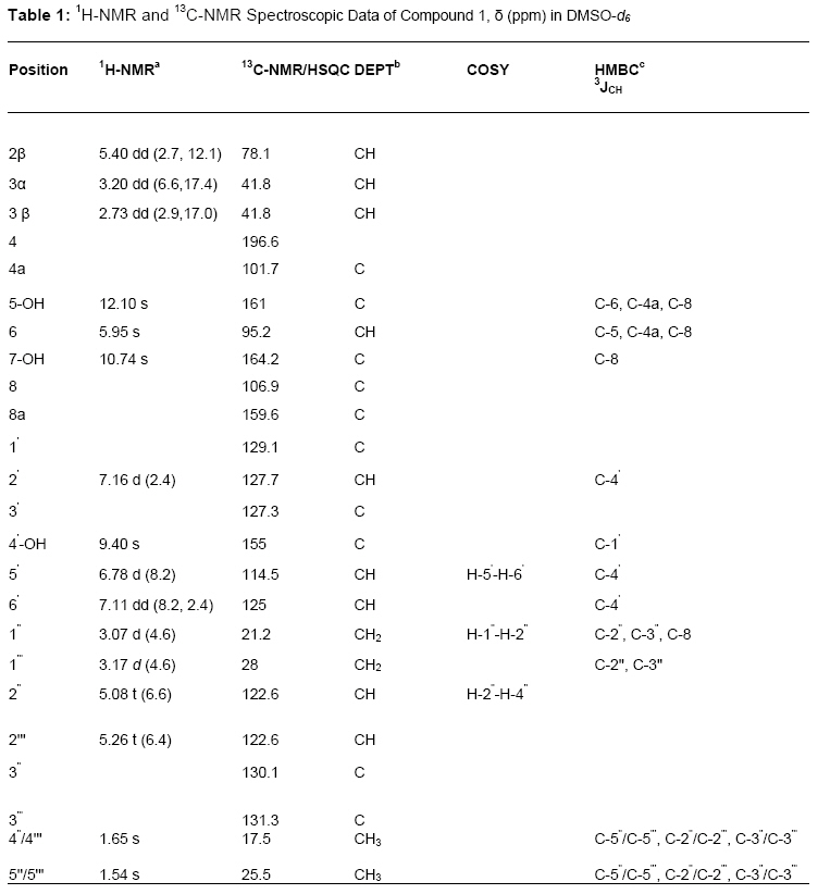
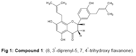
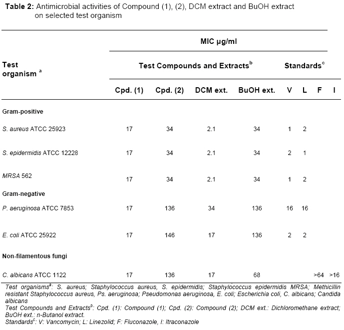
Purpose: To carry out a bioactivity guided fractionation and isolation of the antimicrobial constituent(s) of the roots of Flemingia strobilifera against some bacteria and fungi. Key words: Flemingia strobilifera, Flemingiaflavanone (8, 3'-diprenyl-5, 7, 4'-trihydroxy' flavanone), Genistin (5, 4-dihydroxy isoflavone 7-O-glucoside), MeOH (Methanol), BuOH (Butanol), Antimicrobial activity, MIC (Minimum inhibitory concentration). INTRODUCTION Flemingia strobilifera (R.Br.), an important medicinal plant, commonly known as Kusrunt and belongs to the Leguminosae family 1, 2. The plant is found in Sind, Rajputana, Bengal, South India and Andamans3. The roots of this plant have been indigenously used in epilepsy and hysteria and the leaves are reported to be used as vermifuge4. Previous phytochemical investigations reported various chalkones4, 5, flavonoid glycosides6, aurone glycosides7 and epoxy chromenes8. This paper reports the isolation and structural elucidation of a new flavanone from the roots of Flemingia strobilifera characterized as 8, 3 'diprenyl-5, 7, 4 '-trihydroxy flavanone (1) that is being reported for the first time from the genus Flemingia along with two known compounds and their antimicrobial activity. Experimental General Melting point was determined on a Buchi Melting Point B-540 (Switzerland) apparatus. U.V was performed in U.V-Perkin Elmer double beam U.V Spectrophotometry (Germany). I.R spectra were recorded on a Jasco FT/IR 410 (U.S.A) in KBr. NMR spectra were measured in Bruker 400 MHz Ultrashield, Advance 400 (Germany) spectrometer, using TMS as internal standard. NMR experiment included the HSQC, HMBC, and COSY pulse sequences. Coupling constants (J values) were given in Hz. Quatro Micro Mass; Waters (U.S) was used to record Mass experiment. Silica gel used for column chromatography was normal phase 60-120 mesh size while TLC was carried out on silica gel GF254 sheets (Merck, Germany). Plant material The roots of F. strobilifera were collected from forests of Shann Power House, Joginder Nagar, (Distt Mandi) Himachal Pradesh in October 2006. The identity of the plant material was verified by Dr. B. Naag Ex-Botanist Research Institute of Ayurveda, Joginder Nagar, (H.P) and Dr. H.B Singh, Head, Raw Materials Herbarium and Museum, NISCAIR, New Delhi and a voucher specimen number NISCAIR/RHMD/Consult/06/757/74 was deposited at the Herbarium of National Institute of Science Communication and Information Resources, New Delhi. Extraction and isolation The root (2.94 Kg) of F. strobilifera were airdried, ground and extracted with five liters of methanol for 24 h by maceration. The mark left was repeatedly extracted five times similarly, for complete extraction. The MeOH extract was evaporated in rota-vapor to yield a semisolid (1000 g), 900 g of which was suspended in five liters of water and partitioned with ten liters of DCM for five times and also partitioned with ten liters of n-BuOH for five times to yield 18 g and 34 g of extracted material, respectively. The DCM fraction (16 g) was column chromatographed over silica gel using petroleum ether (PE) and ethyl acetate (EtoAc), step gradient as eluents to yield compound 1 and 3. The PE and EtoAc (85:15) fractions were collected and these fractions (203.9 mg) were further chromatographed using DCM and MeOH (95:5) over silica gel to yield compound 1, (6.7 mg) and compound 3 (81.8 mg), β- sitosterol-D glucoside was eluted from PE: EtoAc (40:60) eluents. The n-BuOH fraction (25 g) was subjected to column chromatography on silica gel eluted with chloroform (CHCl3): MeOH (85: 15) to yield compound 2, (58.5 mg). Antimicrobial activity method The minimal inhibitory concentration (MIC) of extracts and isolated compounds were determined by the broth microdilution method according to National Committee for Clinical Laboratory Standards guidelines9 as well as for non-filamentous fungi in 96-well microtitre plates with MHB (Muller Hinton broth) made in-house. 96-well microtitre plates contained the antimicrobial agents in serial twofold dilutions from 136 to 0.53 µg/ml, depending on the antimicrobial agent being tested. Inocula were prepared in MHB from cultures grown on tryptic soya agar. The final concentration was 1 × 10⁵ CFU/ml. All microtitre plates were prepared in duplicate and incubated at 35°C for 24 hrs. The susceptibility of the standard drugs vancomycin, linezolid, fluconazole and itraconazole were defined as the lowest concentration of drug that resulted in total inhibition of microbial growth. The MIC was defined as the minimum inhibitory concentration of the extract or compound that resulted in total inhibition of microbial growth. RESULTS The DCM fraction was column chromatographed over silica gel using PE and EtoAc to yield compound 1 and 3. The n-BuOH fraction was column chromatographed over silica gel using CHCl3: MeOH to yield compound 2. Compound 1 showed Rf value of 0.52 in DCM: MeOH (95:5) & showed Rf value of 0.58 in CHCl3: MeOH (95:5) solvent system. It gave orangish yellow colour with 10% methanolic sulfuric acid and pink colour with Shinoda confirmed the presence of flavanone. Compound 2 showed Rf value of 0.61 in CHCl3: MeOH (70:30) solvent system and showed Rf value of 0.52 in PE: EtoAc (75:25) solvent system. It gave yellow colour with 10% methanolic sulfuric acid and greenbrown colour with alcoholic FeCl3. Compound 3 showed Rf value of 0.50 in EtoAc: MeOH: H2O (10: 1: 0.5) and showed Rf value of 0.54 in CHCl3: MeOH (80:20) solvent system and gave purple colour with 10% methanolic sulfuric acid and also showed positive Molisch test with formation of violet ring. The antimicrobial activity of compound 1 has shown the most significant activity against Gram-positive, Gram-negative bacteria and fungi. Compound 2 showed moderate activity against Gram-positive, Gram-negative bacteria and fungi.Compounds 2 and 3 are known and identified as Genistin (2) and β -sitosterol-D glucoside (3) by comparisons of their spectral data (U.V, I.R, NMR and MS) with those reported previously10, 11, 12, 23, 24.The compound 1 was isolated for the first time from the plant and showed MIC of 17µg/ml against Staphylococcus aureus, Staphylococcus epidermidis, Methicillin resistant Staphylococcus aureus, Pseudomonas aeruginosa, Escherichia coli and Candida albicans. DISCUSSION Compound 1 Compound 1 designated as 8, 3'-diprenyl-5, 7, 4 '-trihydroxy flavanone was obtained as pale yellow crystals. Its molecular formula was established as C25 H28 O5 due to parent ion at m/z 408 [M] + and [M + 1] at 409. The I.R spectrum showed strong absorptions at 1630cm-1 (chelated C=O group) and 3300cm-1 (OH). The characteristic U.V absorption bands [λmax MeOH nm = 228 sh, 293,339 + NaoMe, 248, 285, 333; + AlCl3, 221, 316, 392; + NaoAc, 287, 297,334 + H3 BO3 ] suggested a flavanone structure. That was confirmed by the detection of 1H-NMR signals characteristic at δH 5.40 (1 H, dd, J = 12.1, 2.7 Hz, C2 -βH), 3.20 (1H, dd, J = 17.4, 6.6Hz, C3 -αH), 2.73 (1H, dd, J = 17.0, 2.9 Hz, C3 -βH) attributed to the flavanone C-ring protons and at δC 78.1 and 41.8 in its 13C-NMR spectrum (Table 1). It also indicated the presence of two-prenyl unit at δH 1.65, 1.54 [(each 6 H) s (CH3 × 4)], 3.07, 3.17 (each 2H, d, J = 4.6 Hz, Ar-CH2 -CH = × 2), 5.08, 5.26 (each 1H, t, J = 6.6 Hz, CH2 -CH = × 2), three hydroxyl groups [δH 12.00, 6.20 and 5.32 (each 1H, s) which shifted in DMSO-d6 to δH 12.10, 10.74 and 9.40]. Aromatic protons at δH 5.95 (1H, s) were assigned to the H-6 in A ring13. A characteristic ABX system at δH 6.78, 7.16, and 7.11 indicated the presence of a C-3 , 4 disubstitution on the B-ring moiety. Positive U.V shifts after the addition of sodium acetate and aluminium chloride indicated that the three hydroxyl groups at C5 , C7 and C4' were free and therefore the prenyl group in the A ring must be at C-814. Since the 1H-NMR spectrum (B-ring) of compound showed ABX type proton signals of the aromatic ring, the prenyl group in the B-ring must be located at C-3’. These data indicated the substitution pattern of the A ring was 5-hydroxy substituted and 8-prenylated, and that of the B ring was 4 -hydroxylated and 3 - prenylated and the later three signals were assigned to the C-5', C-2' and C-6' protons, respectively from their chemical shifts and coupling patterns. The 13C-NMR spectrum of 1 showed 25 carbon atoms that were classified as four methyl carbons at δC 17.5/17.5, 25.5/25.5 (C4''/4''' and C-5 ''/5'''), three methylene carbons at δc 21.2, 28 (C-1 ''/C-1''' ), 41.8 (C-3), seven quaternary carbon at δc 106.9, 159.6, 101.7, 130.1, 129.1, 127.3, 131.3 (C-8, C-8a, C-4a, C-3'', C-1', C-3', C-3'''), seven methines at δc 78.1, 95.2, 122.6, 127.7, 125.0, 114.5, 122.6 (C-2, C-6, C-2 '', C-2 ', C-6', C-5', C-2''') and three- hydroxylated carbons at δc 161.0, 164.2, 155.0 (C-5, C-7, C-4), with one carbonyl carbon at 196.6 using distortion less enhancement by polarization transfer (DEPT 90º and 135º) spectral analysis. This was confirmed by the HMBC experiment; long-range correlations were observed between the following protons and carbons: 5-OH and 6, 4a, 8-C; 7-OH and 8-C; 4 '-OH and 3', 1'-C. In the HMBC spectrum (Fig.1), the proton at δH 5.95 (1H, s, H-6) was correlated with C-5, 4a, 8, (δC 161, 101.7, 106.9), suggesting that one prenyl unit was located at C-8 and the other prenyl unit will be attached at C-3 of the ABX system of ‘B’ ring. The H-5' proton of Ring-B showed correlation with H-6' in 1H-1H COSY spectrum and H-1'' showed correlation with H-2'' in 1H-1H COSY spectrum. The absolute configuration at C-2 was established as S by comparing the optical rotation value with literature data of euchrestaflavanone A15. This is the first report of a flavanone (1) in a Flemingia species. The related 8, 3-diprenyl-5, 7, 4-trihydroxy flavanone (1) (Euchrestaflavanone A) is found in Euchresta japonica15 , Sophora moorcroftian16 , Lupinus luteus17, Euchresta formonsa18 , Azadirachta indica19and Lespedezaflavanone B is found in Lespedeza davidii20 Glycyrrhiza glabra21 22. From the above discussion the structure of 1 was concluded to be (S) - 8, 3 -diprenyl-5, 7, 4'-trihydroxy flavanone. 8, 3 -diprenyl 5, 7, 4 -trihydroxy flavanone (1): Green-brown with FeCl3, Pink colour with Shinoda (Mg-HCl), Pale yellow crystal; m.p 155ºC; U.V λmax [MeOH, nm (log ε)]: 228 sh, 293, 339; + NaOMe, 248, 285, 333; + AlCl3, 221,316, 392; + NaOAC, 287, 297, 334 (+ H3BO3); IR (KBr) cm–1: 3300(OH), 1630 (C=O), 1000, 1500 (arom C=C), 1390, 1370 (CH3). 1H-NMR (400 MHz, DMSO-d6) δH and 13C-NMR (400 MHz, DMSO-d6) δc given in Table 1. MS m/z (rel.int): 409 [M+H]+ C25H28O5 (100), 408 [M]+ (20). Genistin (2): Green-brown with FeCl3, No Pink colour with Shinoda (Mg-HCl), Yellow colour after spray with 10% MeOH-H2 SO4. Properties and spectra were identical to those reported earlier10, 11. β-sitosterol-D glucoside (3): Violet ring formation with Molisch reagent. Purple colour after spray with 10% MeOH-H2 SO4 , obtained as white crystals. Properties and spectra were identical to those reported earlier12. Compound 1 and 2 were tested for its in vitro antimicrobial activity by measuring their MIC 9 against selected test organisms (Table 2). 8, 3'-diprenyl 5, 7, 4 '-trihydroxy flavanone (1) showed the higher activity against Grampositive, Gram-negative bacteria and fungi. Genistin (2) showed moderate activity against Gram-positive and Gram-negative bacteria, and fungi. The DCM and n-BuOH fractions of the plant also showed potent activity against these selected test organisms. Compound (3), already known compound and its activity have been reported in the literature23, 24. Vancomycin and Linezolid showed potent activity against Gram-positive bacteria in comparison to Gram-negative bacteria. Fluconazole and Itraconazole were active at MIC of>64 and >16 µg/ml respectively against fungi. CONCLUSION The present study has identified the isolation and characterization of a new flavanone for the first time from the Flemingia species. The antimicrobial activity of compound (1) has shown the most significant activity against Gram-positive, Gram-negative bacteria and fungi. Compound (2) showed moderate activity against Gram-positive, Gram-negative bacteria and fungi. The DCM and n-BuOH extracts of the plant also showed potent activity against these selected test organisms. The antimicrobial activity of these compounds and extracts has not been reported earlier. ACKNOWLEDGEMENTS The authors are thankful to Dr. C. K. Katiyar, Dr. Anil Kanaujia, Dr. Steve Thomas, Mr. Rajeev Duggar, Dr. C. P. Gupta and Herbal Drug Research Division, Ranbaxy Research Laboratories, for providing the required facilities for the completion of this work. REFERENCES
© Pharmacotherapy Group, Faculty of Pharmacy, University of Benin, Benin City, Nigeria. The following images related to this document are available:Photo images[pr08008f1.jpg] [pr08008t2.jpg] [pr08008t1.jpg] |
| |||||||||
