 |
| About Bioline | All Journals | Testimonials | Membership | News |
|
||||||
|
||||||
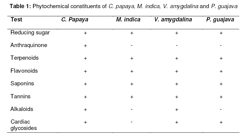
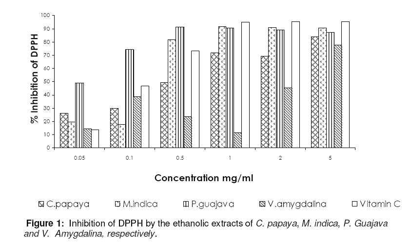
Tropical Journal of Pharmaceutical Research, Vol. 7, No. 3, September, 2008, pp. 1019-1024 Research Article Phytochemical Screening and Antioxidant Activities of Some Selected Medicinal Plants Used for Malaria Therapy in Southwestern Nigeria GA Ayoola1*, HAB Coker1, SA Adesegun2, AA Adepoju-Bello1, K Obaweya1, EC Ezennia1, TO Atangbayila1 1Department of Pharmaceutical Chemistry, Code Number: pr08022 Abstract Purpose: Oxidative stress has been shown to play an important role in the development of anaemia in malaria. Indeed, increase in total antioxidant status has been shown to be important in recovery from malaria. The antioxidant activities of four medicinal plants traditionally used in the treatment of malaria in southwestern Nigeria were determined. Key words: Carica papaya, Magnifera indica, Psidium guajava, Vernonia amygdalina, Antioxidants, Malaria, DPPH, Oxidative stress. INTRODUCTION Malaria is a global disease prevalent in the tropics caused by Plasmodium species. It is one of the oldest and greatest health challenges affecting 40% of the world’s population. It affects 300-500 million people and kills 1.5-2.7 million people annually1 . High mortality rate is reported in children and pregnant women, also the disease has a negative impact on the economy of prevalent countries2 . In Nigeria, malaria is endemic throughout the country. World health Organization (WHO) has estimated malaria mortality rate for children under five in Nigeria at 729 per 100,0003 Nigeria’s Ministry of Health reported in April 2004 that malaria is responsible for one out of ten deaths in pregnant women and costs the Federal Government of Nigeria over one billion naira annually3 . One of the major reasons for the development of anaemia in malaria seems to be oxidative stress4-6 . The immune system of the body is activated by infections, including malaria, thereby causing the release of reactive oxygen species. In addition to this, the malaria parasite also stimulates certain cells to produce reactive oxygen species thereby resulting in haemoglobin degradation5,7 . Indeed, depressed level of plasma antioxidants has been shown in Plasmodium falciparum-infected children and it has been suggested as a possible contributor to the morbidity and mortality of malaria1 . Increased resistance of malaria parasites to the commonly used antimalarial drugs have been reported, and hence the need to intensify research in the area of development of new antimalarial drugs especially from medicinal plants. A review of the medicinal plants used in the southwestern part of Nigeria for the treatment of malaria indicates that a rich flora diversity exists in Nigeria8 . The present study aims to investigate the free radical scavenging activities of some of the commonly used medicinal plants in southwestern Nigeria. The following plants were selected for investigation: C. papaya, P. guajava, V. amygdalina, and M. indica. The decoctions of the leaves of C. papaya, P. guajava, V. amygdalina and a decoction of the stem bark of M. indica are commonly used in the traditional treatment of malaria in southwestern Nigeria. MATERIALS AND METHODS Collection and identification of plant materials Fresh leaves of C. papaya, P. guajava, V. amygdalina and the stem bark M. indica were collected from the premises of Lagos University Teaching Hospital, Lagos, Nigeria in January 2007. The plants were identified by Dr S.A. Adesegun of the Department of Pharmacognosy, Faculty of Pharmacy, University of Lagos, Lagos, Nigeria. Extraction of plant materials The plant materials (leaves of C. papaya, P. guajava, V. amygdalina and the stem bark of M. indica) were air-dried at room temperature (26˚C) for 2 weeks, after which it was grinded to a uniform powder. The ethanol extracts were prepared by soaking 100 g each of the dry powdered plant materials in 1 L of ethanol at room temperature for 48 h. The extracts were filtered after 48 h, first through a Whatmann filter paper No. 42 (125mm) and then through cotton wool. The extracts were concentrated using a rotary evaporator with the water bath set at 40˚C. The percentage yield of extracts ranged from 7–19%w/w. Phytochemical screening Phytochemical screening were perfomed using standard procedures9-10 . Test for reducing sugars (Fehling’s test) The aqueous ethanol extract (0.5 g in 5 ml of water) was added to boiling Fehling’s solution (A and B) in a test tube. The solution was observed for a colour reaction. Test for anthraquinones 0.5 g of the extract was boiled with 10 ml of sulphuric acid (H2SO4) and filtered while hot. The filtrate was shaken with 5 ml of chloroform. The chloroform layer was pipette into another test tube and 1 ml of dilute ammonia was added. The resulting solution was observed for colour changes. Test for terpenoids (Salkowski test) To 0.5 g each of the extract was added 2 ml of chloroform. Concentrated H2S04 (3 ml) was carefully added to form a layer. A reddish brown colouration of the interface indicates the presence of terpenoids. Test for flavonoids Three methods were used to test for flavonoids. First, dilute ammonia (5 ml) was added to a portion of an aqueous filtrate of the extract. Concentrated sulphuric acid (1 ml) was added. A yellow colouration that disappear on standing indicates the presence of flavonoids. Second, a few drops of 1% aluminium solution were added to a portion of the filtrate. A yellow colouration indicates the presence of flavonoids. Third, a portion of the extract was heated with 10 ml of ethyl acetate over a steam bath for 3 min. The mixture was filtered and 4 ml of the filtrate was shaken with 1 ml of dilute ammonia solution. A yellow colouration indicates the presence of flavonoids. Test for saponins To 0.5 g of extract was added 5 ml of distilled water in a test tube. The solution was shaken vigourously.and observed for a stable persistent froth. The frothing was mixed with 3 drops of olive oil and shaken vigourously after which it was observed for the formation of an emulsion. Test for tannins About 0.5 g of the extract was boiled in 10 ml of water in a test tube and then filtered. A few drops of 0.1% ferric chloride was added and observed for brownish green or a blue-black colouration Test for alkaloids 0.5 g of extract was diluted to 10 ml with acid alcohol, boiled and filtered. To 5 ml of the filtrate was added 2 ml of dilute ammonia. 5 ml of chloroform was added and shaken gently to extract the alkaloidal base. The chloroform layer was extracted with 10 ml of acetic acid. This was divided into two portions. Mayer’s reagent was added to one portion and Draggendorff’s reagent to the other. The formation of a cream (with Mayer’s reagent) or reddish brown precipitate (with Draggendorff’s reagent) was regarded as positive for the presence of alkaloids. Test for cardiac glycosides (Keller-Killiani test) To 0.5 g of extract diluted to 5 ml in water was added 2 ml of glacial acetic acid containing one drop of ferric chloride solution. This was underlayed with 1 ml of concentrated sulphuric acid. A brown ring at the interface indicated the presence of a deoxysugar characteristic of cardenolides. A violet ring may appear below the brown ring, while in the acetic acid layer a greenish ring may form just above the brown ring and gradually spread throughout this layer. Determination of antioxidant activity The radical scavenging activities of the plant extracts against 2,2-Diphenyl-1-picryl hydrazyl radical (Sigma-Aldrich) were determined by UV spectrophotometry at 517 nm. Radical scavenging activity was measured by a slightly modified method previously described12,13 . The following concentrations of the extracts were prepared, 0.05, 0.1, 0.5, 1.0, 2.0 and 5 mg/ml in methanol (Analar grade). Vitamins C was used as the antioxidant standard at concentrations of 0.02, 0.05, 0.1, 0.2, 0.5 and 0.75 mg/ml. I ml of the extract was placed in a test tube, and 3 ml of methanol was added followed by 0.5 ml of 1 mM DPPH in methanol. A blank solution was prepared containing the same amount of methanol and DPPH. The radical scavenging activity was calculated using the following formula: % inhibition = {[Ab-Aa]/Ab} x 100 ...............(1) where Ab is the absorption of the blank sample and Aa is the absorption of the extract RESULTS Phytochemical screening of plant materials The phytochemical screening of the plants studied showed the presence of flavonoids terpenoids, saponins and tannins (Table 1), M. indica, V. amygdalina and P. guajava showed the absence of anthraquinones. M. indica and P. guajava tested negative for the presence of alkaloids and only M. indica tested negative for the presence of cardiac glycosides (Table 1). Radical scavenging (antioxidant) activity IC50 of 0.04, 0.31, 0.58, 2.30 and 0.054 mg/ml were recorded for P. guajava, M. indica, C. papaya, V. amygdalina and Vitamin C, respectively (Figure 1). DISCUSSION Phytochemical screening of the plants revealed some differences in the constituents of the four plants tested. C. papaya tested positive for all the phytochemicals tested; M. indica showed the absence of anthraquinones, alkaloids and cardiac glycosides; V. amygdalina tested positive for all except anthraquinones while P. guajava tested positive for all except Anthraquinones and alkaloids. All the plants exhibited potent antioxidant activity. The presence of flavonoids and tannins in all the plants is likely to be responsible for the free radical scavenging effects observed. Flavonoids and tannins are phenolic compounds and plant phenolics are a major group of compounds that act as primary antioxidants or free radical scavengers 14 . The DPPH test provides information on the reactivity of the test compounds with a stable free radical. DPPH gives a strong absorption band at 517nm in visible region. When the odd electron becomes paired off in the presence of a free radical scavenger, the absorption reduces and the DPPH solution is decolourised as the colour changes from deep violet to light yellow. The degree of reduction in absorbance measurement is indicative of the radical scavenging (antioxidant) power of the extract. The crude extract of P. guajava appeared to be as potent as Vitamin C with a maximum inhibition of 91% at 0.5mg/ml which is comparable to 95% for vitamin C at the same concentration. M. indica was six times less potent than the standard with a maximum inhibition of 91% at 1 mg/ml, followed by C. Papaya which was eleven times less potent (than vitamin C) with a maximum inhibition of 83.8% at 5mg/ml. V. amygdalina was the least potent (42 times less potent than the standard) showing a maximum inhibition of 77.7% at 5 mg/ml. This study suggests that these plants possess antioxidant activities which can counteract the oxidative damage induced by the malaria parasite. This may be one of their mode of action in malaria therapy. CONCLUSION Extracts from C. papaya, M. Indica, V. amygdalina and P. guajava showed varying antioxidant (free radical scavenging) activities when compared to vitamin C in the following order: V. amygdalina < C. papaya < M. indica < Vitamin C ≤ P. guajava. The results suggest that the antioxidant activity of these plants may contribute to their claimed antimalarial property. ACKNOWLEGEMENT We thank Mr P. Ojobor of the Central Research Laboratory; Mr T.I. Adeleke of the Pharmacognosy Department; Mrs.Y.A. Bashorun, Mr Olatunji and Mr Olajide of Pharmaceutical Chemistry Department, all of the University of Lagos, for technical support. We also thank Ms J.O. Ashamu for assistance in the preparation of this manuscript. REFERENCES
© Pharmacotherapy Group, Faculty of Pharmacy, University of Benin, Benin City, 300001 Nigeria. The following images related to this document are available:Photo images[pr08022f1.jpg] [pr08022t1.jpg] |
| |||||||||
