 |
| About Bioline | All Journals | Testimonials | Membership | News |
|
||||||
|
||||||
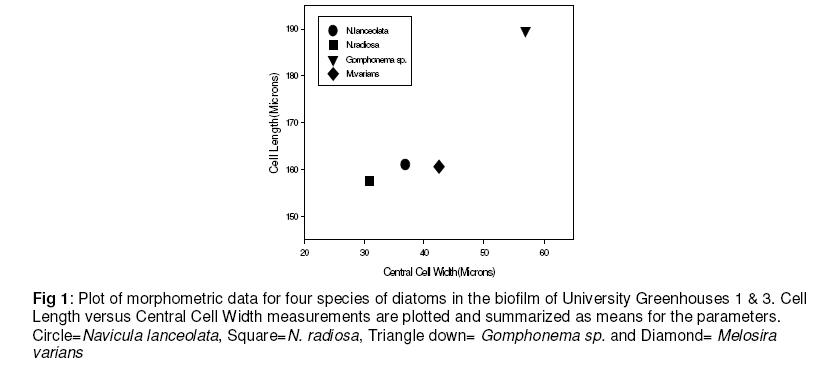
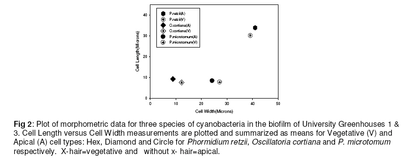
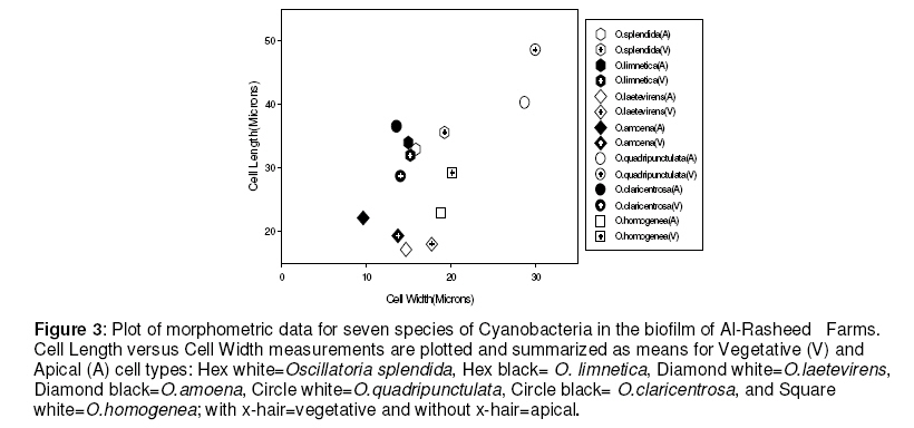
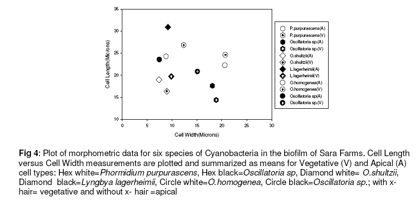
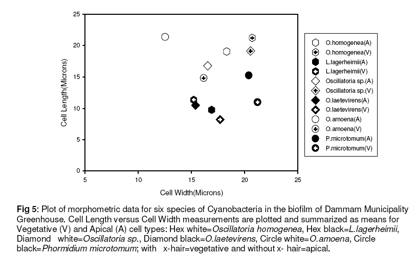
Tropical Journal of Pharmaceutical Research, Vol. 7, No. 3, September, 2008, pp. 1043-1049 Research Article Characterization of Biotic and Abiotic Profiles of Greenhouse Evaporative Cooling System Fouling AA Nuhu*1 and S Ahmad2 1Biology Programme, Code Number: pr08025 Abstract Purpose: To characterize the microorganisms influencing greenhouse evaporative cooling system fouling as a prelude to designing a sustainable remediation scheme that would assist in reducing the heavy financial loss incurred by stakeholders due to deterioration of cooling pads. Key words: Cyanobacteria, Diatoms, Biofilms, Morphometric, Evaporative cooling INTRODUCTION In arid regions, such as Saudi Arabia, where temperatures could some times exceed 40◦C, greenhouses are often equipped with evaporative cooling systems to provide good condition for workers as well as to prevent desiccation of plant materials. The efficiency of evaporative cooling pads in such systems greatly depends on how freely air can pass through them1 . This efficiency is frequently thwarted by the daunting challenge of algal and bacterial growth which shortens their lifespan, leading to decreased cooling potentials of such pads2 . This problem causes a lot of economic damage to the stakeholders. Biofouling is a phenomenon used to describe the nuisance deposition and formation of biofilms on solid supports3 . Biofilms are heterogeneous and are found in diverse environments4 . They are often characterized based on their complex community relationships, morphological differences, genetic diversity, or complex polymeric materials. The diverse extra cellular polymeric materials allow the microorganisms to perform various functions including attachment to solid platforms5 . Nearly all kinds of microorganisms, ranging from algae to bacteria, can infest and cause clogging and degradation of materials including the evaporative cooling pads. Even though evaporative cooling system fouling can result from trapping of debris, silt, salt and scale by the cooling pads, the presence of biofilms makes the process a very daunting and complex one. Bacterial biofilms often contain photosynthetic cyanobacteria. These are very small and usually unicellular, though they can also form colonies large enough to be seen with the naked eyes. Also found among biofilms is an important group of eukaryotic algae known as diatoms. They have a penchant for aggregation and sinking made possible through the production of mucilage or heavy resting spores6 . MATERIALS AND METHODS Sample collection and treatment Samples of biofilm on evaporative cooling system pads from the King Fahd University of Petroleum and Minerals (KFUPM) greenhouses, Sara Farms in Al-Kharj, Al-Rasheed Farms in Aziziya (Dhurma) and Dammam Municipality Greenhouse (DMG) were collected by scrapping with clean forceps. Sampling was done randomly both from the inside and outside surfaces having different degrees of light exposure. Samples were fixed with 5% formaldehyde solution and stored in stoppered bottles at 5◦C of a Biotronette Plant Growth Chamber. Sample preparation for light microscopy The sample was prepared for light microscopic examination according to standard protocols7 . Briefly, thin sections of the cooling system pads above were removed by forceps on to a microscope slide and then covered with a cover slip. Drops of glycerin were added to the edge and allowed to sip inside by capillarity. The slide was then observed under the Zeiss light microscope at 40 X. Visible microorganisms were captured using the attached digital camera TKC 1380. Sample preparation for scanning electron microscopy After fixing the samples in 5% formaldehyde solution, they were subjected to different concentrations of alcohol, 25%, 50%, 75% and 95%, for dehydration, followed by acetone in the final step consecutively, as earlier described8 . The samples were then goldcoated and examined in SEM Jeol JSM-5900 LV operating at 20kV. Different portions of the images were analyzed for mineral clues using energy-dispersive X-ray spectroscopy. Morphometrics of cyanobacteria and diatoms For cyanobacteria, cell width and cell length of Apical (A) and Vegetative (V) cells were measured using QWIN image analysis software; for diatoms frustules, apical(A), central (C) and lateral length(L) of identified organisms were measured-the ratios of C/L and A/C were also calculated. For each measurement, 25 replicates were taken and their mean and standard deviation calculated. The mean cell length values were then plotted against the mean cell width values. Water quality examination Water samples from the various greenhouses were collected and analyzed for pH, conductivity and total dissolved solids (TDS). This was aimed at determining the effect that any differences in composition of the water samples might have on the development of biofilms, as well as on the enhancement of deposition. Conductivity and pH were estimated using Pasport PS-2116 Conductivity sensor and PS-2102 pH sensor respectively. All measurements were carried out at room temperature (25◦C). The respective TDS of each water sample was then calculated from its conductivity value. RESULTS Different genera of cyanobacteria and diatom including Phormidium, Oscillatoria, Lyngbya, Navicula, Melosira and Gomphonema were identified and characterized at different conditions of total dissolved solids and pH. TDS values of 2641 ppm (conductivity, 5282 µS/cm; pH 8.13) for Al-Rasheed, 2327.5 ppm (conductivity, 4655 µS/cm; pH 8.04) for Sara , 3352 ppm (conductivity, 6704 µS/cm; pH 7.08) for Dammam Municipality Greenhouse (DMG) and 61.5 ppm ( conductivity, 123 µS/cm; pH 6.88) for KFUPM were obtained at 25◦C. The results for the morphometric presentation of these various organisms are given on Figures 1, 2, 3, 4, and 5, which were summarized and plotted as means of different cell parameters characteristic of the identified microorganisms. DISCUSSION Our findings revealed that the various greenhouses were rich in different species of cyanobacteria and less in species of diatoms. This was possibly due to the high degree of solar irradiation to which our greenhouses are exposed year round. Though diatoms can acclimatise to certain light levels by varying the ratio of diadinoxanthin: chlorophyll a9 , they are however more efficient in low irradiances10 , which was not the case with our reference sites. This limited occurrence and distribution of diatoms was further substantiated from the results of our subsequent study and phylogenic analysis, even as these revealed the presence of Shingomonads, Acidobacteria and Flexibacteria (unpublished data). We observed from the scatter plot in Figure 1 that all the diatoms identified in this study have a C/L ratio of ≤ 0.3 and A/C ratio of less than 1; however, the more the value tends to 1, the higher the likelihood of the microorganism assuming a rectangular shape, as depicted by Melosira varians. It is noteworthy to indicate that with the exception of Melosira varians, all other species of diatoms identified have A/C ratio of less than 0.5, necessitating the microbes to take other conformations such as rhombic and lanceolate, whether or not they are isopolar or heteropolar, isobilateral (symmetrical) or dorsoventricular (asymmetrical)11 . On the other hand, Figure 2 shows the parameters in the University greenhouses for the identified cyanobacteria, with cell length between 6.67 and 37.48 microns as against between 9.98 and 43.57 microns for the cell width. Conversely, Al-Rasheed Farms recorded the range of 15.73-55.83 microns for cell length as against 17.99-32.80 microns for cell width (Figure 3). Figure 4 depicts the cell parameters for the cyanobacteria in Sara Farms; 12.37-33.52 microns for cell length and 5.40-22.64 microns for cell width. Dammam Municipality greenhouse followed somewhat the same trend; while the range of 7.56-23.13 microns was recorded for cell length, 10.43-22.84 microns was registered for the cell width of cyanobacteria therein (Figure 5). Moreover, analysis of the water used for evaporative cooling at the various locations revealed an acidic pH and a low TDS value for King Fahd University of Petroleum and Minerals (KFUPM) as against alkaline pH values and the presence of high values of TDS for the other greenhouses. This difference may be partly due to the hardness of ground water used for evaporative cooling in the latter case. As a result, this may be expected to contribute to the deterioration of cooling pads due to the trapping of precipitated salt in the extracellular polymeric substances produced by the organisms. It may also account for the difference in values of TDS and pH. Analysis of SEM distribution of cyanobacteria and diatoms at images using energy-dispersive X-ray the various greenhouses, as some spectroscopy further confirmed the presence cyanobacteria can be very adaptive to higher of mineral salts in those biofilm samples. Coccoid and colony-forming filamentous cyanobacterial biofilms have been reported on salted portion of limestone buildings14 . Using transmission electron microscope, Konhauser et al.15 have shown that mineralization of the Cyanobacterium calothrix species took place entirely on the extra-cellular polymeric substance (EPS) of the bacterium. The most commonly used material for greenhouse cooling pads is corrugated cellulose fabric, which is susceptible to deterioration over time. In addition to the characterized Cyanobacteria, we observed the presence of white-rot fungus, Phanerochaete chrysosporium, in the biofilm sample from Sara Farm. This fungus is capable of developing biofilms and producing hydrolytic enzymes even on ceramic and polysulphone membranes16 . In an experiment to assess the degrading potential of P. chrysosporium, it was found that the fungus was faster in forming biofilms when compared to other fungi, and effectively achieved reduction in the lignin content of pulp mill wastewaters by about 71%17 . Since most of the cooling pads in this part of the globe (Saudi Arabia) are made of cellulose, it may be disturbing to note that P. chrysosporium has the potential for enhancing crystalline cellulose degradation by cellulases18 . Although air-conditioning and refrigeration systems can also be employed in achieving greenhouse cooling, it is unfortunate that the installation and operating costs of these systems make them less favorable in greenhouse applications2 . CONCLUSION Various organisms, including cyanobacteria and diatoms, which have been implicated in the biofilms of evaporative cooling systems at different locations in Saudi Arabia, were characterized with the aid of morphometric parameters. Identifying the constituent microorganisms would aid in designing a sustainable remediation scheme that is expected to reduce the heavy financial cost incurred by stakeholders due to frequent replacement of evaporative cooling pads during routine maintenance. ACKNOWLEDGMENT The authors gratefully acknowledge Mr. Mushabbab Assiri for helping with SEM. The work presented here was supported by College of Sciences, KFUPM, through a research Grant for a project titled “New techniques for characterization and remediation 2005/6”. REFERENCES
© Pharmacotherapy Group, Faculty of Pharmacy, University of Benin, Benin City, 300001 Nigeria. The following images related to this document are available:Photo images[pr08025f5.jpg] [pr08025f3.jpg] [pr08025f2.jpg] [pr08025f1.jpg] [pr08025f4.jpg] |
| |||||||||
