 |
| About Bioline | All Journals | Testimonials | Membership | News |
|
||||||
|
||||||
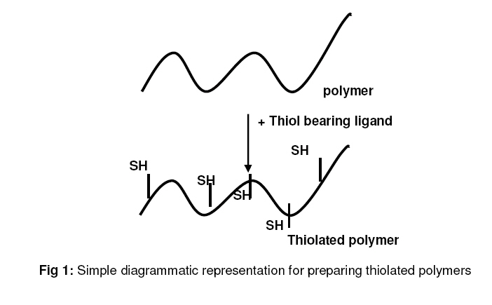
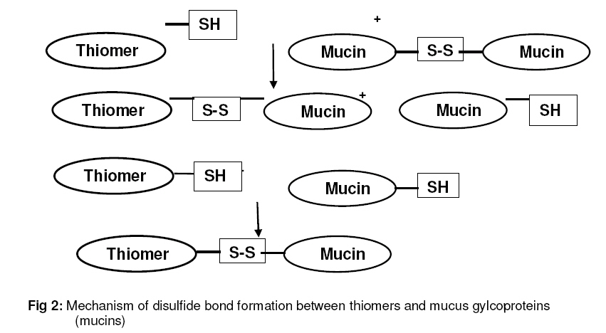
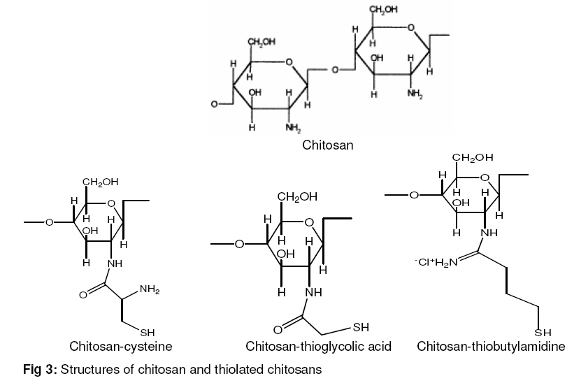
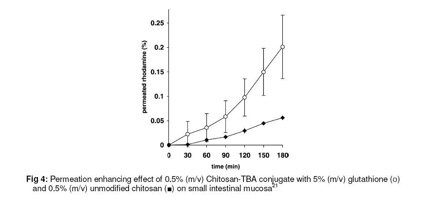
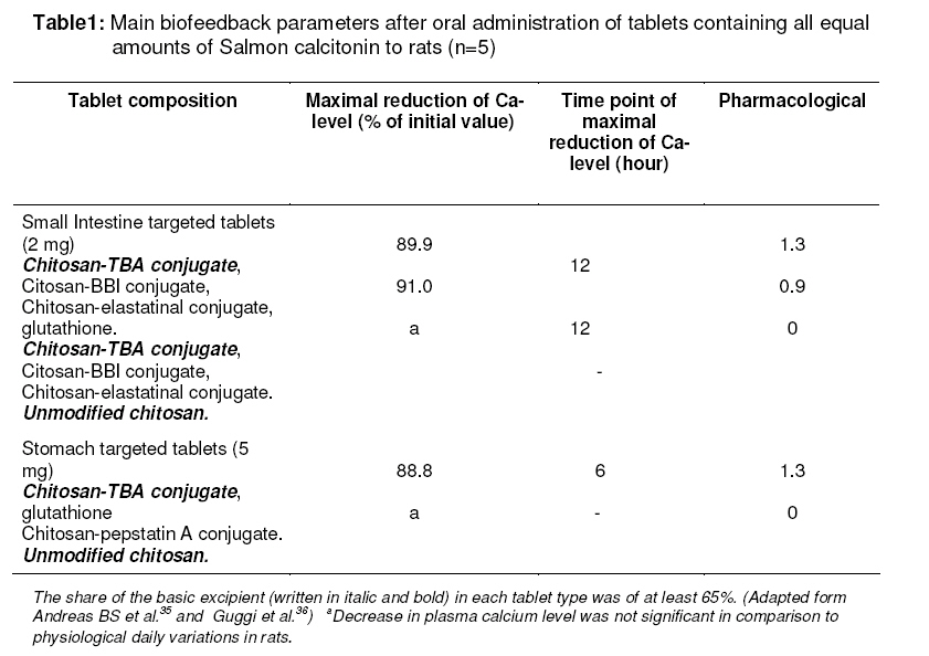
Tropical Journal of Pharmaceutical Research, Vol. 7, No. 3, September, 2008, pp. 1077-1088 Review Article Thiolated Chitosans: Novel Polymers for Mucoadhesive Drug Delivery – A Review SA Sreenivas1* and KV Pai2 1Department of Pharmaceutics, KLES’s College of Pharmacy, Vidyanagar, Hubli, Karnataka. Code Number: pr08029 Abstract Chitosan is a natural polycationic copolymer consisting of glucosamine and N-acetylglucosamine units. The polymer has valuable properties as a biomaterial because it is considered to be biocompatible, biodegradable and non-toxic. The purpose of this review article is to provide detailed information about thiolated chitosans which are gaining popularity because of their high mucoadhsiveness and extended drug release properties. The derivatization of the primary amino groups of chitosan with coupling reagents bearing thiol functions leads to the formation of thiolated chitosans. Various properties of chitosan are improved by the immobilization of thiol groups. Due to the formation of disulfide bonds with mucus glycoproteins, mucoadhesiveness is augmented. The permeation of paracellular markers through mucosa can be enhanced by utilizing thiolated instead of unmodified chitosan. Moreover, thiolated chitosans display in situ gelling features due to the pH-dependent formation of inter-as well as intra-molecular disulfide bonds. This latter process provides, strong cohesion and stability of carrier matrices, being based on thiolated chitosans. The in situ gel formation within the pH range of 5 to 6.8 makes the application of thiolated chitosans on vaginal, nasal and ocular mucosa also possible. Thiolated chitosans can guarantee prolonged controlled release of embedded therapeutic ingredients. Key words: Thiolated chitosan,Thiomers, Mucoadhesion, Permeation enhancement, In situ gelation INTRODUCTION Mucoadhesive drug delivery systems Mucoadhesion can be defined as the ability of synthetic or biological macromolecules to adhere to mucosal tissues such as the mucosa of the small intestine. Since the early 1980s, the concept of mucoadhesion has gained considerable interest in pharmaceutical technology. If this concept can reach its full potential, it might open the door for novel, highly efficient dosage forms especially for oral drug delivery. Mucoadhesive drug delivery systems promise several advantages that arise from localization at a given target site, prolonged residence time at the site of drug absorption, and an intensified contact with the mucosa increasing the drug concentration gradient1 Hence, uptake and consequently bioavailability of the drug may be increased and frequency of dosing reduced with the result that patient compliance is improved. Various natural and synthetic polymers have been discovered as mucoadhesive excipients. Their mucoadhesive properties can be explained by their interaction with the glycoproteins of the mucus, based mainly on non-covalent bonds such as ionic interactions, hydrogen bonds and van der Waals forces2 . Chitosan The biopolymer chitosan is obtained by alkaline deacetylation of chitin, which is one of the most abundant polysaccharides in nature. Shell wastes of shrimp, lobster and crab are the main industrial sources of chitin3 Chitosan is a polysaccharide consisting of copolymers of glucosamine and N-acetylglucosamine. The primary amino group accounts for the possibility of relatively easy chemical modification of chitosan and salt formation with acids. At acidic pH, the amino groups are protonated, which promotes solubility, whereas chitosan is insoluble at alkaline and neutral pH4 . Because of its favorable properties, such as enzymatic biodegradeability, non-toxicity and biocom-atibility3 chitosan has received considerable attention as a novel excipient in drug delivery systems, and has been included in the European Pharmacopoeia since 2002. So far, chitosan has been utilized in various fields of pharmaceutical technology, including the formulation of controlled release dosage forms such as tablets, gels and microspheres, as mucoadhesive and/or permeation enhancing excipient for oral, nasal, ocular and buccal drug delivery5-8 and in non-viral gene delivery9,10 . Thiolated chitosans Recently, it has been shown that polymers with thiol groups provide much higher adhesive properties than polymers generally considered to be mucoadhesive. The enhancement of mucoadhesion can be explained by the formation of covalent bonds between the polymer and the mucus layer which are stronger than non-covalent bonds. These thiolated polymers (see Fig. 1), known as thiomers, interact with cysteinerich subdomains of mucus glycoproteins via disulfide exchange reactions11 or via simple oxidation process as shown in Fig. 2. To further enhance the solubility of chitosan and to improve its mucoadhesive and/or permeation enhancing properties, various derivatives such as trimethylated chitosan12 , mono-N-carboxymethyl chitosan13 , N sulfochitosan14 and chitosan-EDTA conjugates15 were developed. A further modification is based on the immobilization of thiol bearing moieties on the polymeric backbone of chitosan. To date, three different thiolated chitosan derivatives have been synthesized: chitosan-thioglycolic acid conjugates16,17 chitosan-cysteine conjugates18 and chitosan-4-thio-butyl-amidine (chitosan TBA) conjugates19. These thiolated chitosans have numerous advantageous features in comparison to unmodified chitosan, such as significantly improved mucoadhesive and permeation enhancing properties18-21 . The strong cohesive properties of thiolated chitosans make them highly suitable excipients for controlled drug release dosage forms19,22. Moreover, solutions of thiolated chitosans display in situ gelling properties at physiological pH values17 . It is the aim of this review to provide an overview about different thiolated chitosan derivatives that have been synthesized so far, as well as their characterization and optimization utilizing various in vitro test systems. The performance of thiolated chitosan in in vivo studies, providing proof of their applicability in peroral peptide delivery systems, will be discussed as well. Synthesis of thiolated chitosans The primary amino group at the 2-position (Fig. 3) of the glucosamine subunit of chitosan is the main target for the immobilization of thiol groups. As shown in Fig. 3 sulfhydryl bearing agents can be covalently attached to this primary amino group via the formation of amide or amidine bonds. In case of the formation of amide bonds the carboxylic acid group of the ligands cysteine and thioglycolic acid reacts with the primary amino group of chitosan mediated by a water soluble carbodiimide2 . The formation of disulfide bonds by air oxidation during synthesis is avoided by performing the process at a pH below 5. At this pH-range the concentration of thiolate anions, representing the reactive form for oxidation of thiol groups, is low, and the formation of disulfide bonds can be almost excluded. Alternatively, the coupling reaction can be performed under inert conditions. In the case of the formation of amidine bonds, 2-iminothiolane is used as a coupling reagent19 . It offers the advantage of a simple one step coupling reaction. In addition, the thiol group of the reagent is protected against oxidation because of the chemical structure of the reagent. Orientating studies with all these thiolated chitosans showed that a degree of modification of 25–250 mmol thiol groups per gram chitosan leads to the highest improvement in the mucoadhesive and permeation enhancing properties. The amount of immobilized thiol groups in reduced and oxidized form can be determined via Ellman’s17 reagent with and without previous quantitative reduction of disulfide bonds with borohydride23 . PROPERTIES OF THIOLATED CHITOSANS Mucoadhesive properties The improved mucoadhesive properties of thiolated chitosans are explained by the formation of covalent bonds between thiol groups of the polymer and cysteine rich subdomains of glycoproteins in the mucus layer24 . These covalent bonds are supposedly stronger than noncovalent bonds such as ionic interactions of chitosan with anionic substructures of the mucus layer. This theory was supported by the results of tensile studies with tablets of thiolated chitosans which demonstrated a positive correlation between the degree of modification with thiol bearing moieties and the adhesive properties of the polymer2,20. These findings were confirmed by another in vitro mucoadhesion test system where the time of adhesion of tablets on intestinal mucosa was determined. The contact time of the thiolated chitosan derivatives increased with increasing amounts of immobilized thiol groups2,19 . With chitosan-thioglycolic acid conjugates a 5–10-fold increase in mucoadhesion in comparison to unmodified chitosan was achieved. The mucoadhesive properties of chitosan-TBA (chitosan-4-thio-butyl-amidine) conjugates were even further improved. One explanation for this phenomenon can be given by the theory that chitosan-TBA conjugates additionally increased mucoadhesive properties due to improved ionic interactions between the additional cationic amidine substructure of the conjugate (see Fig. 3) and anionic substructures within the mucus layer. Tensile studies with chitosan-TBA conjugates of low, medium and high molecular mass (150, 400 and 600 kDa) furthermore indicated that medium molecular mass thiolated chitosans displayed relatively, the highest mucoadhesiveness. Utilizing a medium molecular mass chitosan-TBA conjugate displaying 264 mM thiol groups per gram polymer led to a more than 100-fold improvement in mucoadhesion compared to unmodified chitosan. This represents the greatest progress made so far in the development of mucoadhesive polymers20. Permeation enhancing effect In 1994 Illum et al showed the permeation enhancing capabilities of chitosan for the first time6 Chitosan is able to enhance the paracellular route of absorption, which is important for the transport of hydrophilic compounds such as therapeutic peptides and antisense oligonucleotides across the membrane. Various studies carried out on Caco-2 cell monolayers demonstrated a significant decrease in the transepithelial electrical resistance after the addition of chitosan25-27 . The mechanism underlying this permeation enhancing effect seems to be based on the positive charges of the polymer which interact with the cell membrane resulting in a structural reorganization of tight junction-associated proteins28 . In the presence of the mucus layer, however, this permeation enhancing effect is comparatively lower, as chitosan cannot reach the epithelium because of size limited diffusion and/or competitive charge interactions with mucins29 . Nevertheless, these results obtained on Caco2 cell monolayers could be confirmed by in vivo studies, showing an enhanced intestinal absorption of the peptide drug, buserelin, in rats due to the co-administration of chitosan hydrochloride30 . The permeation enhancing effect of chitosan can be greatly improved by the immobilization of thiol groups. The effect of thiolated chitosans has been shown in various permeation studies in Ussing type chambers using freshly excised intestinal mucosa18 . The uptake of fluorescence-labeled bacitracin, for instance, was improved 1.6-fold utilizing 0.5% of chitosan-cysteine conjugate instead of unmodified chitosan18 . In another study, the permeation enhancing effect of chitosan-TBA, in comparison to the permeation enhancing effect of unmodified chitosan, was shown. The uptake of the cationic marker compound, rhodamine-123 was 3-fold higher in the presence of thiolated chitosan than in unmodified chitosans21 . The likely mechanism responsible for this improved permeation enhancement has been attributed to the inhibition of the protein, tyrosine phosphatase. This enzyme seems to be involved in the opening and closing process of the tight junctions. Tyrosine phosphatase is responsible for the dephosphorylation of tyrosine subunits of occludin, representing an essential transmembrane protein of the tight junctions. When these tyrosine subunits of occludin are dephosphorylated, the tight junctions are closed. In contrast, when these tyrosine subunits are phosphorylated, the tight junctions are opened. The inhibition of tyrosine phosphatase by compounds such as phenylarsine oxide, pervanadate or reduced glutathione leads consequently to phosphorylation and opening of the tight junctions31-33 . In contrast to the stable but toxic tyrosine phosphatase inhibitors phenylarsine oxide and pervanadate, the inhibitory effect of glutathione is lower as it is rapidly oxidized on the cell surface, loosing its inhibitory activity34 . Due to the combination of reduced glutathione with thiolated chitosans, however, the oxidation of the inhibitor on the membrane can be restricted, as thiomers are capable of reducing oxidized glutathione31 . Thiolated chitosans as matrices for controlled drug release Chitosan represents, primarily due to its mucoadhesive properties, a valuable tool for non-invasive drug delivery5 . The longer residence time of formulations based on mucoadhesive polymers at the absorption site is believed to contribute to an increased absorption rate of the incorporated drug. However, such an enhanced bioavailability can be achieved only if a controlled release of the active agent out of the formulation is provided. Thiolated chitosans also display, besides their strong mucoadhesive and permeation enhancing properties, excellent cohesive properties. The reduced thiol functions on the chitosan backbone enable thiolated chitosans not only to form disulfide bonds with mucus glycoproteins, but also to form inter-as well as intra-molecular disulfide bonds. Such a crosslinking of the polymeric chains20 results in a high stability of drug carrier systems based on thiolated chitosans (Fig. 4). The cohesion and stability of a drug delivery system over the intended duration of drug liberation is often a substantial requirement for a controlled release. The usefulness of thiolated chitosans as carrier matrices for controlled drug release was demonstrated with model drugs, such as clotrimazole 19-22 and salmon calcitonin36-37 . Clotrimazole is well-established as an antimycotic drug in the treatment of vaginal infections. In order to improve its therapeutic efficacy, a sustained release of the drug over a period of several days might be highly beneficial. The release of clotrimazole out of matrix tablets based on either chitosan-thioglycolic acid conjugate or chitosan-TBA conjugate was quantified. Both thiolated chitosan tablets remained stable during the whole period of the experiment (6 hours) and no disintegration could be observed. However, only the chitosan-TBA conjugate was able to guarantee a significant delay in drug release, compared to unmodified chitosan, leading to a sustained release over a much longer time period19-22 . Furthermore, the release profile of salmon calcitonin out of matrix tablets based on the chitosan-TBA conjugate was determined. A pseudo zero order release profile of salmon calcitonin over the first 8 hours was observed in simulated intestinal fluid. During the experiment the tablets swelled continuously, maintaining good cohesiveness and releasing the active agent via a controlled diffusion process. These release studies, in which a peptide drug was liberated from a thiolated chitosan matrix system, permit information concerning the chemical events within the formulation to be gained. Strong unintended interactions between the polymeric matrix system and the peptide drug could be excluded, based the according to this controlled and sustained release profile37 . Both studies confirm that, controlled drug release out of thiolated chitosan drug carrier systems can be achieved. In situ gelling properties Rapid clearance from the site of drug action is one important factor that limits the efficacy of drugs administered to the ocular, nasal and vaginal mucosa. It is widely accepted that limiting the clearance by increasing the viscosity of a drug formulation will result in increased bioavailability of these drugs. A very promising strategy to obtain drug formulations of sufficient viscosity is based on in situ gel formation. The formation of a gel at the site of drug delivery combines the advantages of a solution, which can be easily administered, with the favorable viscoelastic properties of a gel, providing a prolonged residence time of the formulation. The sol–gel transition occurs in the physiological environment as a result of physicochemical changes, such as changes in the pH38 , temperature38,39 or electrolyte concentration40,41 . Thiolated chitosans display in situ gelling properties due to the oxidation of thiol groups at physiological pH-values, which results in the formation of inter-and intramolecular disulfide bonds. This crosslinking process can be observed within the pH range of 5–6.8. The in situ gelling behavior of thiolated chitosans was characterized in vitro by rheological measurements. The sol–gel transition of thiolated chitosans at pH 5.5 was completed after 2 hours when highly crosslinked gels were formed. In parallel, a significant decrease in the thiol group content of the polymers was observed, indicating the formation of disulfide bonds17,19 . The rheological properties of unmodified chitosan remained constant over the whole observation period. Rheological investigation of thiolated chitosans furthermore demonstrated a clear correlation between the total amount of polymer-linked thiol groups and the increase in elasticity of the formed gel. The more thiol groups were immobilized on chitosan, the higher was the increase in elastic modulus in solutions of thiolated chitosan17,19 . Thiolated chitosan derivatives, therefore, seem to be promising new excipients for liquid or semisolid formulations, which should stabilize themselves once applied on the site of drug delivery. The in situ gel formation within the pH range of 5 to 6.8 makes the application of thiolated chitosans on vaginal, nasal and ocular mucosa plausible. IN VIVO STUDIES: PROOF OF CONCEPT The potential of thiolated chitosans for the oral administration of hydrophilic macromolecules could meanwhile be shown by various in vivo studies35,36 . As model drug, for instance, salmon calcitonin was utilized, which is a peptide drug of cationic net charge and a molecular mass of 3.2 kDa. Salmon calcitonin is used for the treatment of chronic bone diseases37,42. It is currently marketed in nasal spray and injectable forms, both having the drawback of low patient acceptance. A higher patient compliance should be achieved by the application of an oral delivery system for this drug. However, the oral bioavailability thus far obtained is too low to permit therapeutic employment43 . Therefore, this peptide was regarded as a challenging model drug for testing the potential of thiolated chitosans. Different drug carrier matrices, comprising chitosan-TBA conjugate as substantial polymeric excipient and containing equal amounts of salmon calcitonin and optionally the permeation mediator, reduced glutathione, were developed. In order to avoid an enzymatic degradation of the peptide drug in the gastrointestinal tract, chitosan-enzyme inhibitor conjugates were added. All compounds were homogenized and directly compressed to tablets. To enteric-coated tablets targeted to the small intestine, a chitosan-BBI(Bowman-Birk inhibitor) conjugate44 and a chitosan-elastatinal conjugate45 were added. Furthermore, an alternative strategy was evaluated, focusing on targeted drug release and absorption in the stomach. Tablets targeted to the stomach contained chitosan-pepstatin A conjugate36 which should avoid pepsinic digestion of salmon calcitonin. In order to prevent mucoadhesion in the oral cavity and oesophagus, these tablets were coated with a triglyceride. The different tablets were orally given to rats and the plasma calcium level was monitored as a function of time. Pharmacological efficacy was calculated on the basis of the area under the reduction in plasma calcium levels of the oral matrix tablets versus intravenous injection. The main biofeedback parameters after application of the drug carrier matrices for the oral delivery of salmon calcitonin are shown in Table 1. In vivo studies showed no statistically significant (P<0.05) reduction of the plasma calcium level caused by salmon calcitonin, which was orally given in solution. Furthermore, no significant effect was observed after oral administration of tablets comprising the peptide drug and unmodified chitosan, although the native polymer is reported to be mucoadhesive and to exhibit permeation enhancing effect for hydrophilic macromolecules46 (see Table 1). Table 1 shows that the presence of the chitosan-TBA conjugate is essential for calcitonin absorption, since only tablets based on, thiolated chitosan caused a decrease in plasma calcium level of more than 5% for several hours. The increased absorption of the peptide, when embedded in a thiolated chitosan matrix, occurred due to the properties of the polymer derivative: the high stability and cohesiveness can provide a sustained release of the peptide47 , while the mucoadhesive features should lead to a prolonged residence time of the dosage form at the site of absorption. Moreover, the combination of thiolated chitosan with the permeation mediator, reduced glutathione, seems to have an impact on the bioresponse of orally given calcitonin. The significantly higher pharmacological efficacy of thiolated chitosan tablets containing glutathione in comparison to corresponding tablets without glutathione (see Table 1) indicates that glutathione contributes to the drug absorption process. These results are in good agreement with in vitro results demonstrating that thiomers show strong permeation enhancing effect, which can be further improved by the addition of glutathione31 . Therefore, the high in vivo efficacy of thiolated chitosans can be additionally raised by the use of glutathione. Among all thiolated chitosan formulations, stomach targeted tablets based on chitosan-TBA conjugate with the addition of both glutathione and chitosan-pepstatin A conjugate, showed the strongest effect. They led to a decrease of the plasma calcium level of more than 10% for at least 12 hours, thus demonstrating the validity of systemic peptide delivery via the stomach. Moreover, a faster and more reproducible onset of action was obtained by this novel approach35,36 . According to these results, the applicability of thiolated chitosans for the oral administration of other peptide drugs seems also likely and is the subject of ongoing studies. FUTURE TRENDS Non-invasive peptide delivery The incorporation of peptide drugs exhibiting a cationic net charge in anionic mucoadhesive polymers on the one hand leads to a strong reduction in the mucoadhesive properties and, on the other hand, may hinder drug release as a result of strong ionic interactions between the therapeutic ingredient and the polymeric network. Consequently, cationic therapeutic peptides or peptidomimetics such as calcitonin or desmopressin need to be embedded in cationic or non-ionic mucoadhesive polymers. As non-ionic polymers cannot provide sufficient high mucoadhesion and thiolated chitosans display comparatively the highest mucoadhesive properties among cationic polymers, this type of thiomer seems to be a favorable tool for the oral administration of cationic hydrophilic macromolecules. Apart from oral delivery systems thiolated chitosans seem to be useful also for other non-invasive routes of peptide drug administration. In particular, the nasal, vaginal, buccal and ocular mucosas are interesting targets. Production of micro-and nano-particles Microparticles based on chitosan disintegrate very rapidly unless they are combined with multivalent anionic compounds such as sodium sulfate48 or alginate leading to stabilization by an ionic cross-linking process. Due to the addition of such multivalent anionic compounds, however, the mucoadhesive properties of chitosan are strongly reduced. In contrast, microparticles that are based on thiolated chitosan do not disintegrate. Because of the formation of disulfide bonds within the polymeric network, microparticles are strongly stabilized49 . Consequently, a controlled drug release out of thiolated chitosan microparticles can be provided. In contrast to the addition of multivalent anionic compounds, the immobilization of thiol groups on chitosan leads to strongly improved mucoadhesive properties. Tissue engineering A further interesting application of thiolated chitosans is their use in tissue engineering. The expanding field of tissue engineering applications has accelerated the need for materials which are tissue compatible, biodegradable and with mechanical properties similar to the target tissues. Biodegradable and biocompatible polymers have been attractive candidates for scaffolding materials because they degrade as the new tissues are formed, eventually without inflammatory reactions or toxic degradation. Recently, Kast et al demonstrated the biodegradability of thiolated chitosan, paving the way for its use as novel scaffold material50 . Further, studies in this direction were performed with L-929 mouse fibroblasts seeded onto chitosanthioglycolic acid sheets. The results of this study showed that thiolated chitosan can provide a porous scaffold structure guaranteeing cell anchorage, proliferation and tissue formation in three dimensions50 . Due to the in situ gelling properties, it seems possible to provide a certain shape of the scaffold material by pouring a liquid thiolated chitosan51 cell suspension in a mold. Furthermore, liquid polymer cell suspensions may be applied by injection forming semi-solid scaffolds at the site of tissue damage. Since low concentrated aqueous solutions of thiolated chitosan remain liquid when stored under inert conditions and are rapidly gel under access of oxygen, they seem to be promising candidates for such applications. Coating of stents Another promising application of thiolated chitosans is their use as coating material for stents. Polymer-coated drug-eluting stents are a potential technique to achieve high local tissue concentrations of an effective drug at the precise site and at the time of vessel injury. First orientating studies demonstrated that by simply dipping the stent in a thiolated chitosan solution and drying it on air, a stable coating could be achieved. During the drying process, cross-linking of chitosan by the formation of disulfide bonds due to air oxidation, takes place. The polymeric network is thereby stabilized on the stent. The chitosan coating should allow sustained release of incorporated drugs such as anti-inflammatory agents or agents avoiding cell proliferation. Recently, it was shown that stents can be successfully coated with thiolated poly(acrylic acid) and that sustained release of a model peptide drug out of this thiomeric coating can be provided. Similar results can be expected for thiolated chitosans but have to be verified by ongoing studies. CONCLUSION The chemical modification of chitosan via derivatization with various reagents bearing sulfhydryl functions causes a dramatic change in the polymer’s properties. Mucoadhesiveness and cohesiveness are strongly improved. A comparatively stronger permeation enhancing effect is provided which can be further raised by the combination of thiolated chitosans with the permeation mediator, glutathione. Furthermore, thiolated chitosans display in situ gelling features and facilitate controlled drug release. Due to these advantageous features thiolated chitosans have been successfully used for peroral administration of peptide drugs. They seem to represent a promising new generation of polymeric excipients, in particular for the noninvasive administration of hydrophilic macromolecular drugs. REFERENCES
© Pharmacotherapy Group, Faculty of Pharmacy, University of Benin, Benin City, 300001 Nigeria. The following images related to this document are available:Photo images[pr08029f2.jpg] [pr08029t1.jpg] [pr08029f1.jpg] [pr08029f4.jpg] [pr08029f3.jpg] |
| |||||||||
