 |
| About Bioline | All Journals | Testimonials | Membership | News |
|
||||||
|
||||||
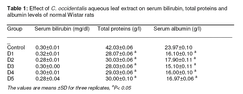
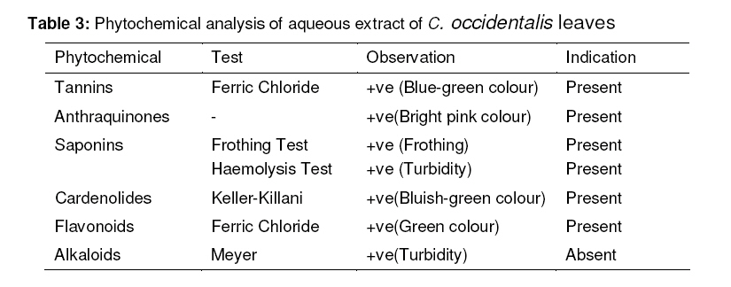
Tropical Journal of Pharmaceutical Research, Vol. 7, No. 4, December, 2008, pp. 1137-1142 Research Article Effects of Cassia occidentalis aqueous leaf extract on biochemical markers of tissue damage in rats AA Nuhu1* and R Aliyu2 1 Biology Programme, Abubakar Tafawa Balewa University, ATBU-Bauchi, Bauchi, Nigeria, Received: 14 June 2008 Revised accepted: 19 August 2008 Code Number: pr08036 Abstract
Purpose: Among the Hausas of Northern Nigeria, it is claimed by local (traditional) healers that the infusion of C. occidentalis leaves is used as a cure for hepatitis. This study was designed to evaluate the possible negative effects of the aqueous extract of this herb on serum total proteins, albumin, bilirubin, alanine amino transferase (ALT), aspartate amino transferase (AST) and alkaline phosphatase (ALP), as part of a larger study undertaken by these authors to ascertain the hepatoprotective potential of the plant extract. Key words: C. occidentalis, Liver, Serum proteins, Transaminases, Hypoproteinaemic effects INTRODUCTION Traditional medicine, making use of herbs in different preparations, is greatly relied upon especially by rural dwellers, for the treatment of various ailments: traditional doctors or healers are the dispensers of such concoctions. Chandan and others have observed that the extracts of the plant Boerhaavia diffusa shortened hexabarbitone ‘sleeping time’ in rats and decreased the levels of bilirubin, ALT and AST in CCl4 induced rat liver damage1 . Previously, postand pre-treatment of rats with the leaf and stem extracts of Melothria maderaspatana did hasten recovery from CCl4- or ethanol induced liver damage2 . Occidentalis species belongs to the genus Cassia and the Family Caesalpiniaceae. It is called Stink Weed, Stinking or Negro Coffee. In Hausa, it is known as “Rai dore”. It is an erect herb, commonly found by road sides, in ditches and waste dumping sites. Local healers in Jos, Nigeria, have reported that the infusion of the leaves of C. occidentalis is used as an effective treatment for hepatitis. The potential of the leaf extract of C. occidentalis may be related to its antioxidant activity. The extract contains flavonoids which are powerful antioxidant polyphenolic compounds. Torell et al.3 and Faure et al.4 have shown that flavonoids inhibit peroxidation of polyunsaturated fatty acids in cell membranes. Moreover, results have shown that flavonoids from Helichrysum genus inhibit the formation of superoxide ions and hydroxy radicals, which are two strong peroxidation agents5 . For a productive usage, positive effects of any drug have to outweigh its negative ones. This work is aimed at assessing some of the possible negative effects that the aqueous extract of C. occidentalis leaves may have when used in the treatment of hepatitis. MATERIALS AND METHODS Plant material Fresh leaves of C. occidentalis were collected from different locations around the University of Jos main campus in August, 2002. The plant for this study was identified by Mr. Abdul Kareem of the Federal College of Forestry, Jos, Nigeria. Preparation of C. occidentalis aqueous leaf extract 4.5 g of the fresh leaves above was coarsely ground in a mortar. Thereafter, it was gradually, but continuously, sun-dried for two days. The dried material was further crushed into fine powder using mortar and pestle. 3.89 g of the powder was placed in an Erlenmeyer flask containing 250 ml of water and left to infuse for 24 hours following the description given by the traditional healers. Subsequently, it was filtered using Whatman filter paper No. 1. For determining the yield from the extraction procedure, the residue was then dried to a constant weight at 70oC. 0.5 g of powdered potash (of high quality and obtained from Terminus market, Jos) was added to the filtrate to reduce its acidity. The pH of this filtrate was determined with a Pasport PS-2102 pH meter and after which it was stored in a stoppered brown bottle for subsequent evaluation. Phytochemical screening The aqueous extract of C. occidentalis leaves was screened for tannins, anthraquiones, saponins, cardenolides, flavonoids and alkaloids according to the methods described by Trease and Evans6 . Experimental animals and treatment 18 male albino rats of 120g average weights, which were maintained on a standard feed (Pfizer, Lagos) and water ad libitum, were used. Approval for experimentation with these animals was granted by the Ethics Committee on Animal Experimentation (University of Jos) in conformity with Helsinki Declaration. The animals were divided into six groups of three each, namely, D1, D2, D3, D4, D5 and D6 (control group). Groups D1:D5 received 1 ml of the extract orally by intubation as a oncetreatment and sacrificed at the termination of the treatment periods of: 24 h (D1), 48 h (D2), 72 h (D3), 96 h (D4), and 120 h (D5). The control group was sacrificed after chloroform anaesthesia along with the last treatment group, D5. Blood samples were collected, by incision of jugular veins, in clean plastic centrifuge tubes without anticoagulant and allowed to stand for 15 min. After coagulation, blood samples were centrifuged at 2,000 rpm for 10 min. The clear supernatants were obtained as the sera using micropipette. Determination of serum albumin concentration Serum albumin was determined colorimetrically using bromocresol green (BCG): Three test tubes were labeled as test, standard and blank respectively. Serum (0.1 ml) was added to the test, 0.1 ml of standard albumin was added to standard and 0.1 ml of water to the blank. BCG (0.8 ml) reagent was then added to each of the test tubes. Subsequently, the tubes were incubated at 37° C for 15 minutes and the absorbance of the test and standard were read at 640 nm against the blank and albumin concentration derived. Determination of serum bilirubin This was performed based on the modified method of Malloy and Evelyn7 : 0.4 ml of serum and 3.6 ml of water were added to each of two test tubes labeled A and B. 1.0 ml of diazo reagent was added to the tube A and 1.0 ml of blank solution (15 ml of Conc. H2 SO4 in 1 liter H2 O) was added to tube B. Both tubes were mixed immediately. After five minutes, the absorbance of A was measured spectrophotometrically at 540 nm using B as blank. This absorbance was termed E1. Absolute methanol (5.0 ml) was then added to each tube and mixed. After a further 30 minutes, the absorbance of A was read using B as a blank. This was termed E2 (for total bilirubin). The absorbance of a standard bilirubin solution (0.016 mg/100 ml) was read at 540 nm using water as the blank. This was termed Es. Total serum bilirubin (expressed as mg/100 ml) was calculated as follows: (E2- Ablank ) x 0.016 x 100/ 0.4 Es Determination of serum alkaline phosphatase Four test tubes were labeled as test, blank, standard and Sblank respectively. 1.0 ml of bicarbonate buffer and 1.0 ml of the substrate, disodium phenyl phosphate, were added to both the test and blank samples. The buffer (1.1 ml) was added to both the standard and Sblank . To the Sblank was also added 1.0 ml distilled water, while 1.0 ml phenol standard was added to the standard. 1 N NaOH (0.8 ml) was then added to the blank, standard and Sblank . All the four tubes were then incubated for 15 minutes at 37 oC. The addition of 0.8 ml NaOH to the test followed the incubation. To all the tubes were then added 1.2 ml sodium bicarbonate, 1.0 ml potassium ferricyanide. The red/ pink colour absorbance was then measured in a spectrophotometer at 510 nm using the appropriate blank to set to zero. King Armstrong unit was calculated per 100 ml as: K.A. /100 ml = Absorbance of Test x 100/ (0.1 x Absorbance of Standard.) Determination of total serum proteins by Biuret method 5.0 ml of Biuret reagent was added to 0.1 ml of serum. This was mixed and warmed at 37°C for 10 minutes followed by cooling. Similarly, 0.1 ml of the protein standard (BSA) was added to 5.0 ml of Biuret reagent. Absorbance of both the standard and test mixtures was taken at 540 nm against a blank containing 5.0 ml Biuret reagent in 0.1 ml of water. Total protein content of the serum was determined using the formula: Absorbance of Test x Concentration of Standard/ Absorbance of Standard Determination of AST and ALT AST and ALT were assayed colorimetrically according to the method of Reitmann and Frankel8 . AST: The buffered substrate (0.5 ml, pH 7.4), containing L-aspartic acid (13.3 g) and α-ketoglutaric acid (0.146 g), was incubated at 37 oC for 3 minutes and 0.1 ml of serum was added with thorough mixing. This was then re-incubated at 37 oC for 60 minutes. Subsequently, 0.5 ml of 1 mM 2, 4dinitrophenyl hydrazine was added and the mixture was left to stand at room temperature for 20 minutes. After this time, 5. 0 ml of 0.4 N NaOH was added to stop the reaction. The tube contents were mixed and left to stand for 10 minutes at room temperature. Thereafter, the absorbance was taken in a spectrophotometer at 510 nm using a blank solution in which 0.1 ml distilled water replaced the serum. ALT: The same procedure was used as applied to AST except that ALT substrate was substituted for AST substrate and incubation was for 40 minutes. Concentrations of both enzymes were graphically extrapolated using different concentrations of the standard. Statistical analysis Results were expressed as mean ± S.D. Statistical analysis was performed using Student’s t-test and p-values < 0.05 were considered statistically significant. RESULTS The results obtained for serum total proteins, albumin and bilirubin for the six groups of animals are presented in Table 1. The results for AST, ALT and ALP are listed in Table 2, while the findings in respect of phytochemical screening, showing the presence/ absence of different constituents in the aqueous extract of C. occidentalis, are presented in Table 3. The final pH of the extract was 6.05 and the extraction yield was 88% equivalent to the concentration of 1.37%. DISCUSSION Hypoproteinaemia is the deficiency of protein in the plasma, partly due to dietary insufficiency or excessive excretion. The extract of C. occidentalis (concentration 1.37%, pH 6.05) has displayed such effect, as indicated by the significant decrease (p<0.05) in the total serum proteins of all the treatment groups when compared to the control group (Table 1). Since albumin is the chief protein of the plasma and other serous fluids, any effect that negatively affects albumin content would be expected to have a deleterious impact on total plasma proteins as in massive hepatic necrosis, chronic cirrhosis and other disorders with significant destruction or replacement of liver cells. Inadequate amino acid supply as in protein caloric malnutrition or malabsorption can also lead to hypoproteinaemia. The observed decrease in the total serum proteins might have resulted from the first-pass febrile shock experienced by the experimental animals following administration of the extract. Roper has shown that this type of condition could be transient and albumin level may revert to normal9 . Albumin serves in the maintenance of osmotic pressure of the blood and body fluids, and transport of inorganic anions, fatty acids and drugs10 . Therefore, decrease in serum albumin level would affect the metabolism of these substances that are transported by it11. Experiments performed with extract of C.occidentalis revealed the possibility of mild hyperbilirubinaemic effect as observed from the insignificant increase in the bilirubin levels of the treatment group D1 as compared to the control (p>0.05). Hyperbilirubinaemia is often the first and sometimes the only manifestation of liver disease12 . Impaired hepatic bilirubin clearance due specifically to reduced uptake or possible competition for binding to 2-protein or ligandin, is associated with the administration of drugs such as rifanpicin, bunamiodyl, flaraspidic acid and probanecid, but plasma concentration rapidly reverts to normal following discontinuous use of the offending drug13 . Another possible explanation for the mild hyperbilirubinaemic effect of the extract might be as a result of the hemolytic effect of its saponin content (Table 3). Haemolysis may give rise to mild hyperbilirubinaemia. Although the plasma bilirubin level increases lineally in relation to bilirubin production, the level may still be near normal in subjects with a 50% reduction in red cell survival, provided that hepatic bilirubin clearance is normal13 . Generally, therefore, the extract has no significant negative effect on bilirubin level. From the results obtained in Table 2, it can be shown that C.occidentalis aqueous leaf extract brought about significant increase in the levels of ALT, AST and ALP in the Wistar strain rats. Highest values were obtained with group D1 for ALT and AST while D2 gave the highest increase for ALP. ALT is a more specific marker of the liver than AST14 . High levels of this enzyme in the serum may indicate cardiac infarction, muscle injury and hepatic necrosis9 . Alkaline phosphatase, ALP, is a plasma and endoplasmic reticulum membrane-bound enzyme15 . Transient increase of this enzyme may be noticeable in all types of liver drug, apart from the dose, is also required in problems. determining the degree of toxicity, Based on the results obtained, the extract of toxicological studies on the extract are far C. occidentalis may be slightly toxic. However, from being complete. In this regard, since the since knowledge of pharmacokinetics of a rats used in our study were not malnourished as they were maintained on a normal chow ad libitum, the observed hypoproteinaemia might be due to liver damage. This can be confirmed by liver histology in subsequent study. CONCLUSION Hypoproteinaemic effects and increase in ALT, AST and ALP were indications that the crude extract of C. occidentalis leaves may be slightly toxic as concoction for liver ailments. ACKNOWLEDGEMENT The authors wish to express their appreciation to Mr. Abdul Kareem of the Federal College of Forestry, Jos, for the identification and classification of the plant for this study. Our gratitude goes to the personnel of Biochemistry and Chemistry Laboratories of the University of Jos, Chemical Pathology Laboratory, NVRI Vom and Chemistry and Agricultural Laboratories, ATBU Bauchi, where aspects of this work were carried out. The work was supported by ATBU Postgraduate Fellowship. REFERENCES
© Pharmacotherapy Group, Faculty of Pharmacy, University of Benin, Benin City, 300001 Nigeria. The following images related to this document are available:Photo images[pr08036t1.jpg] [pr08036t3.jpg] [pr08036t2.jpg] |
| |||||||||
