 |
| About Bioline | All Journals | Testimonials | Membership | News |
|
||||||
|
||||||
Tropical Journal of Pharmaceutical Research, Vol. 7, No. 4, December, 2008, pp. 1143-1149 Research Article In-vitro Antimicrobial and Antitumor Activities of Stevia Rebaudiana (Asteraceae) Leaf Extracts Sathishkumar Jayaraman *, Muthu Saravanan Manoharan, Seethalakshmi Illanchezian Life Teck Research Centre, Vadapalani, Chennai – 600026, India Received: 02 July 2008 Revised accepted: 26 August 2008 Code Number: pr08037 Abstract
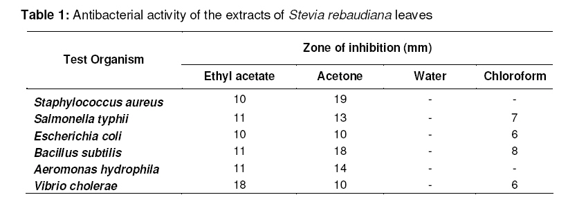
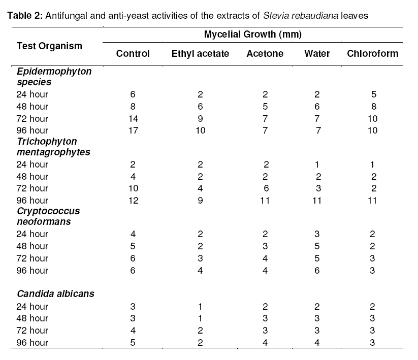
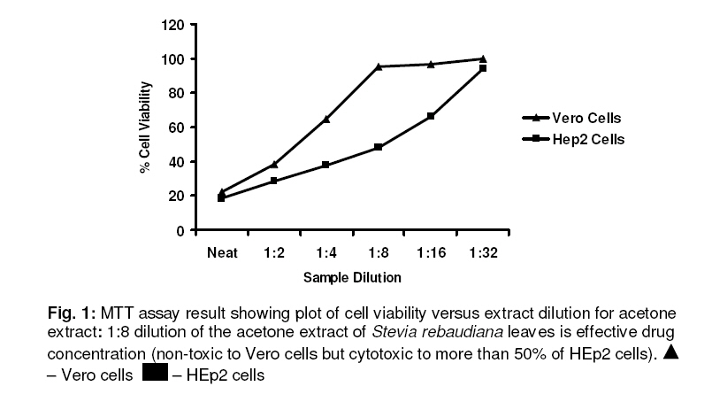
Purpose: The purpose of the study is to evaluate the antimicrobial and antitumor activities of Stevia rebaudiana (Asteraceae) leaf extracts. Key words: Stevia rebaudiana; Antibacterial; Antifungal; Antitumor; HEp2 cells; MTT assay INTRODUCTION There is a continuous and urgent need to discover new antimicrobial compounds with diverse chemical structures and novel mechanisms of action due to an alarming increase in the incidence of new and reemerging infectious diseases and development of resistance to the antibiotics in current clinical use1 . The screening of plant extracts has been of great interest to scientists in the search for new drugs for greater effective treatment of several diseases2 . Therefore, plant extracts and phytochemicals with known antimicrobial properties can be of great significance in therapeutic treatments3-5 . The medicinal value of plants lies in some chemical substances that produce a definite physiological action on the human body. The most important of these bioactive compounds of plants are alkaloids, flavanoids, tannins and phenolic compounds6 . Many plant leaves have antimicrobial principles such as tannins, essential oils and other aromatic compounds7,8 . In addition, many biological activities and antibacterial effects have been reported for plant tannins and flavanoids9-11 . Plants have an almost limitless ability to synthesize aromatic substances, most of which are phenols or their oxygen-substituted derivatives12 . These compounds protect the plant from microbial infection and deterioration13 . Some of these phytochemicals can significantly reduce the risk of cancer due to polyphenol antioxidant and antiinflammatory effects. Some preclinical studies suggest that phytochemicals can prevent colorectal cancer and other cancers14-16 . One of the potent members of the Asteraceae family is Stevia rebaudiana (commonly referred to as Honey leaf, Candy leaf and Sweet leaf). It is rich in terpenes and flavanoids. The phytochemicals present in Stevia rebaudiana are austroinullin, βcarotene, dulcoside, nilacin, rebaudi oxides, riboflavin, steviol, stevioside and tiamin17 . Stevia has important industrial uses in beverages, energizers as well as medicinal uses such as low uric acid treatment, vasodilator cardiotonic, anesthetic and antiinflammatory. The present study was carried out to evaluate the antimicrobial and antitumor activity of Stevia rebaudiana leaves extracted using various solvents. MATERIALS AND METHODS Test organisms Escherichia coli, Bacillus subtilis, Staphylococcus aureus, Salmonella typhi and Vibrio cholerae were used to test antibacterial activity while Candida albicans, Cryptococcus neoformans, Trichophyton mentagrophytes, Epidermophyton species were used to assess anti-yeast and antifungal activities. All the stock cultures were obtained from Microbial Type Cell Culture (IMTECH, India). Plant material Stevia rebaudiana leaves were obtained from Anna medicinal farm, Chennai. The leaves were washed with sterile water, dried in shade, finely powdered & stored in air tight bottles. Preparation of plant extract 25 g of air-dried powder of Stevia rebaudiana leaves was immersed in 100 mL of organic solvent (ethyl acetate, acetone, chloroform) and water separately in a conical flask. It was incubated at room temperature for 48 hour at 150 rpm in an orbital shaker. The suspension was filtered and concentrated to dryness at 40°C in hot air oven. The extract was dissolved in 0.25% Dimethyl Sulphoxide (DMSO, Merck) to a concentration of 100 mg/mL. Assay for antibacterial activity Preparation of inoculum Stock cultures were maintained at 4 oC on nutrient agar (HiMedia) slants. Active cultures for experiments were prepared by transferring a loopful of culture to 10 mL of nutrient broth (HiMedia) and incubated at 37 oC for 24 hours for bacterial proliferation. Agar-well diffusion method Agar well bioassay was employed for testing antibacterial activity of Stevia rebaudiana leaves18 . Each extracts were made to a final concentration of 50 mg/mL. 24 hour old cultures of test organisms (0.05 mL) were seeded onto Mueller Hinton agar (HiMedia) plate and uniformly spread with a spreader. Wells (5mm) were made in the agar plate with a sterile cork borer. The plant extract was introduced into the well and the plates were incubated at 37 oC for 24 hours. The antibacterial activity of the plant extract was determined by measuring the diameter of the inhibition zone. Controls contained only DiMethyl Sulfoxide (DMSO). The antibacterial assay for each of the extracts against all microorganisms tested was performed in triplicates. Assay for antifungal activity Potato dextrose agar (HiMedia) was prepared and 1 mL (50 mg/mL) of plant extract was added to the medium. After solidification loopful of culture was placed in the centre of the plate. Controls contained only DMSO. All the plates were incubated at 25 oC for 18 days. The growth of the fungal cultures was measured and compared with the respective control plates. The antifungal assay for each of the extracts against all microorganisms tested was performed in triplicates. Cell viability assay Vero cells (African green monkey kidney cells) obtained from King Institute of Preventive Medicine, Chennai, India was used to determine the non-toxic dose of the plant extract. The cells were grown in a 24-well plate in Eagle’s Minimum Essential Medium (HiMedia) supplemented with 10% fetal bovine serum (Gibco Laboratories) and antibiotics (streptomycin, penicillin-G, kanamycin, amphotericin B). About 1 mL cell suspension (105 cells/mL) was seeded in each well and incubated at 37 oC for 48 hour in 5% CO2 for the formation of confluent monolayer. The monolayer of cells in the plate was exposed to various dilutions of the plant extract. The cell viability was measured using MTT assay19 with MTT (5 mg/mL) and DMSO. This tetrazolium salt is metabolically reduced by viable cells to yield a blue formosan product measured at 540nm spectrophotometerically19 .Controls were maintained throughout the experiment (untreated wells as cell control and diluent treated wells as diluent control). The assay was performed in triplicate for each of the extracts. The mean of the cell viability values was compared to the control to determine the effect of the extract on cells and % cell viability (Vero cells) was plotted against concentration of the plant extract. Antitumor assay The antitumor assay was performed on Human laryngeal epithiloma cells (HEp2) obtained from King Institute of Preventive Medicine, Chennai, India with non-toxic dose of the plant extract and its dilutions. The cell viability was measured using MTT assay as described above. Controls were maintained throughout the experiment (Untreated wells as cell control and diluent treated wells as diluent a control). The assay was performed in triplicates for each of the extracts. The mean of the cell viability values was compared to the 4 control to determine the effect of the extract on cells. A graph was plotted against the % cell viability (HEp2 cells) Vs dilution of the plant extract. The minimum concentration of plant extract that was non-toxic to Vero cells but toxic to HEp2 cells was recorded as the effective drug concentration. RESULTS The antibacterial activities of the solvent extracts of Stevia rebaudiana showed significant variations as shown in Table 1. Among the four extracts tested, acetone extract had greater antibacterial potential, followed by ethyl acetate extract and then the other extracts. The largest zones of inhibition were observed for acetone extract against Staphylococcus aureus (19 mm) and Bacillus subtulis (18 mm). Ethyl acetate extract was very effective against Vibrio cholerae (18 mm). Chloroform and water extracts were either slightly effective or ineffective against the test organisms, respectively. The antifungal and anti-yeast activities of the solvent extracts of Stevia rebaudiana also varied significantly among the test organisms as shown in Table 2. All the four extracts inhibited the growth of Epidermophyton species. All the extracts had inhibitory effect on the growth of Cryptococcus neoformans except the water extract. All the extracts inhibited the growth of Trichophyton mentagrophytes but as the incubation time was prolonged, the ethyl acetate extract showed higher inhibitory activity against Trichophyton mentagrophytes than the other extracts.With regard to antitumor activity, the MTT assay for acetone extract of Stevia rebaudiana treated cells showed that 1:8 dilution was the effective drug concentration (Fig 1 This extract concentration was nontoxic to Vero cells and also caused more than 50% cytotoxicity to HEp2 cells. DISCUSSION Stevia rebaudiana leaf extracts demonstrated antibacterial, antifungal, anti-yeast and antitumor activity. To the best of our knowledge, there is no previous reported work on the antimicrobial and antitumor activity of Stevia rebaudiana, except that of Tadhani and Subhash20 who also studied antimicrobial activity of Stevia rebaudiana leaves. The antibacterial activity of the acetone extract of Stevia rebaudiana leaves was higher than that of the other extracts. The acetone extract showed greater activity against Gram-positive organism than against Gram-negative organism. The higher antibacterial activity of the acetone and ethyl acetate extracts may be due to the greater solubility of the extract in these organic solvents21 . The inhibitory activity (measured by zone of inhibition) of chloroform extract was not pronounced against Bacillus subtulis (8 mm), Salmonella typhi (7 mm), Escherichia coli (6 mm), respectively, and non-existent against Staphylococcus aureus. The water extract of Stevia rebaudiana leaves was practically ineffective against the test organisms. This finding is similar to that of Tadhani and Subhash20 who also recorded very low antibacterial activity for water extracts of Stevia rebaudiana leaves. Several workers22-24 have reported that water extracts do not have much activity against bacteria. It should be noted, however, that growth media also seems to play an important role in the determination of antibacterial activity25 . All the extracts were active against Epidermophyton species and Candida albicans. The ethyl acetate extract showed high activity against Trichophyton mentagrophytes and Epidermophyton species, and this may be due to the greater stability of the active principles in the solvent over a longer period of time. The aqueous extract of Stevia rebaudiana showed no pronounced antitumour activity but the acetone and ethyl acetate extracts of Stevia rebaudiana were more cytotoxic to HEp2 cells. Acetone extracts showed the highest cytotoxic activity followed by ethyl acetate and chloroform extracts. MTT assay was used to evaluate cytotoxicity based on metabolic reduction of MTT. On treatment with Vero cells, 1:2 and 1:4 dilutions of the acetone extract showed cytotoxicity but there was no apparent cytotoxicity at 1:8 dilution. Further dilutions also had no toxic effects on Vero cells. 1:2 and 1:4 dilutions were cytotoxic to HEp2 cells, whereas 1:8 dilution caused more than 50% cytotoxicity and also cessation of cell growth. Further dilutions had less effect on the viability of the cancerous cells. Thus, the 1:8 dilution of the acetone extract of Stevia rebaudiana is non-toxic to the normal cells and also has both anticancer and anti-proliferative activities against the cancerous cells. CONCLUSION This study points to the probable antimicrobial and antitumor potentials of some solvent extracts of Stevia rebaudiana leaves. There is a need for further investigation of this plant in order to identify and isolate its active anticancer principle(s). The results of the study will also need to be confirmed using in vivo models. ACKNOWLEDGEMENT We thank Life Teck Research Centre, Chennai, for providing us the facilities and requisite support for this work. We also express our thanks to Dr. K. Rajagopal for his critical review of this paper. REFERENCES
© Pharmacotherapy Group, Faculty of Pharmacy, University of Benin, Benin City, 300001 Nigeria. The following images related to this document are available:Photo images[pr08037t2.jpg] [pr08037t1.jpg] [pr08037f1.jpg] |
| |||||||||
