 |
search
for |
| About Bioline | All Journals | Testimonials | Membership | News |
|
||||||
|
||||||
Tropical Journal of Pharmaceutical Research, Vol. 8, No. 1, February, 2009, pp. 3-9 Research Article Oct-4 expression maintained stem cell properties in prostate cancer-derived CD133+MDR1+ cells Satyanarayana Rentala1,2 and Lakshmi Narasu Mangamoori1* 1 Centre for Biotechnology, Institute of Science and Technology, Jawaharlal Nehru Technological University, Received: 7 August 2008 Revised accepted: 15 October 2008 Code Number: pr09002 Abstract
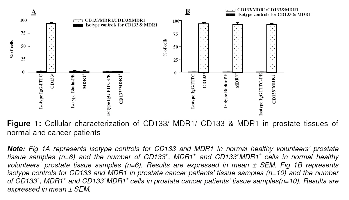
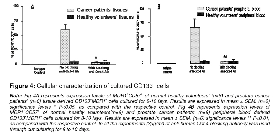
Purpose: CD133+ (prominin-1), a 5-transmembrane glycoprotein, has recently been considered an important marker that represents the subset population of cancer stem-like cells. The purpose of the present study is to isolate cancerous stem-like cells from normal healthy volunteers and prostate cancer patients (CD133+ ) which also express MDR1 and to ascertain the influence of Oct-4 on ‘stem-ness’ and differentiation of these CD133+ cells towards epithelium. Keywords: Prostate cancer, Cancer stem-like cells, Oct-4, CD133, Multi-drug resistance1 (MDR1) INTRODUCTION Prostate cancer is one of the leading causes of cancer-related deaths in males. Radiotherapy and chemotherapy play significant and crucial roles in clinical prostate cancer treatment to achieve prolonged patient survival. Autopsy studies show that many older men (and even younger men) who died of other diseases also had prostate cancer that never caused a problem during their lives. These studies showed that seven or eight out of ten men had prostate cancer by the age of 80 years1 . To improve the patient survival rate, investigation to elucidate the mechanism of tumorigenesis of prostate cancer is needed. Recent data have demonstrated that tumors contain a small subpopulation of cells, i.e., cancer stem-like cells (CSCs) or cancerinitiating cells (CICs), which exhibit a selfrenewing capacity and are responsible for tumor maintenance and metastasis. Stem cells have been isolated by their ability to efflux Hoechst 33342 dye2 and are referred to as the ‘‘side population (SP)’’. Our previous publication11 showed MDR1 in stem cells, enhanced the migration and homing of cultured bone marrow stem cells. A transmembrane glycoprotein, CD133 (prominin-1) was first recognized in CD34+ progenitor populations from adult blood, bone marrow, and fetal liver cells. Recently, CD133 has been considered an important marker to represent the subset population of CSCs in leukemia, brain tumors, retinoblastoma, renal tumors, pancreatic tumors, colon carcinoma, prostate carcinoma, and hepatocellular carcinoma. However, the gene regulation mechanisms in maintaining the self-renewal and drug resistant properties in putative cancer stem-like cells of prostate tumors are still unclear. Oct-4, a member of the family of POU-domain transcription factors, is expressed in pluripotent embryonic stem (ES) and germ cells. Oct-4 activates transcription via octamer motifs, and Oct-4 binding sites have been found in various genes, including fgf4 (fibroblast growth factor 4) and pdgfαr (platelet-derived growth factor α receptor). This suggests that Oct-4 functions as a master switch during differentiation by regulating the pluripotent potentials of the stem cell, and Oct-43-5 plays a pivotal role in mammalian development. In this study, CD133-positive cells (CD133+) were isolated from normal volunteers’ (NV) and prostate cancer patients’ tissue samples (PC) and investigated for their ability to differentiate into prostate cancer epithelial cells bearing CD57. MATERIALS AND METHODS All normal and prostate cancer tissues were kindly donated by Drs. Satish Kumar and S. N. Chary (Urologist) from those obtained from volunteers in NRI Hospital, Krishna (Dist), Andhra Pradesh, India. The study was approved by Institutional animal ethics committee (IAEC), India. Isolation of CD133+ population from prostate cancer biopsies The study was approved by the Institutional Ethics Committee/Institutional Review Board of NRI hospital. Biopsy samples from both normal (n=6) and prostate cancer patients (n=10) were digested with Trypsincollagenase mixture (Sigma) to dissociate cells from tissues. From this fraction, cells were selected for CD133 expression (Miltenyi Biotech) using magnetic beads as described by the manufacturer and were analyzed for the presence of CD133 (AC133, Miltenyi Biotech) and MDR1 (Sigma-Aldrich). The purity of CD133+ cells in the sorted samples was routinely greater than 92% and nearly all these cells also expressed MDR1. Western blot analysis for Oct-4, CD133 and MDR1 The isolated cells from normal and prostate cancer patients’ biopsies were lysed by adding SDS sample buffer containing 1 mM orthovanadate, and the samples were processed for Western Blotting. The expression patterns of Oct-4, MDR1 (both from Sigma-Aldrich) and CD133 (Miltenyi Biotech) were detected in normal and prostate cancer patients’ biopsies. Isolation of CD133+ population from peripheral blood samples Mononuclear cells were obtained from normal and prostate cancer patients’ buffy coats, on a Ficoll gradient (Biocoll 1077, Sigma). From this fraction, cells were selected for CD133 expression (Miltenyi Biotech) using magnetic beads as described by the manufacturer and were analyzed for the presence of CD133 (AC133, Miltenyi Biotech) and MDR1. The purity of CD133+ cells in the sorted samples was routinely greater than 94% and nearly all these cells also expressed MDR1. The above method is also used for isolation of CD133+ cells from prostate tissues. Culturing of CD133+ cells The above isolated cells from peripheral blood as well as from prostate tissues, were cultured on plates coated with fibronectin (50 µg/ml) in Iscove’s modified Dulbecco medium (IMDM), BSA (2 mg/ml), bovine transferrin (200 µg/ml), human insulin (10 µg/ml), hydrocortisone (2 µM), hepatocyte growth factor (HGF) (20 ng/ml) and GM-CSF (20 ng/ml). Cells were cultured in serum-free basal medium (Himedia) for 8-10 days after which half of the medium was replaced with fresh medium on alternate days. The cultured cells were trypsinzed and analyzed for various markers like CD133 and MDR1. In some experiments we used blocking rabbit anti-human Oct-4 antibody to block Oct-4 antigen in cell culture. The medium containing the antibody and growth factors was also replaced with fresh medium on alternate days. Flow cytometry About 5 x 10⁵ freshly isolated cells from patient biopsies and peripheral blood samples were stained by incubating with 50 µl of diluted primary antibodies at 4oC for 45 min. The cells were washed and stained with secondary antibody and then again washed and fixed with 0.2% paraformaldehyde. The antibodies used in this study were biotinylated anti-CD133 (Miltenyi Biotech, Labmate (Asia) Pvt., Ltd, India), mouse anti-human MDR1 (Clone F4, Sigma Aldrich), rabbit anti-human Oct-4 (Sigma Aldrich), anti-CD57 (BD Biosciences – Pharmingen. Clone NK-1, labeled with FITC), anti-biotin-PE (Miltenyi Biotech), streptavidin-FITC (Pharmingen), and isotype control antbodies were also procured from Pharmingen. The cells were analyzed on a customized BD FACS Caliber (BD Biosciences, San Jose, CA) using filters of 530/28 BP for FITC and 575/26 BP for PE with a 488-nm argon laser to excite both PE and FITC. RESULTS Isolation and characterization of prostate cancer-derived CD133-positive cells Using the magnetic bead method, we isolated CD133+ cells from tissue samples of six normal healthy volunteers and ten prostate cancer patients. To characterize our isolated NV-CD133+ and PC-CD133+ cells, FACS was used for the detection of the expression profile of cell surface markers. The high percentage (97%) of CD133+ (PC-CD133+) subset was isolated from the PC tissues (Fig 1). Peripheral blood derived CD133+ cells were isolated from normal healthy volunteers and prostate cancer patients. This isolation also gave us 97% pure population of CD133+ cells (Fig 2). Surprisingly prostate cancer patients’ tissues and peripheral blood samples derived CD133+ cells showed 80-90% also positive for MDR1 (Figs 1 and 2). We also found that in CD133+ cell fraction, MDR1+ cells were significantly less in tissues of normal nondiseased volunteers than prostate cancer patients (Figs 1 and 2). Blotting analysis for Oct-4, MDR1 and CD133 Western blot results suggested that the expression level of Oct-4, self-renewal and stem-ness protein in PC-CD133+ cells was significantly up-regulated than that in normal healthy volunteers (Fig 3A). To validate this finding, we examined the expression of Oct-4 in peripheral blood also. The amounts of Oct-4 protein in isolated PC-CD133+ cells were significantly more than in normal healthy volunteers (Fig 3B). Role of Oct-4 Expression in CD133+ differentiation towards prostate epithelium To investigate whether Oct-4 expression plays a role in maintaining self-renewal or cancer stem-like properties in PC-CD133+cells, we used the rabbit anti-human Oct-4 blocking antibody (3μg/ml), from day 1 to 8-10 days of culture of tissue and peripheral blood derived CD133+ cells of both normal healthy volunteers and prostate cancer patients. We found that treatment of blocking antihuman Oct-4 antibody in CD133+ cell differentiation could significantly interfere with the capabilities of differentiation towards prostate epithelium bearing CD57. However, this was not the case with NV-CD133+ differentiation (Figs 4A and B). Statistical analysis Data were analyzed by one–way ANOVA followed by Dunnett’s t-test using computerized Graph Pad Instat® version 3.05 (Graph Pad software, U.S.A.). DISCUSSION Oct-4 has been suggested as one of four major factors that render the reprogramming capability3-5 of adult cells into germlinecompetent-induced pluripotent stem cells. Recently, Oct-4 transcript can be consistently detected in human embryonal carcinomas, testicular germ cell tumors6-9 , seminomas, and bladder carcinomas. The expression of Oct-4 has further been shown in human breast cancer stem-like cells10 , suggesting that its expression may be implicated in self-renewal and tumorigenesis via activating its downstream target genes. Here we report the isolation of CD133-positive cells (PC-CD133+) from clinical tissue samples and peripheral blood samples. PC-CD133+ cells showed strong stem-ness and differentiation in vitro and in vivo (Figs 1, 2, and 4). PC-CD133+ cells also displayed significant expression profiles of Oct-4 and MDR1 (Fig 3). We also demonstrated that Oct-4 regulated differentiation into prostate epithelium expressing CD57 in PC-CD133+ (Fig 4). Indeed, Oct-4 functions as a master switch during differentiation31–33 by regulating the pluripotent potential in stem cells. Using the blocking antihuman Oct-4 blocking antibody in PC-CD133+ cells in culture, our data showed that the treatment of Oct-4 blocking antibody can block the differentiation of PC-CD133+ cells towards epithelial cells bearing CD57 (Fig 4). To our knowledge, this is the first study to show that Oct-4 expression plays a crucial role in maintaining self-renewal and cancer stem-like properties in PC-CD133+ cells. The property of resistance to chemotherapy (MDR1) and irradiation treatment is the major clinical criterion to characterize ‘‘cancer stem-like cells (CSCs)’’. The existence of cancer stem-like cells may explain why conventional anti-cancer therapies1,12,13 are able only to suppress or shrink a tumor but often cannot completely eradicate it, resulting in eventual recurrence. These results indicate that the up-regulated expression of Oct-4 in PC-CD133+ cells may contribute to the development of chemo-radio resistance in patients with prostate cancer. Recent studies have revealed that the human ABCG2 transporter is a P-glycoprotein that causes multidrug resistance (MDR1)13,14 including mitoxantrone, doxorubicin, and topoisomerase I inhibitors of irinotecan, topotecan, and 7-ethyl-10-hydroxycamptothecin (topoisomerase inhibitor) and gefitinib (an inhibitor of EGF receptor) in patients with lung cancer. Monzani and colleagues15 further showed that cancer stem-like cells derived from the melanoma cell line highly coexpressed CD133 and MDR1 markers with enhanced tumorigenic potential. In this study, we found that PC-CD133+ cells highly coexpressed with MDR1 showing resistance to conventional treatment methods compared with NV-CD133+ (Figs 1 and 2). Interestingly, a significant down-regulating of MDR1 expression and differentiation marker CD57 expression of PC-CD133+ were observed when the antihuman Oct-4 blocking antibody treatment was given (Fig 4). Thus, more studies are needed to investigate whether over-expression of Oct-4, CD133, and/or MDR1 play a role in progression of prostate cancer. CONCLUSION We demonstrated that PC-CD133+ cells display a higher Oct-4 expression with the ability to self-renew and may represent a reservoir with differentiation potentials for progression of prostate cancer. The MDR1 expression of PC-CD133+ cells in vitro and in vivo is partially due to preferential activation of Oct-4 gene expression. In addition, these data support that the up-regulated expression of Oct-4 play an important role in the tumorigenesis of patients with prostate cancer. REFERENCES
© Pharmacotherapy Group, Faculty of Pharmacy, University of Benin, Benin City, 300001 Nigeria. The following images related to this document are available:Photo images[pr09002f4.jpg] [pr09002f3.jpg] [pr09002f1.jpg] [pr09002f2.jpg] |
| |||||||||
