 |
| About Bioline | All Journals | Testimonials | Membership | News |
|
||||||
|
||||||
Tropical Journal of Pharmaceutical Research, Vol. 8, No. 1, February, 2009, pp. 27-31 Research Article The effect of Garcinia kola extract on lipopolysaccharide-induced tissue damage in rats Tebekeme Okoko*, Solomon Ndoni Biochemistry Division, Department of Chemical Sciences, Niger Delta University, PMB 71, Wilberforce Island, Bayelsa State, Nigeria. Received: 1 June 2008 Revised accepted: 6 October 2008 Code Number: pr09005 Abstract
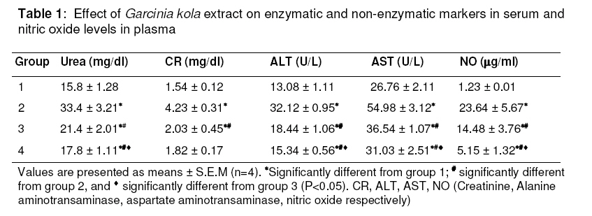
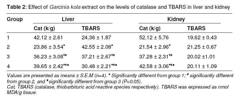
Purpose: To study the effect of the crude methanol extract of Garcinia kola seed on lipopolysaccharide (LPS)-induced tissue damage in rats Keywords: Garcinia kola, lipopolysaccharide, creatine, urea, hepatotoxicity, nephrotoxicity. INTRODUCTION The use of plants as traditional medicine has been in existence for several years. This practice has been encouraged in recent times due to the less frequent side effects when compared with orthodox medicine. In response to this, the medicinal potentials of a lot of plants have been explored. Garcinia kola Heckel (Gutifera) is a tropical plant which grows in moist forest. The seeds have a bitter taste hence the plant is commonly called bitter kola as such they are consumed as a stimulant1 . Extracts of the seeds have been used traditionally for the treatment of cough, and liver diseases2 . The seeds of the plant have been shown to have various bioactivities in experimental models such as nephroprotective and bronchodilatory effects3,4 . Various complex compounds have been isolated from Garcinia kola such as kolaviron, xanthones, garcinoic acid, garcinal, and tocotrienol5 . Lipopolysaccharide (LPS) is one of the virulence factors of Gram-negative bacteria. Macrophages are among the main targets of LPS and this activity leads to the release of some cytokines and nitric oxide as part of the inflammatory response6 . Recently, Garcinia kola seed extract was shown to reduce the tendency of lipopolysaccharide to activate macrophages in vitro14 however a similar work in an in vivo model has not been reported. This present work looks at the effect of Garcinia kola extract on lipopolysaccharideinduced tissue damage in rats. MATERIALS AND METHODS Chemicals and Reagents A vial of lipopolysaccharide (from E. coli strain 055:B5) was obtained from Sigma Chemicals (USA). All other chemicals were standard laboratory chemicals and reagents of analytical grade and were used without further purification. Buffers and dilutions were prepared in glass-distilled water unless otherwise stated. Animals Sixteen adult male albino rats (Rattus norvegicus) weighing between 110 – 120g were obtained from the Nigerian Institute of Medical Research, Yaba. They were kept in conventional cages in the animal house owned by the Department of Biological Sciences, Niger Delta University, Wilberforce Island. They were freely given water and standard rat chow ad libitum. Approval for the use of the experimental animals was given by the Ethical Committee for Animal Usage of the Faculty of Science, Niger Delta University. Preparation of plant extract The fruits of Garcinia kola were obtained fresh from the Obunagha forest of Bayelsa State, Nigeria in January 2008. The plant was authenticated by Professor B. L. Nyananyo, a taxonomist in the Department of Plant Science and Biotechnology, University of Port Harcourt, Nigeria. A voucher specimen (P.0126) was deposited in the herbarium unit of the Department of Chemical Sciences, Niger Delta University. The seeds were obtained from the fruits and sun-dried after chopping to smaller bits. They were ground and 500 g of the resulting powder was soaked in 1 L of absolute methanol for three days and filtered. The filtrate was later concentrated to dryness using a rotary evaporator set at 40°C ± 5°C. The yield was 36.2%. A 0.2 g/mL extract was later prepared in distilled water and stored at 4°C until required. Treatment of animals The rats were split into four groups (1, 2, 3, and 4). After acclimatizing the rats for six days, rats of groups 2, 3, and 4 were given a single intraperitoneal administration of lipopolysaccharide (1 mg/kg body weight) in order to induce tissue damage. Twenty four hours later, rats of groups 3 and 4 were given 100 mg/kg and 250 mg/kg body weight daily of the extract intragastrically for another 14 days, while group 1 rats were untreated controls. Collection of tissues At the end of the treatment, each rat was given light ether anaesthesia, dissected, the thoracic region opened, blood collected via cardiac puncture and delivered into two tubes -one heparinized and the other containing no anticoagulant. Plasma and serum were collected after allowing the tubes to stand for 10 min, centrifuged and stored at -4°C. The liver and kidney were also excised, washed in ice-cold phosphate buffer (0.2 M, pH 7.5), blotted, and 10 % homogenates were prepared using the same buffer. Perinuclear fractions of both liver and kidney were collected by centrifuging homogenates at 5000 rpm for 20 min. Biochemical analysis The levels of alanine aminotransaminase (ALT), and aspartate aminotransaminase (AST) were determined in serum according to the instructions on commercial kits purchased from Randox (UK). Urea and creatinine levels were also analyzed in serum according to the instructions on commercial kits purchased from QCA (Spain). Nitric oxide was determined in plasma according to the method of Hsieh et al8 . Catalase activity was determined by monitoring the consumption of hydrogen peroxide. Briefly, 1 ml of perinuclear fraction, 1.5 ml of distilled water and 1 ml of phosphate buffer (0.2 M, pH 7.5) were delivered into a test tube. The reaction was initiated by the addition of 0.5 ml of 0.1 M hydrogen peroxide. The amount of hydrogen peroxide consumed was determined by measuring absorbance at 240 nm and the catalase activity was expressed as k/g tissue. Thiobarbituric acid reactive species (TBARS) in tissues were measured as reported9 except that vitamin C was used as the reducing agent and the reaction took place at room temperature. TBARS was expressed as nanomoles of malondialdehyde (MDA) per gram tissue. Statistical analysis Data are presented as mean ± S.E.M. Where appropriate, the data were subjected to a twotailed Students’ t-test using Minitab statistical software (version 14). A confidence level exhibited at P < 0.05 was considered statistically significant. RESULTS The levels of the markers measured in serum and plasma are shown in Table 1. It revealed that the intraperitoneal administration of LPS caused marked elevations in the levels of ALT, AST, urea, creatinine, and nitric oxide. The levels of all these markers were significantly reduced when the Garcinia kola extract was given to the rats. In all cases, the levels of these markers in rats of group 4 are significantly lower than the levels measured in group 3. The catalase activities and the levels of thiobarbituric acid reactive species (TBARS) in the liver and kidney perinuclear fractions are shown in Table 2. LPS caused significant elevation of TBARS and reduction in the level of catalase in liver. These abnormal levels of the catalase and TBARS were brought close to control values when the extract was administered to the rats. However, the changes were more pronounced in group 4 than group 3. A similar result was also observed for catalase in the kidney but LPS did not cause any change in the level of TBARS in the kidney. DISCUSSION The strong correlation between the elevations in ALT, AST, urea and creatinine indicates that tissue damage which may predispose to lipid peroxidation which is associated with the inflammatory response. It has been demonstrated that LPS significantly increases the production of nitric oxide by the activation of macrophages10 . Nitric oxide is a signal molecule in the acute inflammatory response which is derived from the oxidation of L-arginine catalyzed by nitric oxide synthase11 . The results reveal that LPS is both hepatotoxic and nephrotoxic. This may give insight into the pathogenesis of some infectious diseases that are local to the liver and kidney as LPS is a virulence factor of Gram-negative bacteria. The increase in the level of TBARS in the liver following the administration of LPS indicates the activation of the lipid peroxidation system which is often initiated by the abstraction of hydrogen atoms from membrane lipids. Now, it is known that the measurement of TBARS in tissues is a method of choice for monitoring lipid peroxidation which is a major indicator of oxidative stress12 . Although, high levels of TBARS were not observed in kidney when the rats were treated with LPS, the elevations in urea and creatinine confirm the onset of kidney damage. The fact that the methanolic extract of Garcinia kola reduced the damage caused by LPS buttresses the protective role of Garcinia kola in tissues against free radical damage whether from a chemical or microbial source. This work also elucidates some manifestations of the damaging effects of LPS. The protective virtues of Garcinia kola have been ascribed to the presence of biflavonoid compounds5 . Much interest has focused on flavonoids – a group of polyphenols. Flavonoids such as kolaviron have the propensity of improving endothelial function by lowering oxidative stress which predisposes to improvement of coronary circulation13 . Gram-negative bacteria mostly contain (> 90%) of lipopolysaccharide on the surface of the outer membrane and is thought to be the protective wall rendering Gram-negative bacteria resistant to a variety of intrinsic host defence molecules14 . Thus this implies that the reduction in the tissue damage caused by LPS may be attributed to direct interaction with the phytochemicals. CONCLUSION This work further illustrates one of the ways by which gram-negative bacteria can cause damage to tissues and the probable mechanism of the reported antimicrobial properties of Garcinia kola extract. Based on these findings, the controlled consumption of Garcinia kola is recommended. The presence of Garcinia kola in the biosphere is nature’s gift to mankind as a source of abundant nutrients. Progress in this area will be of immense pharmaceutical interest. REFERENCES
The following images related to this document are available:Photo images[pr09005t2.jpg] [pr09005t1.jpg] |
| |||||||||
