 |
| About Bioline | All Journals | Testimonials | Membership | News |
|
||||||
|
||||||
Tropical Journal of Pharmaceutical Research, Vol. 8, No. 1, February, 2009, pp. 53-61 Research Article The effect of chitosan molecular weight on the properties of alginate/ chitosan microparticles containing prednisolone Soheila Honary*, Maryam Maleki and M Karami Department of Pharmaceutics, Faculty of Pharmacy and Pharmaceutical Research Centre, Mazandaran University of Medical Sciences, Sari, Iran Received: 29 November 2008 Revised accepted: 21 October 2008 Code Number: pr09008 Abstract
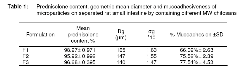
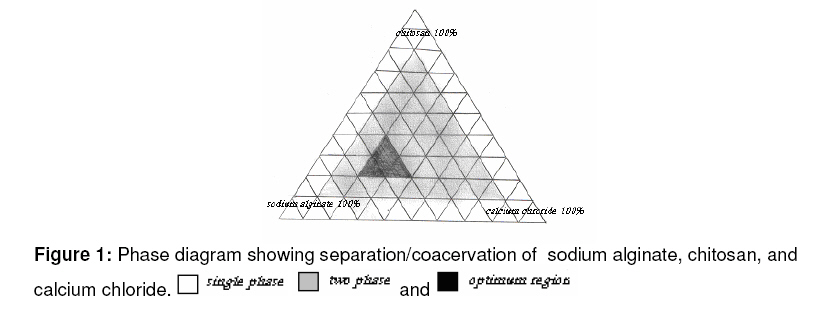
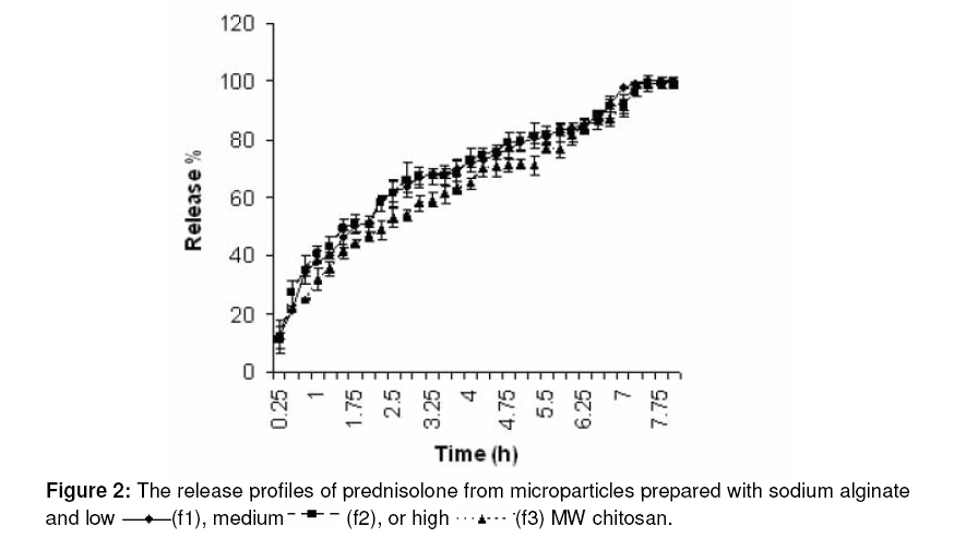
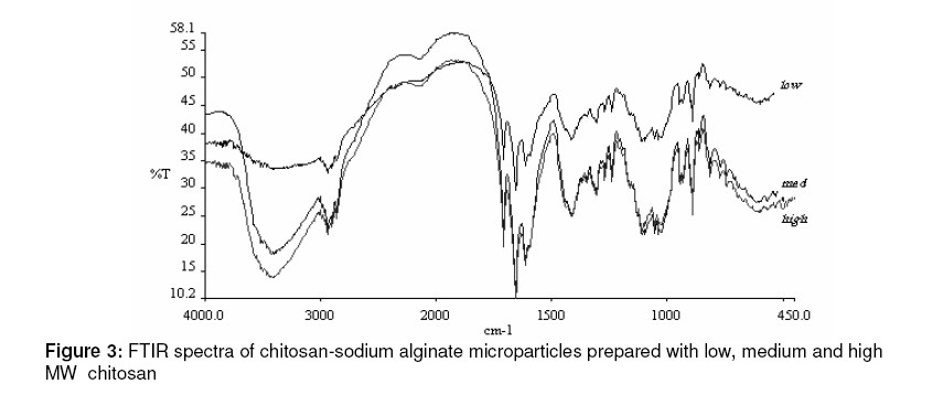
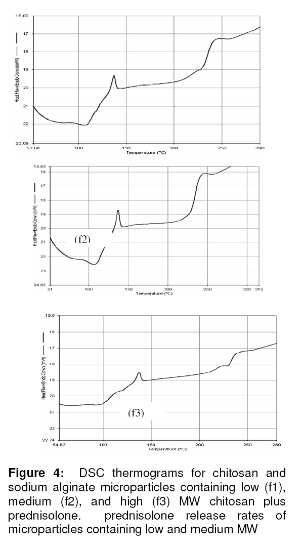
Purpose: The aim of the present study was to investigate the effect of chitosan molecular weight on size, size distribution, release rate, mucoadhesive properties and electrostatic bonding of alginate/chitosan microparticles containing prednisolone Keywords: Chitosan, Microparticle, Alginate, Molecular weight, DSC, FITR. INTRODUCTION Chitosan is a biodegradable and non-toxic hydrophilic polysaccharide with excellent mucoadhesive and permeation enhancing properties. During the last few years, this polymer has been studied and used with the intention of providing microcapsules with a modified drug release profile1,2. Alginate is the name given to a family of linear polysaccharides found in brown algae and is composed of guluronic (G) and manuronic (M) units. Calcium alginate beads are produced by inotropic gelation of alginate in the presence of calcium ions. There has been increasing interest in the study of alginate-chitosan microparticles as carriers for controlled release of proteins and drugs due its biocompatible, biodegradable and mucoadhesive properties. A study on the binding of chitosan to alginate beads using radioactive labeled chitosan showed that binding between the two polymers was markedly increased by reducing the average molecular weight of chitosan and increasing the porosity of the alginate gel1 . The stability of alginate-chitosan capsules was shown to depend strongly on the amount of chitosan bound to the capsules2 . The effect of various factors on protein release from alginatechitosan coacervate microcapsules including alginate, chitosan and calcium chloride concentration, loading rate, chitosan molecular mass and pH of gelation medium was investigated by Vandemberg et al3 . In their study, the size and gelation time of microcapsules were altered to determine their effects on protein retention. A negatively charged surfactant, sodium dioctylsulfosuccinate, was used to prepare microcapsules by ionic interaction with chitosan4 . The effect of deposition temperature on the release rate of indomethacin from chitosan-alginate microcapsules showed that the release rate was significantly reduced by increasing deposition temperature from 20 to 60 ºC5 . Chitosan microspheres have also been used to improve release the behavior of insulin6 . Hollow chitosan-alginate multilayer microcapsules were used as a drug delivery vehicle and the results showed that the encapsulated doxorubicin had higher efficacy than the free drug in terms of tumor inhibition7 . Drugloaded biodegradable microcapsules to control the release rate of ibuprofen fabricated from chitosan and sodium alginate demonstrate that the this system was an effective way to prolong drug release with reduced initial burst8 . The production of microcapsules having a complex coacervate alginate–chitosan membrane and a solid alginate core vary with respect to the applied processes and materials. There are, however, two principally different procedures: a) There is the one-stage procedure in which a complex coacervate membrane is formed at the interface between alginate and chitosan solutions when the alginate solution is dropped directly into chitosan solution9 . This method yields capsules with a complex alginate–chitosan membrane surrounding a liquid alginate core. The core is subsequently gelled either by prior incorporation of calcium chloride in the chitosan solution or by treating the liquid core capsules with calcium chloride after the membrane has been formed. b) There is also a two-stage procedure in which the formation of calcium alginate beads is followed by a membrane forming stage where the microparticles are suspended in a solution of the polycation. In the latter case, the microparticles are often washed with a calcium-free solution to remove excess calcium ions before transferring to the chitosan solution9 . The purpose of the present study was to examine the influence of chitosan molecular weight on the properties of microparticles with an alginate core and chitosan coating and containing prednisolone as an insoluble model drug. This work also focuses on the in vitro evaluation of the mucoadhesive properties of alginate-chitosan microparticles containing prednisolone using rat gut loop. EXPERIMENTAL Materials Low, medium and high molecular weight chitosan, micronized prednisolone, and calcium chloride were purchased from Fluka AG (Switzerland). Low viscosity sodium alginate (viscosity: 250 cps, 2% w/v) was purchased from Sigma–Aldrich Inc. (St. Louis, MO, USA). Other ingredients were of analytical grade, and used as received. Preparation of microparticles Alginate-chitosan microparticles were prepared by complex coacervation using sodium alginate as a gel core. All alginate solutions (2%, w/v) were prepared by dissolving sodium alginate in de-ionized water. Micronized prednisolone powder (2– 4%w/v) was then suspended thoroughly in the alginate solutions by vigorous stirring for 10 min. The two procedures described earlier (see ‘Introduction’) were used9 . In the onestep method, the alginate-prednisolone mixture was directly sprayed into the calcium chloride solution (0.5–1.0%, w/v) containing chitosan (0.5–1.5% w/v). In the two-step method, the alginate-prednisolone mixture was sprayed into calcium chloride solution (0.5–1.0%w/v), followed by a membrane forming step where the prednisolone containing calcium alginate particles were suspended in chitosan solution (0.5– 1.5%w/v). The microparticles were allowed to harden for at 2 h before washing them twice with distilled water and dried at room temperature and the alginate-prednisolone mixture was sprayed into the gelling solutions using two types of spray equipment: Casals 17500 Ripoll (Spain) and Inter Eko 1.5 lit, Pmax, 3 bar and rate, 0.61/min (Czech Republic) were used. The latter was used in the one-step method. Three different molecular weights of chitosan (low, medium, and high) were used to fabricate three microcapsule formulations -f1, f2, and f3, respectively. These microparticles were then evaluated to determine the effect of molecular weight on microparticle properties. Determination of prednisolone content The prednisolone content was determined by first dispersing 100 mg of microparticles in distilled water under sonication Citizen SW 1500 for at least 1 hr. The dispersion were filtered and the supernatant analyzed spectrophotometrically (Genesys 2, USA) at 242 nm. The results are expressed as percent entrapment (Table 1). Particle size determination Measurements of the particle size distributions and mean diameters of the microparticles were carried out with an optical microscope (Nikon E 400 Japan). The overall diameters of at least 300 circular microparticles and/or the mean of two Martin diameters of noncircular microparticles were recorded. Geometric mean diameter and geometric standard deviation were calculated by plotting microparticle size against cumulative % undersize using logarithmic and probability scales, respectively. In vitro release studies Drug release profiles of prednisolone containing alginate-chitosan microparticles were determined using USP dissolution apparatus II (Erweka, Germany). An accurately weighed amount (approx. 30 mg) of microparticles was added to 900 ml distilled water as dissolution medium at 37 ºC and stirred at 50 rpm. At time intervals, samples (5 ml) were withdrawn, filtered and analyzed spectrophotometrically at 242 nm. The release results (in triplicate) were expressed as a percentage. Fourier-transform infrared (FTIR) spectra Individual microparticles were crushed with pestle in an agate mortar. The crushed material was mixed with potassium bromide at 1:100 ratios and compressed to a 2mm semitransparent disk under for 2 min. The FTIR spectra over the wavelength range of 4000–400 cm-1 were recorded using an FTIR spectrometer (Perkin Elmer Spectrum One, Germany). Differential scanning calorimetry Differential scanning calorimetric studies were conducted in a Perkin Elmer differential scanning calorimeter Pyris 6 using an accurate weighed of the sample (10 mg) in a loosely covered aluminum pan and heated from 50 to 250°C at 10°C/min rate with 1min hold at 80°C under N2 parches. An empty loosely covered aluminum pan was used as the reference. Measurement of in vitro mucoadhesive properties Male Wistar rats, with a mean weight of 300 ± 30 g were anesthetized and killed by barbiturate overdose. The small intestine was removed and prepared by the following procedure: Using a syringe filled with physiological saline (NS), it was washed for 10 min at 5–10 ml/min rate and followed by a 20 min wash at 20–30 ml/min rate. At least 500 ml of NS was used to clean the tissue sample and then used immediately in the test10 . Briefly, known quantities (30mg) of chitosan microparticles were suspended in NS and filled into the small intestine (about 10 cm in length) and sealed by knotting it with a fibre. The filled tissue was immersed in NS and incubated at 37°C for 60 min. The microparticle suspension was then removed and the number of microparticles present in the suspension before and after the adhesion study was counted using the microscopic method previously described for particle size determination. The percentage of microparticles that adhered to the tissue was calculated from the difference in the counts. At least five measurements were made for each sample11 . Statistical analysis Experimental results were expressed as mean ± SD. Student’s t-test and one-way analysis of variance (ANOVA) were applied to check significant differences in drug release, mucoadhesive properties, and particle size determination for different formulations. Differences were considered to be statistically significant at p < 0.05. RESULTS A ternary phase diagram for sodium alginate, chitosan and calcium chloride illustrating phase separation/coacervation by the addition of an incompatible polymer is shown in Figure 1. The optimum concentration range required for each compound to form microparticles was identified in the triangular diagram as shown in Figure 1. This region was chosen by comparison of microparticles in terms of particle size, shape, reproducibility, symmetry and absence of free prednisolone crystals. The diagram was then used to select the various ratios of the materials in the formulation. The effects of stirring rate and time, spray apparatus type, distance of spray gun from chitosan solution surface, and the preparation method (one-or two-step) were evaluated. The formulations evaluated were prepared using the one-step method as stated earlier. Prednisolone entrapment Table 1 shows the prednisolone content of each of the formulations and it indicates that there is no statistical difference (p>0.05) in prednisolone content (a measure of % entrapment) between the three formulations. Geometric mean diameter and geometric standard deviation for each formulation are also shown in Table 1. Increase in the molecular weight of chitosan resulted in a decrease in the geometric mean diameter and geometric standard deviation of the microparticles. In vitro prednisolone release The in vitro release data are illustrated in Figure 2. It shows that there is no significant difference (p>0.05) between chitosan. However, highest MW chitosan produced significantly slower release rate than the other chitosans (p<0.05). In general, the FTIR spectrum of blank chitosan-alginate particles showed a broad band around 3500-3100 cm-1 , indicating enhanced hydrogen bonding compared to that of chitosan or sodium alginate alone13 . On the other hand, as shown in Figure 3, this peak is clearly stronger in high and medium MW chitosans than in low MW chitosan. Moreover, the N-H bending vibration of nonacrylated 2-aminoglucose primary amines of chitosan (1570 cm-1 ) and asymmetric and symmetric –C-O stretching at 1407 cm-1 of sodium alginate disappeared, indicating that the (-NH3+) of chitosan reacted with the (– COO -) of alginate13,14 . Absence of these bonds in the FTIR spectra of chitosan/sodium alginate microparticles indicated the formation of an electrostatic bond between them (Figure 3). The DSC studies were also performed to investigate the chitosan-alginate electrostatic interaction. The analysis of the DSC thermogram for the chitosan-sodium alginate particles showed a pair of exothermic and endothermic peaks at 230 ºC and 239 ºC, respectively. The peak at 230 ºC may be related to the breakdown of weak and unspecific electrostatic interactions. The DSC endothermic peak around 239.8 ºC, which was absent from either the chitosan or alginate thermogram, could be ascribed to the formation of an ionic pair between the carboxylate group (-COO-) of alginate and the ammonium group (-NH3 + ) of chitosan15 . Mucoadhesion The adsorption of chitosan-sodium alginate microparticles on rat small intestine was tested by counting the number of the particles adsorbed to the tissue. The results, shown in Table 1, indicate strong interaction between chitosan microparticles and mucous glycoprotein and/or mucosal surfaces, and which, as reported earlier, was dependent upon the method of preparation17 . DISCUSSION The results show that using high MW chitosan under fixed processing conditions produces smaller and more uniform microparticles and this may be attributed to the higher viscosity of the high MW chitosan solution. When the alginate solution is sprayed into the chitosan solution, the spreading properties of alginate droplets would decrease as chitosan solution viscosity increases so that the droplets have less freedom to manouvre and spread, thus resulting in the production of smaller and more uniform microparticles. Usually, smaller particles show a higher release rate, but the reverse is the case here in that although the high MW chitosan generated smaller microparticles than other chitosans, it however produced a slower release rate than the others. It has been stated earlier that low MW chitosan diffuses more rapidly into the alginate gel during the formation of chitosan alginate beads1 . On the other hand, high MW chitosan binds rapidly to the alginate bead surface but shows a more limited diffusion into the gel network; maximum binding was attained at around 4th hour. Although the low MW chitosan showed slow binding rate initially, maximum binding occurred within 24 hours, which is probably the time required to diffuse maximally into the gel bead. The need for diffusion into the gel is probably the underlying reason for the time required for binding to take place; thus, lower diffusion rate requires more time to bind1 . In our study, contact time was limited (3 hr) and so the low MW chitosan did not have enough time to complete diffusion and reach maximum binding, while high MW chitosan produced a stronger bond than others by binding on to the microparticle surface. It has been shown earlier13,14 that there are various interactions (hydrogen binding and electrostatic interaction) between chitosan and alginate. The FTIR spectra of chitosanalginate microparticles prepared with different MW chitosans containing prednisolone showed no difference in electrostatic interaction; however, high and medium MW chitosans demonstrated stronger hydrogen binding than low MW chitosan. Figure 4 shows that the intensity of the pair of exothermic and endothermic peaks at 230ºC and 239 ºC rose with increase in the MW of chitosans. As stated earlier, lack of sufficient time for the diffusion of low MW chitosan into the alginate gel could be the underlying cause of the weaker electrostatic bonding, compared to the high MW chitosan that could bind on the surface of microparticles in a shorter time. This is buttressed by the DSC results. The DSC thermograms show two other peaks: a wide endothermic peak that span from 80 ºC up to 120 ºC and an exothermic peak at 146ºC. The first one can be attributed to the loss of water from microparticles (both surface and crystallization water) while the exothermic process of the second peak may be attributed to the partial crystallization of prednisolone after the loss of water of crystallization at 120ºC16 . Electrostatic interaction apparently plays an important role in the mucoadhesion of microparticles to rat small intestine tissue. It has been shown previously that the amount of chitosan adsorbed on the tissue increased with decrease in cross-linking level17 . Our results also indicate that the mucoadhesion property of the microparticles was dependent on the MW of chitosan and the microparticle size. Table 2 shows that microparticles prepared from high MW chitosan were significantly (p< 0.05) more mucoadhesive than the lower MW chitosans. CONCLUSION This work has demonstrated that when prednisolone alginate/chitosan microparticles were prepared with chitosans of varying MW using a coacervation method, smaller and more uniform microparticles were produced by high MW chitosan. These smaller particles also demonstrated superior mucoadhesive properties and lower release rate than those containing lower MW chitosans. Thus, it is feasible to produce chitosan-alginate microparticles of prednisolone with suitable release characteristics. ACKNOWLEDGEMENT Financial support for this study was provided by Mazandaran University of Medical Sciences Mazandaran, Sari, Iran. REFERENCES
© Pharmacotherapy Group, Faculty of Pharmacy, University of Benin, Benin City, 300001 Nigeria. The following images related to this document are available:Photo images[pr09008f3.jpg] [pr09008f2.jpg] [pr09008t1.jpg] [pr09008f4.jpg] [pr09008f1.jpg] |
| |||||||||
