 |
| About Bioline | All Journals | Testimonials | Membership | News |
|
||||||
|
||||||
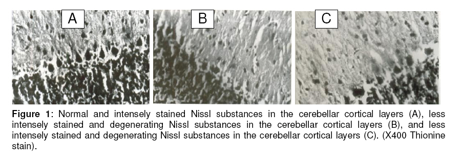
Tropical Journal of Pharmaceutical Research, Vol. 8, No. 2, June 2009, pp. 105-109 Research Article Microstructural Observations on Nissl Substances in the Cerebellar Cortex of Adult Wistar Rats following Quinine Administration Adesina J Ajibade*1, Olusola A Adeeyo1, David A Ofusori2, Thomas K Adenowo3, Olufunto O Ishola4, Ebenezer A Ashamu1 and Spencer C Nwangwu 5 1 Department of Anatomy, Faculty of Basic Medical Sciences, Ladoke Akintola University of Technology, Ogbomoso; Received: 22 September 2008 Revised accepted: 5 December 2008 *Corresponding author Code Number: pr09014 Abstract Purpose: This study assessed some microstructural effects of quinine, commonly used in malaria chemotherapy, especially in chloroquine-resistant and cerebral malaria, on the Nissl substance in the cerebellar cortex of adult Wistar rats using microanatomical studies. Keywords: Quinine, Nissl substances, Intensely stained, Cerebellar cortex, Degenerative changes. Introduction Cerebral malaria remains a major cause of mortality and morbidity in many tropical areas of the world. In spite of improved therapy with ancillary support, mortality remains unacceptably high1. Quinine is an effective chemotherapeutic agent for malaria, especially in chloroquine resistant and cerebral malaria2. It is, however, said to be more toxic than chloroquine3. The drug is extensively metabolized in the liver and only 10% is excreted unchanged in the urine. The cerebellum has three distinct layers and contains five major types of neurons4 . The outermost layer is called the molecular layer, which contains few nerve cells with a finely punctate appearance in transverse section5. The middle layer also called the Purkinje cell layer, is composed of a single layer of Purkinje cell bodies. The deepest layer is the granular layer, which consists of densely packed granule cells6. The cerebellum, like the entire nervous system, is known to be affected by such substances as theobromide, theophylline and caffeine7,8 . Cerebellar injuries have been reported to result from toxins, autoantibodies, structural lesion and inherited cerebellar degeneration9. In view of the reported irreversible toxic effects of quinine on the central nervous system, this work examines microstructural changes in the cerebellar cortex of rats following experimental quinine administration. Materials and Methods Experimental animals Twenty seven mature male albino rats of Wistar strain, weighing between 150g and 190g were used in this study. The rats were fed daily with normal rat pellets purchased from Ladoke Feeds, Ibadan, Nigeria and water was given to the animals ad libitum. All the rats were carefully assessed, screened and confirmed to be healthy during the period of acclimatization. The animals were treated in accordance with the “Guide for the Care and Use of Laboratory Animals” prepared by the National Academy of Sciences and published by the National Institutes of Health10. Drug Quinine hydrochloride powder was procured from Sigma Chemical Company, London and an aqueous solution of the drug equivalent to 0.159%w/v of the quinine base was prepared. This was used as liquid quinine. Experimental design The rats were randomly assigned into three groups. Each group contained nine rats. Group A served as the control and received intramuscular injection of 1 ml of physiological saline (0.9% NaCl). Group B rats were injected intramuscularly with liquid quinine, 16 mg/kg body weight, as a start dose followed by 8 mg/kg body weight 8 hourly for seven days. Group C rats received the same treatment regimen as group B. However, they were subjected to a post-treatment withdrawal period of one week to determine possible withdrawal effects on them before they were sacrificed. Histological procedure Histological study was carried out using the method of Carleton11. These procedures involved dehydration of the cerebellum tissues with graded ethanol concentrations (50%, 70%, 90% and 100%, respectively), clearing in xylene, followed by infiltration in paraffin wax for 2 hr at 56 oC and embedding in paraffin wax for 48 hr. Sections (5 µm thick) were then obtained, using a rotary microtome, subjected to thionine staining procedure and examined under a light microscope. Permanent photomicrographs of the observations were taken, using an Olympus Research Microscope (model BX51). Results The cerebellar cortex of the control rats showed distinct and intensely stained Nissl substance in the cortical layers. The neurons in the three cortical layers appeared normal and were intensely stained (Figure 1A). The neurons were of various shapes and appeared distinct in all the cortical layers. The section of cerebellar cortex in quinine-treated rats (group B) revealed some degenerative changes, which made the neurons indistinct, particularly in the molecular layer. Similarly, the neurons in the molecular layer showed reduced staining intensity compared with the control rats. Some Purkinje cells also showed some degenerative changes with reduced staining intensity in the middle layer. Furthermore, the granule cells in the granular layer beneath the Punkinje cell layer revealed a reduced staining intensity (Figure 1B). Section of cerebellar cortex from group C rats (withdrawal group) showed degenerative changes, which made the neurons indistinct with reduced staining intensity compared with the control rats. The Nissl substances in the Purkinje cells in the middle layer appeared degenerative with reduced staining intensity. The granule cells in the granular layer similarly revealed reduced staining intensity compared with the control (Figure 1C). Discussion Ribosomes, which are intensely basophilic owing to the presence of numerous phosphate groups of constituents, ribosomal ribonucleic acid acting as polyanious, react with such basic stains as methylene blue, toludine blue and haematoxylin. The sites in the cytoplasm that are rich in ribosomes stained intensely with these dyes. These basophilic regions are called Nissl bodies in neurons. The number of Nissl bodies varies according to neuronal types and functional state. They are particularly abundant in large nerve cells such as the motor neurons. The observed neuronal degeneration in association with loss of Nissl bodies with reduced staining intensity of the Nissl substances in the cerebellar cortex of the treated rats in the present study agrees with the findings of Ajibade et al12. Degenerative and vacuolar changes were observed in many large brain stem neurons in mice that were treated with trimethyl chloride; these neurons acquired a chromatolytic character with eccentric nuclei and loss of Nissl substances, which became progressive when studied with electron microscopy13. Fatigue, from over exertion, produced in the brain cells similar changes to those produced by fear, these changes being proportional to the amount of exertion, resulted in exhaustion, and consequently, enormous reduction in Nissl substance14. Similarly, the animals showed neuronal and secondary axonal damage that was most prominent in the cerebellar roof, pontine and vestibular nuclei. The affected neurons showed loss of Nissl substance and shrinkage of the nucleus in dog following intramuscular administration of artemether15. Injury to axons or neuronal exhaustion, resulting from strong or prolonged stimuli, causes a reduction in the number of Nissl bodies. This alteration, which is called chromatolysis, occurs simultaneously with nuclear migration to the periphery of the perikaryon16, and consequently the RNA level is reduced. Chemical and toxic substances affect the Nissl substance thereby influencing their metabolic activity17. Similarly, Martin et al18 reported that neuronal degeneration causes a reduction in Nissl bodies. Neuronal degeneration in quinine-treated rats has already been reported12. The neurotoxic effect of quinine in the present study might have caused neuronal degeneration with a reduction in Nissl bodies; consequently, reduced staining intensity of the Nissl substance in the cerebellar cortex of the treated rats may be due to neuronal pathology, following the neurotoxic effect of quinine. It is possible that quinine had an irreversible effect on the Nissl substances in the cortical layers of the cerebellar cortex, bringing about such microstructural changes in the neurons which manifested as degeneration and loss of Nissl substances with reduced staining intensity of the Nissl substances in the cerebellar cortex of the quinine-treated rats. Degeneration and loss of Nissl substances may consequently affect the synthesis of both structural protein and protein for transport in correlation with neuronal functions. If our findings are extrapolated to man, they suggest that indiscriminate use of quinine may hamper proper coordination of muscular activities and maintenance of posture and equilibrium but this would need to be investigated. Conclusion Administration of an initial dose of 16mg/kg body weight and a further 8mg/kg body weight of quinine to adult Wistar rats for a period of seven days resulted in neuronal degeneration and loss of Nissl substances as evidenced by reduced staining intensity in the Nissl substances of the cerebellar cortex in treated rats. This implies that quinine adversely affected the Nissl substances of the cerebellar cortical layers of the rat model and by extension, may hamper proper coordination of muscular activities and maintenance of posture and equilibrium. Acknowledgment The authors wish to express their profound gratitude to the technical staff of the Anatomy and Cell Biology Department, Obafemi Awolowo University, Nigeria, for their support in the execution of this work. References
© Pharmacotherapy Group, Faculty of Pharmacy, University of Benin, Benin City, 300001 Nigeria. The following images related to this document are available:Photo images[pr09014f1.jpg] |
| |||||||||
