 |
| About Bioline | All Journals | Testimonials | Membership | News |
|
||||||
|
||||||
Tropical Journal of Pharmaceutical Research, Vol. 8, No. 4, Aug, 2009, pp. 317-324 Research Article Anti-Cancer Properties of Diethylether Extract of Wood from Sukun (Artocarpus altilis) in Human Breast Cancer (T47D) Cells Enos Tangke Arung1,2, Britanto Dani Wicaksono1, Yohana Ayupriyanti Handoko1, Irawan Wijaya Kusuma2, Dina Yulia1 and Ferry Sandra1* 1Stem Cell and Cancer Institute, Jalan Ahmad Yani no. 2 Pulomas,
Jakarta, 13210, 2Wood Chemistry Laboratory, Forest Product
Department, Forestry Faculty, Mulawarman University; Jalan KH. Dewantara,
Kampus Gn. Kelua, Samarinda, East Kalimantan 75123, Indonesia. Received: 6 February 2009 Code Number: pr09041 Abstract Purpose: To evaluate the anti-cancer properties of the diethylether
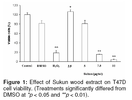
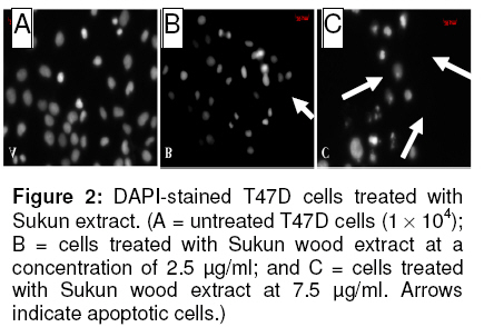
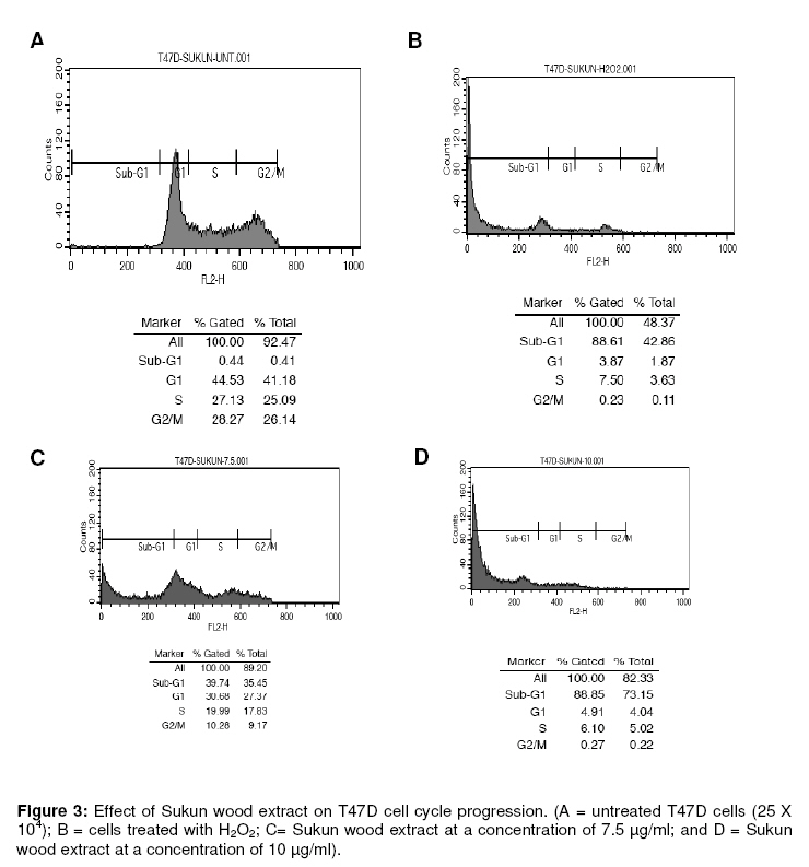
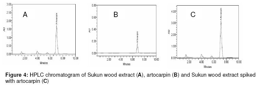
extract of Sukun (Artocarpus altilis) wood. Keywords: Artocarpus altili; Diethylether extract; T47D cells; Anti-cancer; Apoptosis; Sub-G1 formation INTRODUCTION For centuries, people have been using herbs for medicinal purposes. In fact, the use of herbal medicine can be traced back to at least 5000 years. Today, more than 85,000 plant species have been documented for medical use globally1. The World Health Organization (WHO) estimates that almost 75% of the world’s population has therapeutic experience with herbal remedies1. Artocarpus altilis, known as breadfruit, is a widely known food source but is also commonly used as a folk medicine in Indonesia where it is locally called Sukun. Traditionally, the leaves of Sukun are used for the treatment of various kinds of diseases such as liver cirrhosis, hypertension and diabetes2. Scientifically, some biological activities of the extract of this plant have been reported. The methanol/dichloromethane extract from bud covers of sukun was shown to have activity in a cathepsin K inhibition assay3. A study has reported that cathepsin K inhibitors are very effective in preventing bone resorption, and may, therefore, be a potential treatment option for osteoporosis4. The acetone extract of the leaves showed an inhibitory effect on 5a-reductase activity which might make it useful for the selective treatment of benign prostate hyperplasia and prostate cancer5. Sukun extract has also been shown to alleviate the symptoms of diabetes mellitus and urinary problems6. Recently, a study showed the ethylacetate extract of the leaves had cytotoxic effects on some human cancer cell lines, including human lung adenocarcinoma (SPC-A-1 cells), human colon carcinoma (SW-480 cells), and human hepatocellular carcinoma (SMMC-7721 cells)7, thus indicating that the extract might be a potential anti-cancer agent. In this study, we have examined the anti-cancer properties of the diethylether (DE) extract of Sukun (A. altilis) wood. Previous studies have shown that the DE extracts of the wood contain artocarpin, a prenylated flavonoid compound, which has various biological activities such as inhibition of 5a-reductase8,9 and melanin biosynthesis in B16 melanoma cells10. The activity of Sukun wood extract was assessed by investigating cell viability, observing the nuclear morphology of cells and examining the cell cycle process after treatment with Sukun wood extract. EXPERIMENTAL Plant materials The wood of Artocarpus altilis was collected at Saramarinda City, Indonesia in 2006. The plant was identified by Raharjo, BSc. in the Dendrology Laboratory, Forestry Faculty, Mulawarman University, Indonesia and the voucher specimen (AK-CW-2) was deposited in the Wood Chemistry Laboratory, Forestry Faculty, Mulawarman University, Indonesia. Preparation of diethylether (DE) extract The DE extract of A. altilis wood was prepared according to the method previously described by Arung et al.10. The dried heartwood of A. altilis (2.3 kg) was cut into small pieces (approx. 0.5 cm3) and repeatedly extracted with methanol (MeOH) at room temperature. The MeOH extract was concentrated using rotary evaporator under vacuum at room temperature to obtain a final residue (60.6 g). A portion of the extract (43.1 g) was suspended in MeOH/water (1:2) and partitioned with n-hexane, diethyl ether (DE) and ethylacetate (EtOAc). The DE soluble fraction was concentrated under vacuum at room temperature to obtain a final residue (16.3 g) for further tests. The extraction was conducted at the Wood Chemistry Laboratory, Forestry Faculty, Mulawarman University, Samarinda, East Kalimantan, Indonesia. Cell culture T47D cells were obtained from the Indonesian Institute of Sciences Research Centre for Chemistry, Natural Products, Food and Pharmaceuticals Division, Bandung, Indonesia. The cells were grown and maintained in Dulbecco’s Modified Eagle’s Medium (DMEM) with L-glutamine supplemented with 10% v/v foetal bovine serum, sodium bicarbonate, 100 µg/ml streptomycin and 100 U/ml penicillin at 37oC in a humidified atmosphere of 5 % CO2. Cell viability assay To determine cell viability, a microculture tetrazolium teachnique (MTT) assay was performed according to the method previously described by Arung et al11, with minor modifications. The MTT assay provides a quantitative measurement of viable cells by determining the amount of formazan crystals produced by metabolically active cells. Briefly, the cells were seeded into a 96-well plate (5 x 10³ cells per well). After 24 h incubation, the medium was replaced with fresh medium containing Sukun wood extract, dimethyl sulfoxide (1 %v/v DMSO in water), hydrogen peroxide (0.07 %v/v H2O2 in water) or medium alone and the cells incubated for a further 24 h. DMSO (1 %v/v, in water) was used as a negative control since this was used to re-suspend the extract. MTT reagent [3-(4,5-dimethyl-2.thiazolyl)-2,5-diphenyltetrazolium bromide (10 µl) in PBS (5 mg/ml)] was added to each well. The plate was incubated in a humidified atmosphere of 5% CO2 at 37 oC for 3 h, and then the medium was discarded and formazan crystals were dissolved in 1.0 ml of 0.04N HCl. The absorbance of cells was measured at 570 nm with a microplate reader. Control cells were counted with a haemocytometer and used for interpolating the absorbance of MTT assay results. The data were presented as percent of viable cells (%). The data were analyzed with the two-tailed student’s t-test against samples treated with DMSO. Apoptosis measurements and DAPI staining In order to determine the level of apoptosis in cells, DAPI staining was performed as described by Sandra et al12. Briefly, the cells were seeded onto glass slides and treated with Sukun wood extract for 24 h. Untreated and treated cells were rinsed with phosphate buffered saline (PBS), fixed with ice-cold 10% trichloroacetic acid, and further washed with cold 70, 80, 90% and absolute ethanol. The cells were permeabilized with Triton-X (10 %v/v) and stained with 1 µg/ml 4’-6-diamidino-2-phenylindole (DAPI) for 3 min. To reduce the background, the stained cells were washed with PBS, cover-slipped with 90% glycerol and observed under a fluorescence microscope (Zeiss Axio Observer Z1, Göttingen, Germany). Sub-G1 apoptosis assay and flow cytometry The cells were separated into four treatment groups as stated for the cell viability assay above. The cells were analyzed for sub-G1 apoptosis using the method described by Sandra et al12. Briefly, the cells were seeded into a 24-well plate (25 x 104 cells per well) and incubated for 24 h. The medium was then replaced with fresh medium containing Sukun wood extract, DMSO, H2O2 or medium alone. After 24 h, the cells were harvested and suspended in 1 ml of hypotonic fluorochrome solution (50 µg/ml propidium iodide in 0.1% sodium citrate containing 0.1 % Triton X-100). The cell suspensions were placed in the dark by wrapping up the tubes with aluminium foil and incubated at 4 oC for 1-2 h prior to flow cytometric analysis. The propidium iodide fluorescence of individual nuclei was measured using a FACS Calibur apparatus (Benton Dickinson, San Jose, California). High performance liquid chromatographic (HPLC) analysis The wood extract was filtered through a 0.22 µm syringe filter prior to analysis. Spectra were generated on a WatersTM 2487 series HPLC (USA) equipped with a WatersTM 1525 binary HPLC pump and Millenium32 Photodiodide Array Detector (PDA). Data acquisition and treatment were performed with Empower 2 software. The separation was carried out at 25 oC on a 200 mm x 4.6 mm column packed with 5 µm C18 (Symmetry, Waters). The mobile phase was methanol (85%) and water (15%) and delivered isocratically with a flow rate of 1.0 ml/min. The sample volume injected was 20 µl. UV absorbance was measured at 254 nm. Statistical analysis The IC50 (median inhibition concentration) is the concentration of toxic compound that reduces the biological activity by 50 %. The IC50 value was obtained from the MTT assay and calculated using non-linear regression analysis in Microsoft Excel software. The value was expressed as a geometric mean. Differences were considered to be statistically significant when p < 0.05 and p < 0.01. RESULTS Cytotoxic effects of Sukun wood extract in T47D cells Figure 1 shows the cell viability data obtained. There was a moderate decrease (< 20 %) in cell viability when cells were treated with DMSO, the solvent used to re-suspend the extract. T47D cells treated with Sukun wood extract showed a decrease in viability in a concentration dependent-manner. Compared with those treated with DMSO, a significant decrease in cell viability (p< 0.01) was observed for T47D cells treated with 7.5 and 10 µg/ml of extract. IC50 was reached at a concentration of 6.19 µg/ml. Nuclear morphology Based on the MTT assay results, DAPI staining was conducted to investigate whether Sukun wood extract induced apoptosis. Treatment of cells with Sukun wood extract at concentrations of 2.5 and 7.5 µg/ml, respectively, caused nuclear morphological changes compared to normal cells and this may be indicative of apoptosis (compare Figures 2B and C with Figure 2A). Morphological changes observed in the treated cells included cell shrinkage, nuclei that were broken into discrete fragments and cell budding (indicated by arrow) that resulted in cells of various sizes. Higher concentrations of Sukun wood extract appeared to cause more morphological changes, indicating that apoptosis occurred in a concentration-dependent fashion. Sub-G1 apoptosis To understand the mechanism of inhibition of the Sukun wood extract, the effect of Sukun wood extract on cell cycle progression was analyzed by flow cytometry. In this experiment, 2.5, 7.5 and 10 µg/ml of Sukun wood extracts were used. Figure 3 shows that sub-G1 apoptotic content was markedly increased when cells were treated with Sukun wood extract. In the absence of the extract, sub-G1 was formed at 0.44 % (Figure 3A), whereas a concentration of 7.5 µg/ml resulted in 39.74 % of cells being in sub-G1 phase (Figure 3C). This proportion of cells further increased at the extract concentration of 10 µg/ml (88.85%, Figure 3D). This is similar to of the value for cells treated with H2O2 (88.61 %, Figure 3B). Therefore, an increase in the sub-G1 apoptotic fraction may be the major cause of reduced viability of T47D cells. HPLC analysis To investigate the components present in Sukun wood extract, the extract was run in a HPLC. Four peaks were present in the extract and the primary peak appeared at approximately 7 min (Figure 4A). Previous studies have shown that the DE extract of A. altilis contained artocarpin8-10. In this study, therefore, pure artocarpin, from the compound collections atWood Chemistry Laboratory, Forest Product Department, Forestry Faculty, Mulawarman University, Samarinda, East Kalimantan, Indonesia, was run on a HPLC for comparison. Figure 4B shows that artocarpin peak was also produced at approximately 7 min. When the Sukun wood extract was spiked with pure artocarpin (Figure 4C), the main Sukun peak overlapped with the peak of pure artocarpin, indicating that the identity of the main Sukun peak was artocarpin. DISCUSSION Various plant extracts have been evaluated and shown to have cytotoxic or cytostatic effects in cancer cell lines. They include those of Solanum lyratum tested on human colon adenocarcinoma cell line (colo 205)13, Annona glabra on human leukemia cell lines (CEM/VLB)14, Gynostemma pentaphyllum on human lung cancer (A549)15 and Blumea balsamifera on rat and human hepatocellular carcinoma cells (McA-RH7777 and HepG2)16. Previous studies on Artocarpus plants have shown various responses to extracts. Wang et al7 reported that five geranyl dihydrochalcones from the ethylacetate extract of A. altilis leaves had cytotoxic effects on some human cancer cell lines, such as human lung adenocarcinoma (SPC-A-1 cells), human colon carcinoma (SW-480 cells), and human hepatocellular carcinoma (SMMC-7721 cells). The results of our study show that Sukun wood extract has a cytotoxic effect on breast cancer cells (T47D) in a concentration-dependent manner, with an extract IC50 6.19 µg/ml. Our observations of altered nuclear morphology after treatment with the Sukun wood extract (Figure 2B andC) are consistent with previous reports of cells undergoing apoptosis. Kerr et al17 stated that characteristics of cells undergoing apoptosis include the formation of sharply delineated, uniformly fine granular masses adjacent to the nuclear envelope and cytoplasmic condensation; breaking up of the nucleus into discrete fragments surrounded by a double layered envelope; and cell budding to produce membrane-bounded apoptotic bodies. In line with morphological analysis, a sub-G1 apoptosis assay also confirmed that Sukun wood extract induced apoptosis in T47D cells in a concentration-dependent fashion (Figure 3B, C and D). The apoptotic effect of Sukun wood extract may be due to the presence of artocarpin, since this component was found to be the main compound in the extract (Figure 4). A similar finding that artocarpin, which was tested on other breast cancer cells, MCF-7 and MDA-MB-231, showed produced cytotoxicity effect with an IC50 of 3.3 and 3.8 µg/ml, respectively, has also been reported by Wang et al18. CONCLUSION In this study, we found that Sukun wood extract, which primarily contains artocarpin, reduced cell viability by inducing apoptosis and sub-G1 phase formation in human breast T47D cells in vitro. This extract, therefore, has the potential of an anti-cancer agent. However, further study may still be necessary to elucidate the mechanism of apoptosis induction in T47D cells by Sukun wood extract. ACKNOWLEDGEMENT We acknowledge Ms Tjandrawati Mozef of the Indonesian Institute of Sciences Research Centre for Chemistry, Natural Products, Food and Pharmaceuticals Division, Bandung, Indonesia for generously supplying the T47D cells used in this work, and Dr Dyani Lewis of the School of Biological Sciences, Faculty of Science, Monash University, Australia for proof-reading this manuscript. REFERENCES
© Pharmacotherapy Group, Faculty of Pharmacy, University of Benin, Benin City, 300001 Nigeria. The following images related to this document are available:Photo images[pr09041f4.jpg] [pr09041f3.jpg] [pr09041f2.jpg] [pr09041f1.jpg] |
| |||||||||
