 |
| About Bioline | All Journals | Testimonials | Membership | News |
|
||||||
|
||||||
Tropical Journal of Pharmaceutical Research, Vol. 8, No. 5, October, 2009, pp. 433-440 Research Article Alginate-Chitosan Particulate System for Sustained Release of Nimodipine Adhiyaman Rajendran*1 and Sanat Kumar Basu2 1School of Pharmacy and Health Sciences, International Medical University, Kuala Lumpur, Malaysia, 2Division of Pharmaceutics, Department of Pharmaceutical Technology, Jadavpur University, Kolkata, India *Corresponding author: Email: genomic2002@yahoo.com; Tel: 0060-162702470 Received: 27 March 2009 Code Number: pr09056 AbstractPurpose: The aim of this work was to prepare nimodipine-loaded
alginate-chitosan beads for sustained drug release. Key words: Alginate-chitosan beads; Nimodipine; Swelling; Physicochemical characterization; Sustained release. INTRODUCTIONAlginate is a natural biopolymer which forms a hydrogel in the presence of divalent cations such as Ca2+ [1]. The inert environment within the biopolymer network of alginates allows for the entrapment of a wide range of substances [2]. Much attention has been given in recent years to the use of chitosan-alginate polyelectrolyte complex in controlled drug delivery [3]. The use of chitosan has been reported in the literature for coating alginate beads in order to alter the diffusion rate of the encapsulated substances and also as an additive for the bulk modification of the bead structure [4]. Nimodipine (NM) is isopropyl-2-methoxyethyl-1,4-dihydro-2,6-dimethyl-4-(3-nitrophenyl)-3,5 -pyridine dicarboxylate, a dihydropyridine calcium antagonist. It has a short half-life of 1-2 h, and the usual oral dose is 30 to 60 mg to be taken 2 to 4 times a day. Thus, nimodipine is a suitable candidate for oral sustained release drug delivery. In this work, the effect of calcium ion and chitosan in the coagulation fluid on drug release characteristics as well as the release mechanism were evaluated. Also, the physical state of the drug in the beads and its swelling behavior in the bead formulation were assessed. EXPERIMENTAL Materials The following materials were used as received: sodium alginate (low viscosity; viscosity of 2% solution 25 oC, ≈ 250 cps, from SNAP Natural and Alginate Products Limited, Ranipet, India); chitosan (85% degree of deacetylation, molecular weight > 103 kD, from India Sea Foods, Cochin, India); calcium chloride dihydrate (Qualigens, Mumbai, India); and di-sodium hydrogen phosphate anhydrous, potassium di-hydrogen phosphate, sodium acetate, potassium chloride, sodium hydroxide, dichloromethane, ethanol (99%), hydrochloric acid and glacial acetic acid (all from Merck, Mumbai, India). Nimodipine B.P. was obtained as a gift from Cipla Limited, Mumbai, India. Preparation of nimodipine-loaded calcium alginate beads Nimodipine was dissolved in dichloromethane and then slowly dispersed in sodium alginate solution with constant stirring for 3 h, as per the composition outlined in Table 1. The dispersion was added drop-wise to the gelation medium (either 2% or 5% w/v calcium chloride) using a 5 ml hypodermic syringe fitted with a 21o-gauge needle and with the gelation medium stirred constantly at room temperature (25 oC). The beads were left to cure in the gelation medium for 4 h, taken out, washed with distilled water twice and then dried at 30 oC in a dust free chamber until they attained constant weight. Two drug : sodium alginate ratios – 1:3 and 1:4 – were used in the preparation of the beads. The beads were labeled CAB. In another batch labeled ACB-I, drug-loaded alginate-chitosan beads were prepared using chitosan solution (0.5% or 0.75% w/v) containing calcium chloride (2% or 5% w/v) as the gelation medium. The vehicle of the chitosan solution was 3% v/v acetic acid and the pH of the medium was adjusted to 4.5 ± 0.1 with 0.1M sodium hydroxide. Drug: polymer ratio was varied as described above. The gelation medium was prepared 2h before use. The beads were prepared and recovered as described earlier. In another variation, ACB-II beads were prepared as for ACB-I except that the cure time was 2 h instead of 4 h. In a further variation, multilayered (MB) beads prepared as for ACB-II (at a chitosan concentration of 0.75%) were first treated with sodium alginate solution (0.1 % w/v) and then cured in calcium chloride solution (1% w/v) for 30 min at room temperature (25 oC). Table 1: Composition of nimodipine-loaded calcium alginate (Nm-CAB), alginate-chitosan (Nm-ACB-I & II) and multilayer (Nm-MB) beads
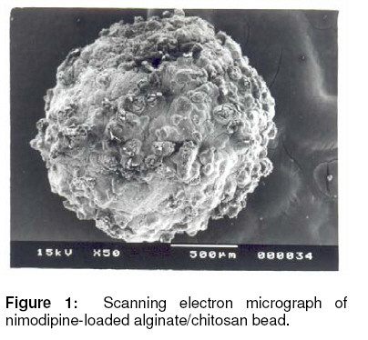
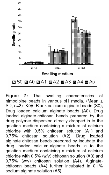
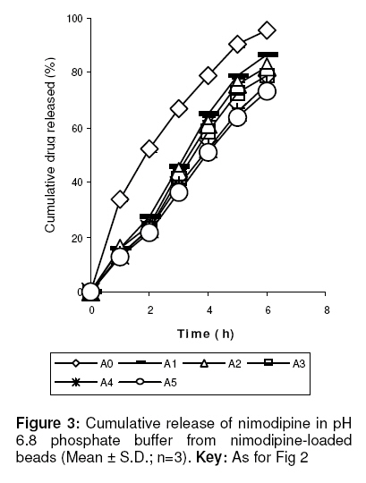
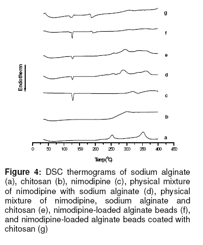
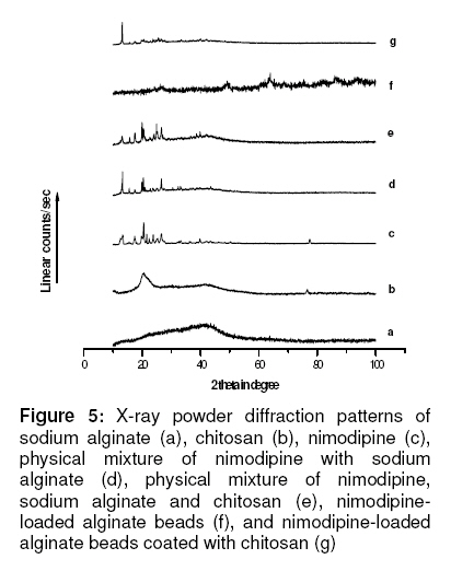
Note: Sodium alginate was 5 %w/v in all formulations; CH = chitosan; CaCl2 = calcium chloride; SA = sodium alginate Morphological characterisation and size of beads The beads were examined by a scanning electron microscope (JEOL JSM – 5200, Japan) operating between 5 - 24 kV. The specimens were mounted on a metal stub (with double-sided adhesive tape) and coated under vacuum with gold in nitrogen atmosphere prior to observation. The bead size of all the formulations was determined by optical microscopy. At least 100 beads were analyzed for each preparation and the mean particle size was calculated. Determination of drug loading An accurately weighed sample (10 mg) of drug-loaded bead was pulverized using mortar and pestle, incubated in 10 ml 0.02M phosphate buffer (pH 6.8) at room temperature for 24 h for complete digestion and the drug extracted with ethanol (99% v/v). The solution was filtered through a filter disc (particle retention: 11μm) and then the filtrate was assayed spectrophotometrically for drug content at 236nm. The same method was utilized to prepare the blank but using unloaded beads. All the experiments were performed in triplicate. Drug loading or incorporation efficiency (%) was calculated using the formula in Eq 1. Experimental drug loading in % (EL) = L/L0 x 100 ……… (1) where L is the actual drug content of the beads and L0 is the weight of the beads. Swelling studies The swelling properties of the calcium alginate, alginate-chitosan and alginate-chitosan multilayer beads were determined in buffer solutions of pH 1.2, 4.5, and 6.8, respectively, at predetermined time intervals. A sample of beads of known weight (10 mg) was placed in a Petri dish containing 20 ml of the buffer solution and allowed to swell at room temperature. At hourly intervals, the beads were removed and weighed. The weight of the swollen beads was determined (after blotting with filter paper to remove surface fluid) on an electronic balance (AB204-S Mettler Toledo), and thereafter returned to the buffer solution. The swelling ratio of the beads was calculated from the formula given in Eq 2 [5]. Swelling ratio= Wt /W0 ………………… (2) where Wt is the weight of the swollen beads and W0 is the initial weight of the beads. Determination of calcium content The calcium content of blank calcium alginate and alginate-chitosan beads, as well as that of nimodipine-loaded beads was determined using the procedure of Takka S et al7. Beads (100mg), accurately weighed, were dissolved in 2 ml of concentrated nitric acid by boiling. The sample was made up to 10 ml with 1 %v/v nitric acid and the calcium content was determined by atomic absorption spectroscopy (Perkin Elmer, model Analysis-200). In vitro drug release studies Each sample for release studies consisted of the beads, equivalent to 20 mg of nimodipine, and filled into a hard gelatin capsule. Drug release studies were carried out, using a USP dissolution rate test apparatus type II (Electro Lab model TDT- 08L), in 600 ml of hydrochloric acid buffer solution (pH 1.2) or phosphate buffer solution (pH 6.8) for 4 h and 6 h, respectively, at 37 ± 0.5°C. The dissolution media also contained 0.3 %w/v of sodium dodecyl sulphate (SDS) to maintain sink conditions for the drug. The apparatus was operated at a stirring speed of 100 rpm. Five (5) ml of the dissolution medium was sampled at predetermined time intervals, and replenished with the same quantity of fresh dissolution medium on each occasion to keep the volume constant. The sample was filtered through a filter disc (particle retention: 11μm) and analyzed for drug content at 236 nm on a spectrophotometer (160-UV-visible Shimadzu spectrophotometer). The release kinetics of nimodipine from the beads were also evaluated using different models, zero order, first order, Higuchi, Peppas-Korsmeyer and Hixon-Crowell. This assessment was carried out with the aid of a program, PCP Disso v 2.08 (Anant Katkar, Poona College of Pharmacy, India). FT-Infrared spectroscopy Individual beads/samples were crushed in a mortar with a pestle. The crushed material was mixed with potassium bromide (Merck IR spectroscopy grade) in a 1:100 proportion and dried at 40 oC. The mixture was compressed to a 12 mm semitransparent disk by applying a pressure of 10 tons for 2 minutes. The FTIR spectra over the wavelength range 4000 to 400 cm-1 were recorded using a FTIR spectrometer (Perkin Elmer 1600 series). Differential scanning calorimetry Differential scanning calorimetry (DSC) nimodipine, sodium alginate, chitosan, as well as nimodipine/sodium alginate physical mixture (PM1), nimodipine/sodium alginate/chitosan physical mixture (PM2),nimodipine-loaded calcium alginate beads (A0) and alginate-chitosan beads-II (A4). The test was carried out using a thermal analysis system (Mettler TA 4000), calibrated with indium as the standard and operated in the temperature range, 30-400 oC. The bead sample (5mg) which was heated at 10 oC/min in an aluminum pan under a nitrogen atmosphere using an empty pan as the reference. The automatically computed onsets of melting point and enthalpy of fusion were noted. X-ray diffraction X-diffraction patterns of the individual materials, physical mixtures and beads were obtained with an x-ray diffractometer (Rich Seifert, model 3000 P) at 30 kV and 15 mA over a range of 10-100 2θ, using Cu Kα radiation wavelength of 1.5405 Ao. In the technique, the cavity of the metal sample holder of the equipment was filled with ground sample powder and then smoothed out with a spatula before the test was run. RESULTS Morphological characteristics and bead size The scanning electron micrograph of a typical drug-loaded alginate bead treated with chitosan is shown in Fig 1. The beads had a spherical shape and showed surface cracks probably caused by partial collapsing of the polymer network during drying. The mean particle size of the beads ranged from 1486.00 ± 0.02 to 1602.00 ± 0.01 µm. Drug loading Drug loading was approx. 23 and 18 % for formulations in which the drug/polymer ratio was 1:3 and 1:4, respectively. Variation in the treatment of the beads did not affect drug loading. Swelling characteristics of nimodipine-loaded beads The results of swelling studies, shown in Fig 2, indicate that swelling varied with the pH of the medium. Maximum swelling ratios were as high as 2, 14 and 36 in media pH of 1.2, 4.5 and 6.8, respectively. The beads did not show any sign of disintegration in the media at pH 1.2 and 4.5 over a period of 8 h. However the swelling ratio of the beads at pH 4.5 was considerably greater than that at pH 1.2. Treatment of the calcium alginate beads with chitosan enhanced swelling at pH of 1.2 and 4.5. On the other hand, the beads began to slowly disintegrate in pH 6.8 medium after 7 h. In vitro drug release of nimodipine At pH 1.2, there was no discernible release of nimodipine from calcium alginate beads and those pretreated with chitosan after 4 h. On the other hand, as Fig 3 illustrates, 95.8 % of nimodipine was released from the untreated beads (formulation A0) at pH 6.8 in 6 h during which it completely disintegrated. Following treatment with 0.75% chitosan, yielding formulation A4, drug release fell to 75.6 %. Further treatment with alginate, resulting in formulation A5, slightly reduced drug release further to 73.1± 1.69%. On subjecting the release data to various models, correlation coefficient (r2) ranged from 0.98 - 0.99 for most of the formulations, except for the Higuchi model where was as low as 0.94 for some of the formulations. Furthermore, in the case of the Peppas-korsmeyer model, the release kinetics were also found to fit the classical power law expression, with n values of 0.57 - 0.58 for the untreated beads, and 0.87 – 0.93 for the treated beads. FT-IR spectroscopy The FT-IR spectra (not displayed) of nimodipine alone manifested major peaks in the wave number range of 1000 to 1400 cm-1, indicating the presence of carboxyl and carboxylate groups, and C-H stretching, appeared at 2981.48 to 2898.38 cm-1. Cross-linking of alginate by Ca2+ resulted in a decrease in the wave number of the carbonyl peak from 1623.7 to 1608.97 cm−1. The principal peaks of nimodipine were observed in the spectra of all the formulations. Differential scanning calorimetric analysis Fig 4 shows the DSC thermograms of nimodipine alone, the polymers, physical mixtures of the polymers and the drug, as well as nimodipine-loaded beads. A sharp endothermic peak corresponding to the melting of crystalline nimodipine was found at 126.8 oC while sodium alginate decomposed at 253.6 oC with a broad exotherm. The degradation exotherm of sodium alginate was absent in formulations A0 (CAB) and A4 (ACB II) beads but an endotherm corresponding to the interaction of alginate with calcium ion was observed at 192.3oC and 187.7oC, respectively. The melting endotherm of nimodipine in PM1 (physical mixture of nimodipine/sodium alginate), and PM2 (nimodipine/sodium alginate/chitosan) appeared at 126.0 and 127.6 oC, respectively. In the bead formulations, the melting endotherm of nimodipine appeared at 125.9 and 123.2 oC for formulations A0 and A4, respectively. X-ray diffraction (XRD) DISCUSSION Bead size was not noticeably enhanced following treatment of the alginate beads with chitosan. Further treatment with alginate also did not alter bead dimensions as would have been expected. At low pH (1.2 and 4.5), bead swelling was minimal. This may be attributed to the inability of the medium to penetrate the beads at pH 1.2, and limited penetration at pH 4.5. On the other hand, the pronounced swelling of the beads in phosphate buffer (pH 6.8) followed by slow disintegration after 7 h might have been influenced by certain factors. It can be said that in the initial phase of the swelling process, the Ca2+ ions present in the polymannuronate units of alginate are exchanged with Na+ ions present in the buffer solution, which ultimately causes chain relaxation and enhances gel formation or swelling. In the later stage of the swelling process, Ca2+ ions bind with the –COO- group of the polyglucuronate units and thus form the tight egg-box structure which also starts to exchange with Na+ ions in the buffer medium because polyglucuronate sequences have a strong auto-cooperative binding of Ca2+ ions. Increase in the proportion of calcium alginate in the drug-loaded beads may led to a corresponding increase in the amount of Ca++, and hence availability of a larger number of binding sites for interaction with anions. It was observed that the amount of calcium ions in formulations (A4, A4a and FA4a) decreased when compared with formulation A0. This indicates that the alginate formed a complex both with calcium ions and chitosan [7]. Poor drug release at pH 1.2 may be attributed to the poor swelling of the alginate beads at acidic pH due to the inability of the dissolution medium to penetrate the beads. However, the dissolution medium readily penetrated the beads at pH 6.8, easily leaching out the drug incorporated in them. Treatment of the alginate beads with chitosan, and then with alginate, substantially reduced drug release. The probable reason is that electrostatic interaction between carboxyl groups of the alginate and the amino group of chitosan produced a compact surface layer that reduced both diffusion of fluid into the beads and the erosion of the beads. Drug release at pH 6.8 was accompanied by rapid swelling and erosion/disintegration of the alginate beads. The drug release data for the various calcium alginate beads fitted into the classical power law expression, and the values of n were around 0.6. This indicates that drug release from alginate beads followed non-Fickian kinetics, due probably to rapid swelling and erosion of the beads. The release data for alginate-chitosan (Nm-ACB-I &II) multilayer beads also fitted well into the power law expression, and the values of n decreased below 1. It is likely that the formation of polyelectrolyte complex membrane reduced the initial swelling and erosion of the beads and shifted the drug release mechanism toward anomalous transport or case II transport, indicating that the drug was diffusing through the beads at the same time as polymer relaxation was taking place. Since the release of the drug from alginate–chitosan beads exhibited a small time lag, the release data were fitted into the modified power law expression, and this yielded excellent linearity. Consequently, it can be concluded that the drug release mechanism was anomalous diffusion type or case II transport. FT-IR and DSC data indicate there was no interaction between nimodipine on the one hand, and chitosan and alginate on the other. For the DSC results, the slight differences observed in the melting endotherms and peak intensities may be attributed to homogeneous dispersion of the drug in the polymers and to variation in drug/polymer ratio. The differences observed between the XRD patterns of the bead formulations and those of the physical mixtures of the drug and polymers was probably due to a decrease in the degree of crystallinity of the drug, which resulted from its dispersion in the polymer matrix [8]. CONCLUSION The study demonstrates that while alginate beads incorporating nimodipine exhibited comparatively rapid drug release at intestinal pH (6.8), treatment with chitosan alone or together with alginate lowered drug release. The bead formulations thus afford an option for achieving modulated release of the drug. REFERENCES
© Pharmacotherapy Group, Faculty of Pharmacy, University of Benin, Benin City, 300001 Nigeria. The following images related to this document are available:Photo images[pr09056f5.jpg] [pr09056f3.jpg] [pr09056f1.jpg] [pr09056f2.jpg] [pr09056f4.jpg] | |||||||||||||||||||||||||||||||||||||||||||||||||||||||||||||||||||||||||||||||||||||||||||||||||||
| |||||||||
