 |
| About Bioline | All Journals | Testimonials | Membership | News |
|
||||||
|
||||||
Tropical Journal of Pharmaceutical Research, Vol. 8, No. 6, December, 2009, pp. 521-530 Research Article Radioprotective and In-Vitro Cytotoxic Sapogenin from Euphorbia neriifolia (Euphorbiaceae) Leaf Papiya Bigoniya*1 and Avtar Chand Rana2 1Radharaman College of Pharmacy, Radharaman Group of Institutes, Ratibad, Bhopal, M.P. 462002, 2B.N College of Pharmacy, B.N Group of Colleges, Udaipur, Rajasthan, India. *Corresponding author: E-mail: p_bigoniya2@hotmail.com; Tel: +91-0755-2477941 (Res), 0755-2896237 (Inst); Fax: +91-0755-2896663 Received: 25 June 2009 Code Number: pr09067 Abstract Purpose:
Euphorbia neriifolia Linn.
(Euphorbiaceae) plant is
traditionally used in the treatment of abdominal troubles, bronchitis, tumours,
leucoderma, piles, inflammation, and enlargement of spleen. The objective of
this study was to evaluate the antioxidant and anticancer activities of a sapogenin
isolate of this plant. Keywords: Euphorbia neriifolia; Euphol; Sapogenin; Antioxidant; Radioprotective; Melanoma; Chromosomal aberration INTRODUCTION Euphorbia neriifolia Linn, belonging to the family, Euphorbiaceae, is found throughout the Deccan Peninsula of India and commonly occurs in the dry hilly rocky grounds of north, central and south India. It is a herb full of spine, and is popularly known as sehund or thohar in Hindi. Ayurveda describes the plant as bitter, pungent, laxative, carminative, improves appetite, as well as useful in abdominal problems, bronchitis, tumours, loss of consciousness, delirium, leucoderma, piles, inflammation, enlargement of spleen, anaemia, ulcers and fever. Its leaves, in the Indian traditional system, are used as aphrodisiac, diuretic, and also in cough and cold, bleeding piles and ano-rectal fistula [1]. Plants of euphorbia species show anticarcinogenic activity due to the presence of several terpenes, anthocyanins, alcohols and steroids; diterpenoid ingenol 3,20-dibenzoate and phorbol 12-tiglate 13-decanoate isolated from Euphorbiaceae plants show antileukaemic activity against the P-388 lymphocytic leukaemia in mice [2]. Euphol, a triterpene alcohol from the roots of Euphorbia kansui, has inhibitory activity against mice skin tumour [3]. E. neriifolia, being widely available in large quantities, is potentially a low-cost source of active therapeutic substances. We have previously reported on the mild CNS depressant, wound healing and immunomodulatory activities of the hydro-alcohol leaf extract [4-6]. Little phyto-pharmacological work, however, has been done on the medicinal application of the leaf. Saponin isolated from the leaf possesses good haemolytic and in-vitro antioxidant activity but it is devoid of antibacterial activity up to 10 mg/ml concentration [7]. Since ethnopharma-cological exploration has shown the traditional use of E. neriifolia, especially its leaf, as antitumour agent, the objective of this study was to isolate sapogenin from the plant’s leaf and study its antioxidant and anticancer activities. EXPERIMENTAL Plant material E. neriifolia leaves were collected from cultivated field hedge plants in the suburban areas of Bhopal (latitude 23.21°, longitude 77.84°, BHOP), Madhya Pradesh, India, in September 2005. The plant was identified with the aid of available literature and authenticated by Dr AP Shrivastava, a taxonomist and Principal, P.K.S Govt. Ayurveda College and Institute, Bhopal, India. A voucher specimen (no. 1085) was deposited in the herbarium of the department. Reagents RPMI 1640 media, foetal calf serum and 1,1 – diphenyl –2-picryl hydrazyl (DPPH) were purchased from Sigma Chemicals Co., St. Louis, USA while phytohaemagglutinin was obtained from Difco, USA. Collagen-coated culture flasks were purchased from Nunc, Denmark. and Neubauer hemocytometer from Feinoptik, Germany. Modified Eagle Minimum Essential Media and deoxyribose were purchased from Himedia, Mumbai; vincristin from Cipla, India, nitroblue tetrazolium from E. Merck, Darmstadt, Germany. Photomicrographs were captured using an Olympus D´60 microscope connected to an Olympus DP-50 digital camera. Extraction, isolation and characterization of compound One kilogram of the dried powder of the leaf was extracted with 3 L of cold ethanol (70 %) by maceration for seven days and the solvent removed under vacuum. Phytochemical investigation of the extract was performed to detect the presence of reducing sugar, tannin flavonoid, alkaloid, saponin, steroid, glycoside and fixed oil [8,9]. The extract was re-suspended in 250 ml of water and 500 ml of chloroform in HCl (50 %v/v) was added to effect acid hydrolysis of its saponins content in order to isolate sapogenins. The chloroform phase was separated and concentrated at < 40 °C to a third of its volume. This phase was exhaustively extracted with water-saturated n-butanol (three times) and the solvent removed under vacuum. The brown dried powder obtained represents the total crude sapogenin (yield: 2.41 %) and it tested positive according to Salkowski and Noller’s test [10]. Characterisation of sapogenin isolate The whole sapogenin isolate was subjected to column chromatography on silica gel using chloroform, a solvent gradient of chloroform/ethyl acetate (80:20, 60:40, 40:60 and 20:80), ethyl acetate and methanol. Five fractions were collected and chromato-graphed on silica gel G plates using CHCl3 : MeOH (50:50). Fraction 3 was subjected to other assessments including Salkowski, Noller’s and Libermann Burchard tests [10]. Its melting point was determined from its DSC thermogram using a Mettler Toledo DSC 821 system in which the sample press-sealed in an aluminium pan with a perforated lid and heated at a rate of 5 ºC/min in a nitrogen environment. The UV spectra of 5 µg/ml sapogenin fraction in chloroform were obtained with a Shimadzu (UV-1700 Pharmaspec) spectrophotometer while the IR spectra were recorded on a Shimadzu (Jasco FTIR-5300) spectrometer using KBr pellet at a scanning speed of 2 mm/sec and with resolution set at 4 cm-1. NMR spectra were determined with a Bruker Deltonics (Avance 300) spectrometer in CDCl3 at 300 MHz and the entire chemical shifts were relative to tetramethyl silane (TMS, d 0.00). The electron-impact mass spectra of the powdered sample was recorded on Shimadzu (QP 5000) spectrometer in CHCl3, injected in HT-8 column, using helium as the carrier gas at a heating rate of 15 ºC/min and scan rate of 1 scan/sec in a scan range of 100-500 Delton. Preparation of extract stock solutions
A stock solution of the total sapogenin (50 mg/ml) was prepared in dimethyl-sulfoxide (DMSO) and the volume made up to 1000 ml (to obtain a 100 mg/ml concentration) with RPMI media (for blood culture) or Eagle’s Modified MEM (for melanoma cell culture). It was then sterilized by filtration through a 0.2 mm membrane filter. Other concentrations, ranging from 40 – 75 mg/ml, were prepared by dilution. The final DMSO concentration of extract media combination was 0.25 %v/v and at this level, no growth inhibitory effects were observed.
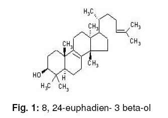
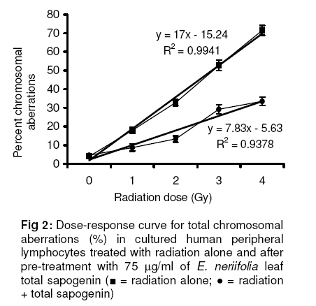
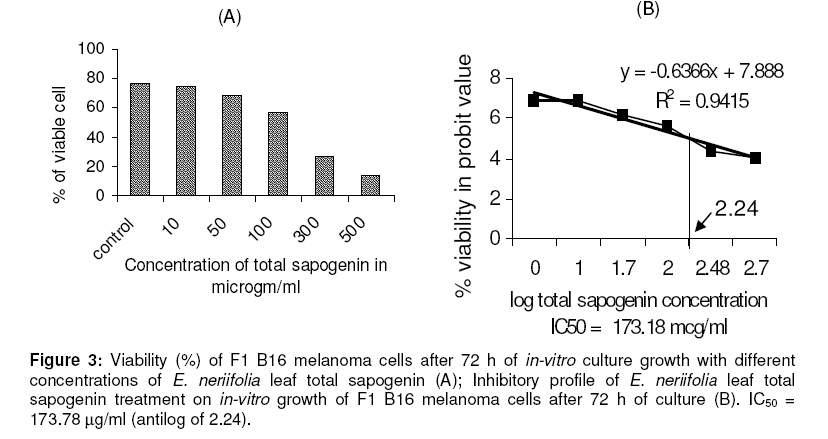
In-vitro antioxidant activities In-vitro antioxidant activities of the total sapogenin were measured in a concentration range of 50 – 1000 mg/ml, using various methods. Reducing power assay The reducing power assay (RPA) involves the oxidation-reduction reaction of potassium ferricyanide and ferric chloride in the presence of the antioxidants. The reducing power of 1 mg sample is equivalent (E) to reducing power of 1 nM ascorbic acid (AS) expressed as ASE/mg assayed following the method of Oyaizu [11]. Different concentrations of the sapogenin and 1nM of ascorbic acid in a final volume of 1ml were taken in different test tubes and mixed with 500 ml of potassium ferricyanide (1%). The mixture was incubated at 50 °C for 20 min. TCA (10%, 50 ml) was added and centrifuged at 600 rpm for 10 min. To the supernatant, 0.5 ml of ferric chloride (0.1%) was added and the absorbance of the resultant violet coloured solution was measured at 700 nm. Hydrogen-donating ability Hydrogen-donating ability (HDA) was measured as the amount of total sapogenin required for inhibiting the formation of 1,1-diphenyl –2-picryl hydrazyl (DPPH) radical by 50 % (IC50 value) according to the method of Hatano et al [12]. Antioxidants react with methanolic DPPH (100 mM) and convert it to 1,1-diphenyl –2-picryl hydrazine. Different concentrations of the sapogenin (200 ml) was added to 3 ml of methanolic DPPH; 20 min later, the amount of DPPH remaining was measured spectrophotometrically at 520 nm against blank. Hydroxyl radical scavenging ability Hydroxyl radical scavenging ability (HRSA) was measured by evaluating the competition between deoxyribose and the test compounds for hydroxyl radicals generated from the Fe3+/ascorbate/EDTA/H2O2 system using the method of Elizabeth and Rao [13]. The reaction mixture contained 100 ml of deoxyribose, 50 ml of FeCl3, 50 ml of EDTA, 100 ml of H2O2 and 100 ml of ascorbic acid. Different concentrations of the sapogenin were added to KH2PO4/ KOH buffer (20 mM, pH 7.4) to make it up to 1 ml. Incubation for 1 h at 37 °C resulted in the formation of thiobarbituric acid reactive substances (TBARS) measured spectrophotometrically at 532 nm following the method of Ohkawa et al [14]. The results were expressed as percent inhibition of TBARS and the amount of sample (mg/ml) producing 50 % antilipid peroxidation was determined. Superoxide radical production The effect of total sapogenin on superoxide radical production (SRP) was evaluated following the nitroblue tetrazolium (NBT) reduction method of McCord and Friodovic [15]. The reaction mixture containing 0.5 mM xanthine as substrate (300 mL), 1 mM EDTA in phosphate buffer (100 mL), 0.05 mM sodium cyanide (100 ml), 0.5 mM xanthine oxidase (20 mL), different concentrations of the sapogenin (20 ml) and 0.1 mM cytochrome C (300 mL) was placed in a 1 cm cuvette and the rate of increase in absorbance at 550 nm was recorded every minute for 5 min. The results were calculated as amount of dismutase required to inhibit the rate of reduction of cytochrome C by 50 % (i.e., to a rate of 0.0125 absorbance unit per minute which is defined as 1 unit of activity) and the amount of sample (mg/ml) producing 50 % reduction of cytochrome C was determined. Effect on radiation-induced chromosomal aberrations in cultured human lymphocytes Fresh whole blood (1.5 ml) was taken in different culture flasks and treated with 40, 55, 75 and 100 mg/ml of total sapogenin in four different sets. One flask in each set was taken as vehicle control and treated with 75 ml of vehicle. All the culture flasks were incubated for 30 min and then each separate set of culture flasks was exposed to 1, 2, 3 and 4 Gy of g-radiation. Triplicate cultures for each individual flask were set up by mixing 0.5 ml of blood with 4.5 ml of RPMI 1640 media. The culture flasks were coded and incubated at 37 °C in a humidified atmosphere of 5 % CO2. The cultures were harvested at 72 h for chromosomal studies. Air-dried preparations of hypotonically treated lymphocytes were made using routine techniques for chromosomal analysis as described by Moorhead et al [16]. Slides were prepared by air drying method and chromosomal aberrations were scored in conventional Giemsa-stained preparations as described by Lioyd et al [17]. Media for blood culturing Blood samples were collected in heparinised sterile glass vials from the median cubital vein of a non-smoking healthy female donor of approximately 25 years. The whole blood (0.5 ml) was incubated with 5 ml of RPMI 1640 media containing 5 % heat-inactivated faetal calf serum, penicillin (100 U/ml), streptomycin (100 mg/ml) and L-glutamine (1.5 mg/ml). An optimum concentration of phytohaemagglutinin 5 mg/ml was used to stimulate the lymphocytes to transform and divide in culture [18]. In-vitro radiation Theratron 780C cobalt teletherapy unit (Theratronics Limited, Canada) was used for radiating whole human blood samples at a dose rate of 1 Gy/min in culture flasks (25 cm3), in a field size of 7 ´ 20 cm2 which can accommodate 5 flasks at a time at a S.S.D (source surface distance) of 90 - 95 cm. In-vitro cytotoxicity assay of the extract on murine F1 B16 melanoma cell line Murine F1 B16 melanoma cell line was used to study the in-vitro anti-cancer activity of total sapogenin. Male C 57 BL/6 mice were used for in-vivo maintenance of cell line. In-vitro harvesting of cells was carried out from a full-grown melanoma site (2.5 to 3 cm) by aseptic transfer to collagen-coated culture flasks in Modified Eagle Minimum Essential Media containing NaHCO3 (220 mg/100ml), which was supplemented with 10 % heat-inactivated faetal calf serum, L-glutamine (300 mg/ml) and 48 mg gentamicin at pH 7.4. The cells were expended in 75 cm2 tissue culture flasks at 37 °C at an atmosphere of 5 % CO2 in air (100 % humidity). A confluent monolayer was detached with 0.1 % trypsin containing 0.02 % EDTA in Ca2+ and Mg2+ free PBS (pH 7.4, 0.01 M) and dissociated into a single-cell suspension for further cell culture. Serially cultured cell lines at P3 to P4 stages were used for cytotoxicity assay. The assay was performed in collagen-coated radiation sterilised cell culture dishes (2 ml capacity, growth surface 60 ´ 15 mm). An amount of the cell culture (2 ´ 104 cells/ml) was seeded and allowed to adhere by incubating for 6 h at 37 °C in 5 % CO2. This method was standardised previously in the laboratory following the method of Umadevi et al [19]. After 6 h, the cells were exposed to different concentrations of total sapogenin for a further 6 h. The range of tested concentrations was from 10 to 500 mg/ml for total sapogenin and from 10 to 500 ng/ml for vincristin as positive control. Triplicate dishes were incubated for 72 h and cell viability was checked every 24 h. The cells were detached by rinsing twice with trypsin, and then palleted by centrifugation; cell viability was measured by tryphan blue dye exclusion test and counted using WBC counting chamber of Neubauer’s chamber. The IC50 (concentration at which cellular growth is inhibited by 50 %) was determined at 72 h. The IC50 values were estimated from a plot of log total sapogenin concentration against percent cell viability. Ethical considerations The experimental protocol for animal studies was approved by the Institutional Animal Ethical Committee (ref no. Animal Eths. Comm./DB/304) prior to carrying out the experiments and the animals were handled as per the ‘WHO guidelines for the care and use of animals in scientific research’. Statistical analysis All data are presented as mean ± SEM. Experimental data were analysed using one-way ANOVA followed by Student’s t-test. P < 0.05 was considered significant. Graph Pad Prism Version 3.02 software was used for statistical calculations. RESULTS Identification of compound The leaf extract (yield: 10.8 %) was positive for reducing sugar, tannins, flavonoids, alkaloids, and triterpenoidal saponin but negative for glycoside and fixed oil. Hydrolysis of the extract followed by extraction with water-saturated n-butanol gave a brown coloured crude sapogenin mixture with a yield 2.4 %. Chromatographic elution of crude sapogenin with chloroform, chloroform/ethyl acetate (80: 20, 60: 40, 40: 60 and 20: 80), ethyl acetate and methanol produced five fractions. The fractions obtained were: Fraction-1 (mixture), Fraction-2 (ENS-1) with Rf value of 0.385, Fraction-3 (ENS-2) with Rf value of 0.360, Fraction-4 (no spot) and Fraction-5 (ENS-3) with Rf value of 0.314. Fraction 3 was positive for Salkowski and Noller’s test and negative for Libermann Burchard test, indicating the presence of triterpene. The residue obtained was carefully crystallized on methanol gives a solid, white crystal (232 mg), m.p. 116 °C. UV d max 270 nm; IR (KBr, cm-1): 3400 & 1030 (3-b-OH), 2923 & 2854, 1637 (-C=C-), 1461 & 1376, 925, 862, 802 & 723; 1H NMR (CDCl3 300 MHz): d 5.43 (1H, t, H-24), 3.29 (1H, m, H-3b) 1.62 (Me-26), 1.77 (Me-27), 0.74 (Me-18), 0.85 (Me-19), 0.91 (Me-28), 1.16 (Me-29), 1.21 (Me-30), 1.12 (Me-21); 13C NMR (CDCl3 75 MHz): d 81.01 (C-3), 135.62 (C-8), 37.26 (C-20), 129.20 (C-24), 20.18 (C-21), 17.52 (C-18), 21.14 (C-19), 18.72 (C-26), 27.32 (C-27), 17.78 (C-28), 26.56 (C-29), 30.14 (C-30); EIMS m/z (%): 426 (M+), 408 (M+-H2O), 297 0.24 % M+-H2O-C8H15) and other fragments suggesting the fragmentation of side chain 111 (16.42 % C8H15), 97 (25.08 % C7H13), 83 (42.96 % C6H11), 69 (70.93 % C5H9), 55 (66.15 % C4H7) and 41 (100 % C3H5). All the data’s were compared with published data for Euphol from Kansui Radix as reported by Lin et al [20]. Thus fraction 3 was characterised as euphol (8, 24-Euphadien- 3 beta-ol; m.p. 116 °C; yield 0.0232%, Fig. 1) based on IR and mass spectrometry. Antioxidant activity The in vitro antioxidant activities of the total sapogenin are depicted in Table 1. Sapogenin, in the amounts used, showed moderate to high free radical scavenging activity and good hydrogen donating ability in relation to a-tocopherol. Effect of the extract on radiation-induced chromosomal aberrations Table 2: Effect of total sapogenin of E. neriifolia leaf on radiation induced chromosomal aberrations in cultured human lymphocytes
Control samples were incubated without any treatment. These data show the effect of the leaf total sapogenin treatment (40 -100 mg/ml) as well as the effect of different doses of radiation (1 - 4 Gy) on chromosomal aberrations in cultured normal non-malignant cells (human lymphocytes). Pre-treatment with 75 mg/ml of total sapogenin fraction reduced total chromosomal aberrations to 33.5 % compared to 71.5 % for radiation treatment (RT) alone at 4 Gy. The slope of the linear dose-response curve for total sapogenin treatment was 7.83, which is significantly (p < 0.001) lower than that of the radiation only treated group (see Figure 2). Cytotoxicity assay on murine F1 B16 Melanoma cell line In-vitro testing of total sapogenin against the murine F1 B16 Melanoma cell line showed 76.6 % cell viability at 10 mg/ml compared to 13.6 % at 500 mg/ml of total sapogenin, with control as 100 % cell viability, as shown in Figure 3(A). Figure 3(B) shows the plot of log total sapogenin concentration against cell viability (probit scale) with a best-fit linear regression curve (slope: 0.9415) superimposed. The assay data show that the IC50 (over a period of 72 h) concentration of total sapogenin that inhibited growth of mouse melanoma cells by 50 % was 173.78 mg/ml compared to 120 ng/ml for vincristin.DISCUSSION
Plants are valuable sources of novel biologically active molecules. Saponins are high molecular weight compounds comprising glycosides with a sugar moiety linked to a triterpene or steroid aglycone. Triterpene saponins, particularly, have been the subject of much interest because of their biological properties. Free radical damage to biosystems is one of the major processes that contribute to degenerative diseases such as cancer and ageing. Free radical scavengers protect cellular DNA against indirect effects of ionizing radiation where hydroxyl radicals are believed to be the primary active species responsible for the damage [21]. The data obtained in this study demonstrate the antioxidant activity of the sapogenin isolated from the leaf extract of E. neriifolia. The good reducing power of sapogenin means that triterpenoidal compounds, especially euphol, are electron donors, and therefore, can act as antioxidants [22]. Hydrogen donating ability is an index of primary antioxidants. DPPH is known to abstract labile hydrogen and the ability to scavenge the DPPH radical is related to the inhibition of lipid peroxidation [23]. Total sapogenin inhibits oxygen derived free radicals such as superoxides and hydroxyl radical in vitro with a relatively moderate potency. Terpenes and bioflavones isolated from Ginko biloba inhibited lipid peroxidase and superoxide anion in hepatocytes that are generally implicated in cell damage [24]. There is a good correlation between antioxidant properties and radioprotection by flavonoids as they could prevent the accumulation of DNA damage induced by UV radiation. Castilla et al [25] has demonstrated the antioxidant as well as radioprotective effects of flavon-3-ol from grape seeds against chromosomal damage induced by x-rays. Peripheral blood lymphocytes (PBL) are extensively used in biomonitoring of populations exposed to various mutagenic or carcinogenic compounds because the sensitivity of this system in detecting chromosome damage induced by exposures of ionising radiation. g-radiation produces morphological changes in lymphocytes by decaying their proliferation, which indirectly defines genomic instability. g-rays generate hydroxyl radicals in cells and induce DNA damage that leads to mutations and chromosomal aberrations [26]. Total sapogenin at a concentration of 75 mg/ml significantly decreased total chromosomal aberration. The results signify that the sapogenins reduced gamma radiation-induced genomic instability by reducing chromosomal aberrations due to the presence of antioxidants. Total sapogenin exhibits cytotoxic activity on murine F1 B16 Melanoma cell line. It showed apparently high IC50 as the cells were exposed to extracts only for a short duration of 6 h. Using conventional cytotoxic tests, the cells were exposed to cytotoxic chemical for 72 h where cytotoxicity may be partly due to cumulative accumulation of the drugs in the culture media. Saponins have many kinds of biological activities such as anti-bacterial, anti-viral, anti-tumour, anti-fertility, anti-inflammatory, anti-hyperlipidemic, anti-hypertension, anti-hyperglycaemic and immunoregulatory, etc. Furthermore, triterpenoids exert physiological activities in the cardiovascular, nervous and, adrenocortical systems as well as enzymatic activity, and therefore, are frequently a subject of research in natural medicine. The total triterpenoidal sapogenin extracted from bamboo (Phyllostachys Sieb. et Zucc) were reported to exhibit pharmacological activities such as anti-free radical, anti-oxidation, anti-tumour and anti-hypertension [27]. Triterpene alcohols such as ursolic and oleanolic acids are said to exert antitumour effect on lung, breast and colon tumours [28]. Topical application of euphol isolated from roots of Euphorbia kansui markedly suppressed the tumour-promoting effect of 12-O-tetradecanoylphorbol-13-acetate in two-stage carcinogenesis in mouse s kin initiated with 7, 12-dimethylbenz[a] anthracene [3].CONCLUSION E. neriifolia Leaf is rich in crude sapogenin, and euphol (0.023 %) was identified as a major constituent. The sapogenin fraction showed antioxidant, radioprotective and cytotoxic activity against malignant melanoma cells. Our study supports the use of E. neriifolia as an antitumour herbal remedy in Indian traditional medicine. This study reports for the first time the potential anticancer properties of E. neriifolia triterpenes. However, the sapogenin content needs to be studied further to isolate other active constituents and to elucidate its in-vivo anticancer activity profile; this further work is in progress in our laboratory. ACKNOWLEDGEMENT The authors are grateful to All India Council of Technical Education, New Delhi, for financial assistance through the award of a National Doctoral Fellowship to one of the authors (PB) (grant no. FD/NDFS/2003-04). We also express our gratitude to Drs P Uma Devi and N Ganesh, and all the research staff of Jawaharlal Nehru Cancer Hospital and Research Center, Bhopal, for their assistance, support and valuable suggestions. REFERENCES
The following images related to this document are available:Photo images[pr09067f2.jpg] [pr09067t1.jpg] [pr09067f1.jpg] [pr09067f3.jpg] | ||||||||||||||||||||||||||||||||||||||||||||||||||||||||||||||||||||||||||||||||||||||||||||||||||||||||||||||||||||||||||||||||||||||||||||||||||||||||||||||||||||||||||||||||||||||||||||||||||||||||||||||||||||||||||||||||||||
| |||||||||
