 |
| About Bioline | All Journals | Testimonials | Membership | News |
|
||||||
|
||||||
Tropical Journal of Pharmaceutical Research, Vol. 8, No. 6, December, 2009, pp. 545-550 Research Article Galangoflavonoid Isolated from Rhizome of Alpinia galanga (L) Sw (Zingiberaceae)SB Jaju1*, NH Indurwade1, DM Sakarkar1, NK Fuloria2, MD Ali3, S Das4 and SP Basu4 1S.N. Institute of Pharmacy, Pusad, Amravati University, M.S -445 204, 2Ram-eesh Institute of Vocational and Technical Education, Knowledge Park–II, Greater Noida, UP 201 306, 3 Department of Pharmacognosy, Jamia Hamdard, New Delhi, 110 062, 4 Department of Pharmaceutical Technology, NIET, Knowledge park –II, Greater Noida, UP, 201 306, India *Corresponding author: E-mail: shivani_jaju@rediffmail.com; Tel: 09911225981 Received: 7 March 2009 Code Number: pr09070 Abstract Purpose: The
purpose of this investigation was to isolate novel flavonoids from Alpinia
galanga rhizomes. Keywords: Alpinia galangal; Methanol extract; Galangoflavonoside; Spectral techniques INTRODUCTION Alpinia galanga (L.) Sw. (Zingiberaceae) is a perennial herb with rhizomatous root stocks and tall leafy stems. It is commonly known as greater galangal [1,2]. This plant is reported to be rich in essential oils such as cineole, methyl cinnamate, myrecene, and methyl eugneol and is also said to contain various flavones such as galangin, alpinin, kampferide and 3-dioxy-4-methoxy flavone [3, 4]. It is known to possess antimicrobial, antioxidant, antifungal, anti-cancer, and gastroprotective activities [5-7]. The present paper reports the isolation of flavone glycoside from the plant and its structural elucidation by spectroscopic methods, including ultraviolet spectroscopy (UV), infrared spectroscopy (IR), nuclear magnetic resonance (NMR) and mass spectrometry (MS). EXPERIMENTAL Plant material The dried rhizomes of Alpinia galanga (Zingiberaceae), collected in Pusad Province of India, were identified by Prof. Anjula Pandey, a taxonomist of the National Bureau of Plant Genetic Resources, Pusa, New Delhi. A voucher specimen, no. EP-542, was deposited in the herbarium of the Natural Medicines Research Center of S.N. Institute of Pharmacy, Pusad, Amravati University.Extraction and isolation Dried, ground rhizome of Alpinia galanga (3000 g) was defatted with 5.5 L of petroleum ether (60-80 °C), and successively extracted with 7.5 L of methanol using Soxhlet apparatus. The methanol extract was evaporated on a waterbath to yield a dark brown solid (35 g), which was subjected to Si-gel column chromatography (100–120 mesh) and eluted with ethyl acetate:methanol (9:1) to give a compound weighing 47 mg. In the Shinoda test [8], 95 % ethanol, HCl and magnesium turning were added to compound AG 11 and boiled for 1 - 2 min. A positive test is indicated by magenta colour. In the lead acetate test [8], lead acetate was added to compound AG 11, and a positive is indicated by yellow colour. Acid hydrolysis of compound AG11 Compound AG11 (20 mg) was dissolved in 5 ml of 2M HCl:methanol (1:1) and refluxed for 1 h. The solvent was evaporated completely under vacuum. The residue was dissolved in water (5 ml) extracted with petroleum ether to separate oleic acid, and extracted with ethyl acetate (3 X 5 ml) to remove flavones and aglycones. The sugars present in aqueous phase were detected by TLC with the aid of standard samples of D-glucose (Rf = 0.12) and L-arabinose (Rf = 0.18) and with n-butanol:acetic acid: water (4:1:5) as the top layer. Melting point and spectroscopic analysis of Isolate Melting point was determined in an open capillary with Perfit melting point apparatus and was uncorrected. IR spectrum was recorded on Jasco FTIR-550 spectrophotometer using a thin film supported on KBR pellets, and values were reported as νmax (cm-1). IH NMR and13C NMR spectra were recorded on a Bruker DPX 300 (Bruker) system using DMSO as solvent. The chemical shift values were reported as values in ppm relative to tetramethyl silane (TMS, δ = 0) as internal standard. The mass spectrum was generated on FAB-JEOL-MS 303 system (JEOL) mass spectrometer operating at 70 eV, by fast atomic bombardment technique, using xenon as the carrier gas. The purity of the isolated compound AG 11 was determined (by observation of a single spot in a UV and iodine chamber) on aluminium TLC sheets coated with Silica gel 60 F254 (0.2 mm thick, Hi Media), using EtOAc–MeOH (9.5:0.5) solvent system. RESULTS Following column chromatography over silica gel using ethyl acetate:ethanol (9:1), a pale yellow crystalline mass of compound (AG 11) was obtained. The compound (AG 11) was positive to Shinoda and lead acetate tests for flavonoid glycosides. The compound (AG 11) was pure as indicated by a single spot with an Rf value of 0.63 in EtOAc–MeOH (9.5:0.5) solvent system. Its melting point was 188-190°C (uncorrected). IR bands occurred at 3510, 3418, 3375, 2926, 1733, 1690, 1652, 1516, 1456, 1228, 1076 cm-1. Positive FAB-MS m/z was 530 (10.5), 433 (20.3), 398 (65.7), 330 (17.8), 281 (7.5), 265 (61.2), 179 (21.5), 163 (32.8), 150 (33.1), 149 (67.2), 134 (39.1), 133 (86.9). 1H-NMR and 13C-NMR data are listed in Table 1. DISCUSSION On the basis of the mass spectral fragmentation pattern, its molecular formula was established as C77H114O44. The characteristic UV spectrum of the molecule: UV λ max MeOH: 276, 315 nm (log ε 5.3, 1.6), λ max MeOH + NaOAc: 276, 314 nm, λ max MeOH l + NaOAC + H3BO3: 275, 310 nm, λ max MeOH + MeONa: 281, 325 nm, and λ max MeOH + AlCl3 + HCl: 279, 370 nm is indicative of a flavone nature. Its IR spectrum showed characteristic absorption bands for hydroxyl groups (3510, 3418, 3375 cm -1) and ester group (1733 cm-1). The 1HNMR spectrum of the compound showed signals at δ 6.67 (br, H-8), δ7.23 (m, H-2’) and δ 7.31(m, H-6’) as well as two (one proton) doublets for C-2’’ methylene protons adjacent to the ester linkage at δ 2.60 (J=11.3 Hz) and δ 2.57 (J=11.3 Hz). A three-proton triplet at δ 0.88 (J=11.3 Hz) was ascribed to C-18’’ terminal primary methyl protons. Signals that appeared at δ 5.36 (2H, m) and δ 5.33 (2H, m) were ascribed to δ C-9’’ and δ C-10’’ vinylic protons, respectively. The remaining methylene protons appeared between δ 1.52-1.08. The anomeric protons Table 1: 1H and 13C NMR data for compound AG 11 (a galangoflavonoside)
Table 2: 1H and 13C NMR data for compound AG 11 (a galangoflavonoside) ..contd
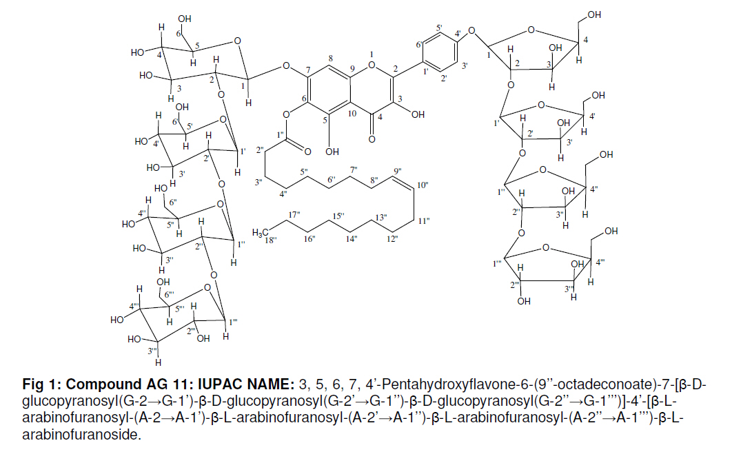
resonated as multiplets between δ 5.01-4.98. The oxygenated methylene protons were present as doublets between δ 3.13-3.03 and as broad signals between δ 3.23-3.20. The other sugar protons appeared in the range of δ 4.20-3.09. The 13C NMR spectrum of the compound exhibited carbon signals for carbonyl carbon at δ 173.81 (C-4), ester carbons at δ 171.01 (C-1’’), vinylic carbons at δ 130.17 (C-9’’) and δ 129.85 (C-10’’), methyl carbon at δ 14.02 (C-18’’), anomeric carbons between δ 104.75-88.53. The signals for flavone ring carbons appeared between δ 166.52-103.58. The signals for other sugar carbons appeared in the range of δ 82.81 - 60.03. The appearance of G-2, G-2’, G-2’’ and G-2’’’ in slightly deshielded region at δ 71.66, 71.66, 71.64,70.71, and 70.79, respectively and A-2, A-2’, A-2’’,A-2’’’ and A-2’’’’ at 78.78, δ 77.67, δ 75.86, δ 77.82 and δ 76.83, indicated a sugar linkage of one C-2 to the anomeric proton of the other sugar moiety. Acid hydrolysis of the compound yielded oleic acid, D-glucose, L-arabinose and the flavones. On the basis of the above discussion, the structure of compound AG 11 has been elucidated as 3, 5, 6, 7, 4’-pentahydroxyflavone-6-(9’’-octadeconoate)-7-[β-D-glucopyranosyl(G-2→G-1’)-β-D-glucopyranosyl(G-2’→G-1’’)-β-D-glucopyranosyl(G-2’’→G-1’’’)]-4’-[β-L-arabinofuranosyl-(A-2→A-1’)-β-L-arabinofuranosyl-(A-2’→A-1’’)-β-L-arabinofuranosyl-(A-2’’→A-1’’’)-β-L-arabinofuranoside. CONCLUSION The present study has isolated and characterized a new flavone, galangoflavonoside, from Alpinia galanga rhizomes for the first time. Further pharmacological investigations are underway to investigate the biological activity of the isolated compound ACKNOWLEDGMENT The authors are grateful to Dr NJ Duragkar, Professor, S. P College of Pharmacy, Nagpur and Dr Rashmi Shukla, Prof. Mumbai Technical Institute of Pharmacy, Mumbai valuable support and suggestions. Thanks are due also to CDRI Lucknow, Indian Institute of technology, Delhi and Arbro Pharmaceuticals, Delhi for assistance in carrying out spectral analyses. REFERENCES
The following images related to this document are available:Photo images[pr09070f1.jpg] | |||||||||||||||||||||||||||||||||||||||||||||||||||||||||||||||||||||||||||||||||||||||||||||||||||||||||||||||||||||||||||||||||||||||||||||||||||||||||||||||||||||||||||||||||||||||||||||||||||||||||||||||||||||||||||||||||||||||||||||||||||||||||||||||||||||||||||||||||||||||||||||||||||||||||||||
| |||||||||
