 |
| About Bioline | All Journals | Testimonials | Membership | News |
|
||||||
|
||||||
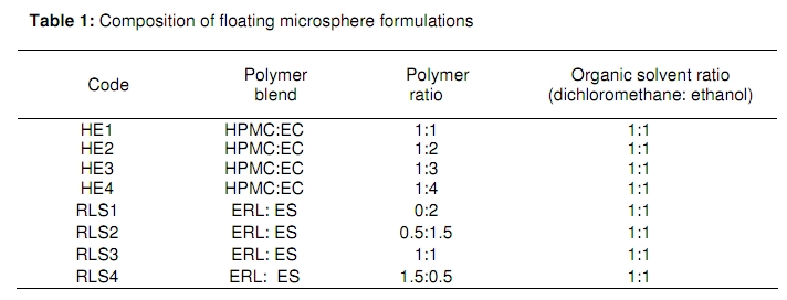
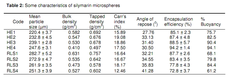
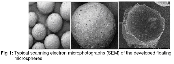
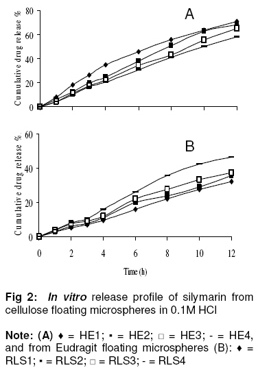
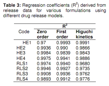
Tropical Journal of Pharmaceutical Research, Vol. 9, No. 1, 2010, pp. 59-66 Research Article Gastroretentive Floating Microspheres of Silymarin: Preparation and In Vitro Evaluation Rajeev Garg and GD Gupta* Department of Pharmaceutics and Pharmaceutical Technology, ASBASJSM College of Pharmacy, Bela, Ropar-140111, India *Corresponding author: E-mail: indianpharmacist@sify.com; Tel: +91-98888-67172 Received: 8 January 2009 Code Number: pr10008 Abstract Purpose: To prepare and evaluate floating microspheres of silymarin for prolonged gastric residence time and increased drug bioavailability. Keywords: Gastroretentive; Prolonged release; Silymarin; Floating microspheres; Ethyl cellulose; Hydroxypropyl methyl cellulose; Eudragit INTRODUCTION Oral sustained drug delivery may be complicated by limited gastric residence time. Rapid gastrointestinal transit can prevent complete drug release in the absorption zone and reduce the efficacy of administered dose since the majority of drugs are absorbed in the stomach or the upper part of the small intestine. Dosage forms that can be retained in the stomach are called gastroretentive drug delivery systems (GRDDS) [1]. Gastroretentive floating drug delivery systems (GRFDDS) have a bulk density lower than that of gastric fluids and thus remains buoyant in the stomach without affecting gastric emptying rate for a prolonged period of time [2]. While the system is floating on gastric contents, the drug is released slowly at a desired rate from the system. Both single and multiple unit systems have been developed. The single unit floating system is more popular but has the disadvantage that its purpose would not be achieved if it fails to float, or is rapidly emptied from the stomach since there is high variability of gastrointestinal transit time [3]. On the other hand, a floating system made up of multiple units may be better suited because they are claimed to reduce intersubject variability in absorption and also lower the probability of dose dumping [4]. Silymarin is a standardised seed extract which is rich in a type of flavonoid compounds known as flavonolignans [5]. The main flavonolignans in silymarin are the isomers, silybin (also known as silibinin), silydianin, and silychristin. Silymarin acts as an antioxidant, scavenger and regulator of the intracellular content of glutathione, cell membrane stabiliser and permeability regulator to prevent hepatotoxic agents from entering hepatocytes. It also acts as a promoter of ribosomal RNA synthesis [6], stimulator of liver regeneration and inhibitor of the transformation of stellate hepatocytes into myofibroblasts - the process responsible for the deposition of collagen fibres, leading to cirrhosis.Silymarin is poorly soluble in water and, therefore, an acidic medium is essential for its dissolution. Its dose is 70 - 140 mg three times a day and has low bioavailability. The low bioavailability of the drug is due to rapid biotransformation in the liver, and has a biological half-life of 6 h. Its reatively short half-life, poor bioavailability and lipophillic nature make it a suitable candidate for gastroretentive drug delivery system. The objective of this work was to develop and characterise gastroretentive floating microspheres of silymarin which, following oral administration, would exhibit prolonged gastric residence time and, hence increase the bioavailability of the drug. EXPERIMENTAL Materials Silymarin was received as a gift from Micro Lab India. Eudragit S 100 (ES100) and Eudragit RL (ERL) were obtained from Rohm Pharma, Darmstadt, Germany while polyvinyl alcohol (PVA), hydrochloric acid (HCl), Tween 80, hydroxylpropyl methylcellulose (HPMC) and ethyl cellulose (EC) were procured from Central Drug House, New Delhi. Dichloromethane and ethanol were purchased from E. Merck (India) Ltd, Mumbai. All the other chemicals used were of analytical grade. Preparation of microspheres Floating microspheres, comprising silymarin and total polymer in a 1:1 ratio, were prepared by emulsion solvent diffusion technique [7] with some modifications. The composition of the microspheres is shown in Table 1. Cellulose microspheres The polymer content was a mixture of HPMC:EC (1 g) in ratios of 1:1 (HE1), 1:2 (HE2), 1:3 (HE3) and 1:4 (HE4). The drug and polymer (in a ratio of 1:1) were dissolved in a 20 ml mixture of dichloromethane and ethanol (1:1) at room temperature. The solution was poured slowly as a thin stream into 150 ml of 0.01 % Tween 80 maintained at 30 - 40 0C. The emulsion was continuously stirred at a rotation speed of 300 rpm for 1 h to allow the volatile solvents to evaporate. The floating microspheres were collected by decantation while the non-floating microspheres were discarded along with polymer residues. The collected micro-spheres were dried overnight in an oven at 40 ± 2 0C and stored in a desiccator containing calcium chloride as a desiccant. Eudragit microspheres The polymer content of the microspheres was a mixture of ES and ERL in ratios of 0:2 (RLS1), 0.5:1.5 (RLS2), 1:1 (RLS3) and 1.5:0.5 (RLS4). The drug and polymer blend (20 ml), in a ratio of 1:1, were dissolved in a mixture of dichloromethane and ethanol (1:1) at room temperature. The solution was poured slowly as a thin stream into 150 ml of 0.2 % sodium lauryl sulphate solution maintained at 30 - 40 0C. The emulsion was continuously stirred at a rotation speed of 300 rpm for 1 h to allow volatile solvents to evaporate. The floating microspheres were collected by decanting, while the non-floating microspheres were discarded along with residues of the polymers. The collected microspheres were dried in an oven overnight at 40 ± 2 0C and stored in a calcium chloride desiccator. Evaluation of silymarin and microspheres Authentication of silymarin Identification of silymarin was by comparison with that of an authentic sample and by verification of the presence of functional groups in its infra-red (IR) spectra. Also, various concentrations of the drug in 0.1M HCl were evaluated by ultraviolet (UV) spectroscopy (Shimadzu –1700) to determine if it would obey Beer’s law. Particle size determination The particle size of the microspheres was determined with an optical microscope under regular polarised light, and mean particle size was calculated by measuring 100 microspheres (n = 3) with the help of a calibrated oculometer [8]. Tapped density Tapping method was used to calculate tapped density. The volume of a weighed quantity of the microspheres was determined, after 100 taps, using a tapped density apparatus (Pharma Chem Machineries, model C-BD 100), as in Eq. 1. DT = MT/VT ……………….……………….. (1) where DT = tapped density, MT is mass of microspheres and VT = volume of microspheres after tapping Carr’s (Compressibility) index This parameter was calculated from bulk density (the ratio of weighed quantity of microspheres to its volume), DP, and tapped density as in Eq 2 [9] Compressibility index = (DT – DP)/DTx100 …... (2) Angle of repose The angle of repose, q, of the microspheres, which measures resistance to particle flow, was determined by the fixed funnel method [10] and calculated as in Eq 3. Tan q = S/D …………………………….... (3) where S = surface area of the free standing height of the microspheres heap and D = diameter of the heap. Scanning electron microscopy Scanning electron microscopy (SEM) studies were performed to determine the porous/hollow nature of the microspheres. Surface morphology of microspheres was also noted.Drug loading The drug content of the floating microspheres was carried out by dissolving the microspheres in a small amount of dichloromethane in a separating funnel and extracting the drugs into 0.1N HCl by evaporating the dichloromethane. Determination of drug loading (n = 3) was carried out at 288 nm spectrophoto-metrically (Shimadzu 1700).In vitro floatability In vitro floatability studies on floating microspheres were carried out using USP XXIV dissolution apparatus II [11]. The microspheres were placed in 0.1M hydrochloric acid containing 0.02 %v/v Tween 80 with the paddle rotating at 100 rpm for 12 h. Tween 80 served to mimic the effect of natural surfactants in the stomach. The floating and the settled portions of the microspheres were recovered separately, dried and weighed. Buoyancy (floatability) was calculated as in Eq 4. Buoyancy (%) = Qf / (Qf + Qs) x 100 .…. (4) where Qf and Qs are the weights of the floating and the settled microspheres, respectively. In vitro drug release studies Drug release studies were carried out in a six-basket USP XXIV dissolution apparatus type I rotating at 100 rpm in 0.1M hydrochloric acid as dissolution medium (900 ml) maintained at 37 ± 0.5 °C. At specific time intervals, up to 12 h, aliquots were withdrawn and analysed at 288 nm spectrophotometrically (Shimadzu 1700) after suitable dilution. The withdrawn volume was replaced with an equal volume of fresh 0.1M hydrochloric acid to maintain sink conditions. All experiments were performed in triplicate. The drug release data were fitted to Zero order (cumulative % drug release versus time), First order (log cumulative % drug retained versus time) and Higuchi models (cumulative % drug released versus square root of time) to assess the kinetics of drug release and determine the release mechanism of the drug from the floating microspheres. Statistical analysis Data of two sets were evaluated by paired t-test and one-way analysis of variance (ANOVA) were applied to check significant difference in drug release from different formulations. p values of less than 0.05 (p < 0.05) were considered to be significant. RESULTS Silymarin authentication The IR spectra showed peaks at 3449.3, 2926.8, 2366.1, 1741.3, 1083.2 and 1027.8 cm-1. which are consistent with the presence of the functional groups of silymarin. Furthermore, the calibration curve of silymarin obeyed Beer’s law in the range of 20 - 55 mg/ml. Physical properties of microspheres The mean particle size of the microspheres significantly increased (p < 0.05) with increasing polymer concentration. Particle size was in the range of 220.4 ± 3.7 to 247.6 ± 3.1 µm for cellulose microspheres and 251.3 ± 3.9 to 282.7 ± 5.2µm for Eudragit microspheres, as shown in Table 2. Angle of repose for cellulose microspheres was between 27.8 and 33.1 º, and 32.2 to 41.3 º for Eudragit microspheres, thus indicating good flow property for both types of microspheres. The findings were supported by Carr’s (compressibility) index, which was < 20 indicating good flow characteristics (Table 2). The production yield of floating microspheres was greater than 65 % for all the formulations. In the floatation test, more than 75 % of cellulose microspheres and 61 % of Eudragit microspheres remained floating at the end of 12 h (Table 2). The encapsulation efficiency of the prepared microspheres was in the range 85.1±2.3 to 94.2±1.4 for cellulose microspheres and 87.7±2.6 to 72.8±3.7 for Eudragit microspheres. Scanning electron microscopy revealed pores on the microsphere as well as a hollow microsphere interior (see Fig 1). In the in vitro drug release studies, the highest cumulative drug released by the cellulose microspheres after 12 h was 70 ± 2.7 % and this was for HE1, i.e., the formulation with the lowest content of ethyl cellulose. Maximum drug released for HE2, HE3 and HE4 was 68.0 ± 1.4, 65.0 ± 1.5 and 58.0 ± 2.2 %, respectively (see Figure 2A). For the Eudragit microspheres, the formulation with the highest content of ERL (i.e., RLS4) showed a maximum drug release of 46.2 ± 1.7 %. Maximum drug release decreased to 37.5 ± 2.2, 35.73 1.5 and 32.2 ± 2.1 % for RLS3, RLS2 and RLS1, respectively, as Figure 2B. DISCUSSION Cellulose microspheres were prepared by gradually increasing EC concentration in combination with a fixed concentration of HPMC in order to assess the effect of polymer (especially EC) concentration on microsphere size. It would be expected that the viscosity of the polymer mixture would increase as polymer concentration rose, resulting in enhanced interfacial tension and hence, formation of larger particles. The mean particle size of Eudragit microspheres was greater than that of cellulose microspheres, and this may be viscosity-related. All the microsphere formulations showed excellent flowability as represented by angle of repose (≤ 35 º), except for RLS4 and this might be due to its high content of ERL. Encapsulation efficiency rose with increase in EC concentration for cellulose microspheres while for Eudragit microspheres, this parameter decreased with increase in ERL content. This could be due to the higher permeability characteristics of HPMC and ERL which would facilitate the diffusion of part of the entrapped drug to the surrounding medium during the preparation of the microparticles. The SEM images in Fig 1 showed the spherical and hollow nature of the microspheres as well as the presence of pores on their surface. Their sphericity contributed considerably to their very good flow properties while their hollow nature would mean lower microsphere density. A large proportion of the microspheres – 61 and 75 % for Eudragit and cellulose microspheres, respectively – remained floating after 12 h. This period was sufficient for up to 70 % of cellulose microspheres (HE1) and 46 % of the Eudgragit microspheres (RLS4) to be released. Thus, prolonged floatation was achieved without apparent gelation. Floatation might have been influenced by the low bulk and tapped densities of the microspheres. Microspheres with the highest levels pf ERL (RLS4) and HPMC (HE1) were least buoyant. It is likely that the surfactant incorporated in the formulations would have increased their wettability and hence, hydration, more than in the other microspheres which had lower levels of the permeable ERL and HPMC. Consequently, the increased amount of absorbed liquid medium replaced the air inside the floating microspheres, thus rendering them less buoyant [12,13]. Silymarin release from cellulose microspheres decreased with increasing EC concentration. Increase in the content of EC (which is less permeable than HPMC) would increase polymer matrix density and thus result in increased diffusional pathlength, leading to a decrease in drug release from the microsphere. Another factor might be that the smaller microspheres formed at low EC concentration had a larger surface area exposed to the dissolution medium, thus, giving rise to faster drug release. On the other hand, the greater the content of ERL, the higher was the rate of drug release from Eudragit microspheres. ES is insoluble in acidic medium and also exhibits low permeability [14]. It is an anionic copolymer of methacrylic acid and methyl methacrylate containing free carboxylic and ester groups. Its very low permeability results from the high intermolecular attraction between its molecules. Hydrogen bonding between the hydroxyl groups of the carboxylic moiety and the carbonyl oxygen of ester group increases the degree of compactness of the polymer and decreases its porosity and permeability. ERL, however, is a copolymer of acrylic and methacrylic acid esters with a low content of quaternary ammonium groups. The ammonium groups present as salts promotes permeability and act as a channeling agent for the entrance of the liquid medium through the floating microsphere wall, causing it to swell. This facilitates the diffusion of the dissolved drug out of the microsphere into the dissolution medium. Thus, by varying the ratio of HPMC and EC in the cellulose microspheres, and ES and ERL in Eudragit microspheres, the rate of release of silymarin can be controlled. The zero order plots of different formulation were found to be fairly linear, as indicated by their high regression values (R2 = 0.9700 - 0.9984). Thus, it seems that drug release from the floating microspheres followed Zero order kinetics. Data based on the Higuchi model usually provide a clue to the diffusion mechanism of drug release from matrix systems such as the floating microspheres developed in this work. R2 values based on the Higuchi model ranged from 0.9680 to 0.9991 (Table 3). As these values were close to 1.0, the drug release mechanism of the developed floating microspheres can be said to be Higuchian and, therefore, matrix diffusion-controlled. CONCLUSION We can conclude that floating microspheres of silymarin prepared with a suitable blend of ES and ERL on the one hand, and HPMC and EC on the other, demonstrate satisfactory release and floating properties. The developed floating microspheres also showed good physicochemical properties. Drug release from the formulations followed Zero order kinetics and the mechanism of drug release was diffusion-controlled. REFERENCES
© Pharmacotherapy Group, Faculty of Pharmacy, University of Benin, Benin City, 300001 Nigeria. The following images related to this document are available:Photo images[pr10008t3.jpg] [pr10008f2.jpg] [pr10008f1.jpg] [pr10008t2.jpg] [pr10008t1.jpg] |
| |||||||||
