 |
| About Bioline | All Journals | Testimonials | Membership | News |
|
||||||
|
||||||
Tropical Journal of Pharmaceutical Research, Vol. 9, No. 5, September-October, 2010, pp. 451-454 Research Article Immunomodulatory Activity of the Methanol Extract of Amorphophallus campanulatus (Araceae) Tuber Mohammad Ali Ebrahimzadeh1, Seyed Mohammad Nabavi1,2 , Seyed Fazel Nabavi1, Bahman Eslami3, Zahra Rahmani4*1Pharmaceutical Sciences Research Center, School of Pharmacy, Mazandaran University of Medical Sciences, Sari, 2Young Researchers Club, Islamic Azad University, Ghaemshahr Branch, Ghaemshahr, 3Department of Biology, 4Islamic Azad University, Ghaemshahr branch, Faculty of Medicine, Mazandaran University of Medical Sciences, Sari, Iran. *Corresponding author: E-mail: zadeh20@yahoo.com; Tel: +98-151-3543081-3; Fax: +98-151-3543084 Received: 9 January 2010 Revised accepted: 23 July 2010 Code Number: pr10052 AbstractPurpose: To investigate the antioxidant and
antihaemolytic properties of the leaves of Laser trilubum grown in Gaduk,
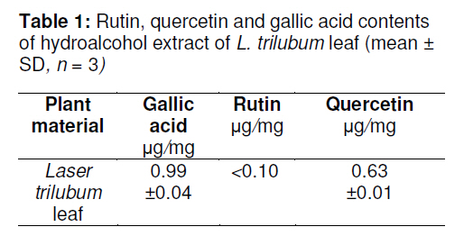
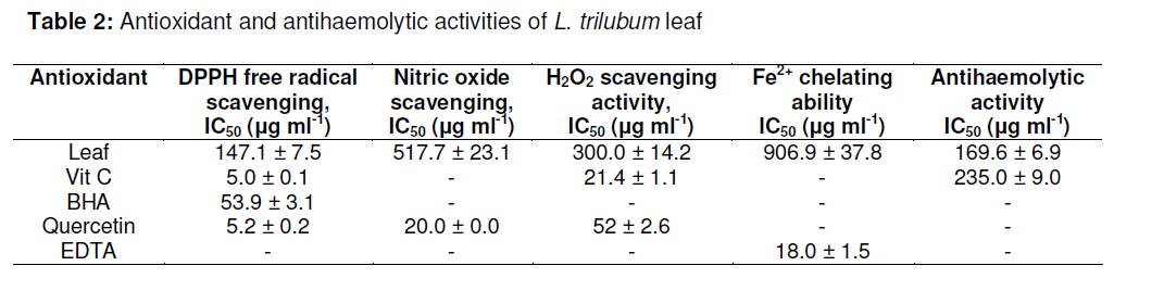
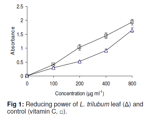
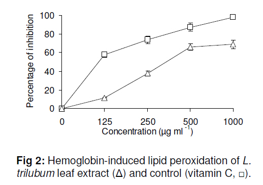
Iran. Keywords: Antioxidant activity, Laser trilobum, Antihaemolytic, Flavonoids, Kefe cumin INTRODUCTIONThe pathology of numerous chronic diseases, including cancer and heart disease, involves oxidative damage to cellular components [1]. Reactive oxygen species (ROS), such as hydrogen peroxide, super oxide anion and hydroxyl radical, capable of causing damage to DNA, have been associated with carcinogenesis, coronary heart disease, and many other health problems related to advancing age [2]. Many endogenous and exogenous defense mechanisms are available in living organisms to limit the levels of ROS and the damage caused by them [3]. Antioxidants terminate direct ROS attacks and radical-mediated oxidative reactions, and appear to be of primary importance in the prevention of these diseases and health problems. Synthetic antioxidants such as butylhydroxyanisole (BHA) or butylhydroxytoluene (BHT) are used to decelerate these processes. However, due to their unstable and highly volatile nature, they have frequently raised some questions about their safety and efficacy ever since their first introduction to the food industry [4]. Consequently, the need to identify alternative natural and safe sources of food antioxidants arose, and hence the search for natural antioxidants, especially of plant origin, has increased in recent years [5]. Laser genus is a member of the Apiaceae family and has only one species in Iran, namely, Laser trilubum that is grown in Gaduk, northern Iran. The antimicrobial effect of L. trilubum extract has been reported [6]. Compositional analysis of the essential oils of the fruit of the plant has revealed abundant levels of limonene and perillaldehyde [6]. The fruits are also rich in potassium, calcium, phosphorus, magnesium, sodium, arsenic and aluminum [6]. A review of available literature shows that there is no scientific report on the antioxidant and antihaemolytic activities of L. trilubum leaf. Flavonoids interactions with cell membranes, which generally serve as targets for lipid peroxidation (LP), constitute an important area of research [7]. Various model membrane systems such as low density lipoprotein (LDL) and red blood cell (RBC) membrane comprising physiologically important membrane protein components, offer a physiologically relevant and a relatively simple system for studying LP [8]. RBC has been used as an in vitro model to study the oxidant/antioxidant interaction since its membrane is rich in polyunsaturated fatty acids, which are extremely susceptible to peroxidation [7]. In recent years, a few interesting studies indicating the protective effect of some plants extracts against oxidative damage in intact RBC membranes have been reported [9-10]. The aim of this study, therefore, was to determine the antioxidant and antihaemolytic activities of the hydroalcohol extract of L. trilubum leaves. EXPERIMENTAL Chemicals and reagentsFerrozine, linoleic acid, trichloroacetic acid (TCA), 1,1-diphenyl-2-picryl hydrazyl radical (DPPH) and potassium ferricyanide were purchased from Sigma Chemicals Co., USA. Gallic acid, quercetin, butylated hydroxyanisole (BHA), ascorbic acid, sulfanilamide, N-(1-naphthyl) ethylenediamine dihydrochloride, ethylenediamine tetraacetic acid (EDTA) and ferric chloride were obtained from Merck, Germany. All other chemicals and reagents used were of analytical grade or purer. Plant materials and preparation of freezedried extractThe leaves of L. trilubum were collected from Gaduk, province of Firouzkooh, northern Iran, in June 2009 at full flowering stage and identified by Dr Bahman Eslami (Department of Biology, Islamic Azad University, Ghaemshahr branch, Iran). A voucher specimen (number 4325) has been deposited at the Sari School of Pharmacy herbarium. The material was dried at room temperature and coarsely ground before extraction. A known amount (250 g) was extracted at room temperature for 24 h by percolation using ethanol/water (600 ml, 70/30 v/v). The extract was separated from the residue by filtration through Whatman number 1 filter paper. This procedure was repeated thrice and the resultant extract was concentrated under vacuum in a rotary evaporator and the residue was then freeze-dried for complete solvent removal. The yield was 15 %. Determination of total phenolic compounds and flavonoidTotal phenol content was determined by Folin-Ciocalteau method [11]. The methanol solution of the extract (0.5 ml, 1.6 mg ml-1) was mixed with 2.5 ml of 0.2N Folin-Ciocalteau reagent for 5 min and 2.0 ml of 75 g l-1 sodium carbonate was then added. The absorbance of the reaction mixture was measured spectrophotometrically (Perkin Elmer, Wellesley, MA, USA) at 760 nm after 2 h of incubation at room temperature. The result was expressed as gallic acid equivalent. Total flavonoid was estimated as previously described [12]. Briefly, 0.5 ml solution of the extract in methanol (1.6 mg ml-1) was separately mixed with 1.5 ml of methanol, 0.1 ml of 10 % aluminum chloride, 0.1 ml of 1M potassium acetate and 2.8 ml of distilled water, and then left at room temperature for 30 min. The absorbance of the reaction mixture was measured at 415 nm in a double beam spectrophotometer (Perkin Elmer). Total flavonoid content was calculated as quercetin from a calibration curve. DPPH radical-scavenging activityStable 1,1-diphenyl-2-picryl hydrazyl radical (DPPH) was used for the determination of free radical scavenging activity of the extract [13]. Different concentrations of the extract (50 -800 µg ml-1) were added, in equal volumes, to the methanol solution of DPPH (100 µM). The mixture was allowed to stand for 15 min at room temperature and the absorbance recorded spectrophotometrically at 517 nm. The experiment was repeated thrice with vitamin C, BHA and quercetin serving as standard controls. IC50 values, which denote the concentration of sample required to scavenge 50 % of DPPH free radicals, was noted. Metal-chelating activityThe ability of the extract to chelate ferrous ions was assessed by a recently published method [1,12]. Briefly, 0.05 ml of 2 mM FeCl2 was added to varying concentrations of the extract (0.2 -3.2 mg ml-1). The reaction was initiated by the addition of 5 mM ferrozine (0.2 ml) and the mixture was shaken vigorously and left to stand at room temperature for 10 min. The absorbance of the solutions was measured spectrophotometrically at 562 nm. Inhibition (%) of ferrozine-Fe2+ complex formation was calculated as {(A0 -A1)/A0} × 100, where A0 is the absorbance of the control and A1 the absorbance of either the mixture containing the extract or the standard solution. EDTA (6.25 -100 µg ml-1) was used as the standard. Assay of nitric oxide-scavenging activityIn this experiment, 1 ml of sodium nitroprusside (10 mM) in phosphate-buffered saline was mixed with varying concentrations of Laser trilubum extract (100 -1600 µgml-1) dissolved in water and incubated at room temperature for 150 min. The same reaction mixture, without the extract but with an equivalent volume of water, served as control. Following the incubation period, 0.5 ml of Griess reagent (1 % sulfanilamide, 2 % H3PO4 and 0.1% N-(1-naphthyl) ethylenediamine dihydrochloride) was added. The absorbance of the chromophore formed was read spectrophotometrically at 546 nm. Quercetin was served as positive control [11]. Hydrogen peroxide scavenging activityThe ability of the extract to scavenge hydrogen peroxide was determined according to the method of Dehpour et al [14]. A solution of hydrogen peroxide (40 mM) was prepared in phosphate buffer (pH 7.4). 1.4 ml of the extract (0.1 -3.2 mg ml-1) in distilled water was added to 0.6 ml of the hydrogen peroxide solution. The absorbance of hydrogen peroxide at 230 nm was determined after 10 min against a blank solution containing phosphate buffer without hydrogen peroxide. Hydrogen peroxide scavenging (%) by the extract and standard was calculated as in Eq 1. % scavenged (H2O2) = (Ao - A1)/Ao × 100 …… (1) where Ao is the absorbance of the control and A1 the absorbance of the mixture containing either the extract or standard. Determination of reducing powerThe reducing power of L. trilubum extract was determined according to the method of Yen and Chen [4,15]. The extract (2.5 ml, 25 -800 µg ml-1) in water was mixed with 2.5 ml of 0.2M phosphate buffer (pH 6.6) and 2.5 ml of 1 % potassium ferricyanide. The mixture was incubated at 50 oC for 20 min and 2.5 ml of 10 % trichloroacetic acid was added to the mixture to stop the reaction. The mixture was then centrifuged at 3000 rpm for 10 min and 2.5 ml of the upper layer of the solution was mixed with distilled 2.5 ml of water and 0.5 ml of 0.1 % FeCl3, and the absorbance was measured spectrophotometrically at 700 nm. Increased absorbance of the reaction mixture indicated increased reducing power. Vitamin C was used as positive control. Antioxidant activity in haemoglobininduced linoleic acid systemThis was determined by a modified spectrophotometry assay [16]. Reaction mixtures (200 µl) containing 40 µl of the extract (125 –1000 mg ml-1 in ethanol), 1 mmol/l of linoleic acid emulsion (40 µl), 70 µl of 40 mmol/l of phosphate buffer (pH 6.5), and 0.0016 % haemoglobin (50 µl), were incubated at 37 ºC for 45 min. Following incubation, 2.5 ml of 0.6 % HCl in ethanol was added to stop the lipid peroxidation. The amount of peroxide was measured in triplicate by the thiocyanate technique by reading the absorbance at 480 nm after colouring with 100 ml of 0.02 mol/l of FeCl2 and 50 ml of ammonium thiocyanate (30 g/100 ml). Vitamin C was used as positive control. Preparation of rat erythrocytesAll the animal experiments were carried out with the approval of the Institutional Animal Ethical Committee. Male Wistar rats with a body weight range of 180 –220 g were housed in individual polypropylene cages and had free access to food and water. The animals were fed with standard diet. They were sacrificed under diethyl ether induced anesthesia and blood was collected by heart puncture in heparinized tubes. Erythrocytes were isolated and stored according to the method described by Yuan et al [16]. Briefly, the blood samples collected were centrifuged (1500 × g, 10 min) at 4 ºC; erythrocytes were separated from the plasma and buffy coat and were washed three times by centrifugation (1500 × g, 5 min) in 10 volumes of 10 mM phosphate buffered saline (PBS, pH 7.4). The supernatant and buffy coats of white cells were carefully removed with each wash. The washed erythrocytes were stored at 4 ºC and used within 6 h for further studies. Assessment of antihaemolytic activity of extractThe inhibition of rat erythrocyte haemolysis by the extract was evaluated according to the procedure described by Ebrahimzadeh et al [17]. Rat erythrocyte hemolysis was performed with H2O2 as free radical initiator. To 100 µl of 5 %v/v suspension of erythrocytes in PBS, 50 µl of the extract at different concentrations (50 –250 µg ml-1 in PBS pH 7.4), which corresponds to 100 –3200 µg of added extract. To this, 100 µl of lM H2O2 (in PBS, pH 7.4) was added. The reaction mixtures were shaken gently in an incubator shaker at 37 °C for 3 h and then diluted with 8 ml of PBS and centrifuged at 2000 × g for 10 min. The absorbance of the resulting supernatant was measured spectrophotometrically at 540 nm to determine the level of haemolysis. Similarly, the erythrocytes were treated with 100 µM H2O2 and without inhibitors (plant extract) to obtain complete haemolysis. The absorbance of the supernatant was also measured at 540 nm. The inhibitory activity of the extract was compared with that of the standard antioxidant, vitamin C. To evaluate haemolysis induced by the extract, the erythrocytes were pre-incubated with 50 µl of the extract (corresponding to 25 µg extract) for 1 h and the level of haemolysis determined. Haemolysis was calculated by determining the haemolysis caused by 100µM H2O2 as 100 %. The IC50 values were calculated from the plots as the antioxidant concentration required for the inhibition of 50% hemolysis. Assay of putative biological active components Gallic acidA Knauer liquid chromatograph system comprising degasser, pump, auto-sampler, thermostatted column compartment, and diode array detector was used. The column used was a C18 reversed phase Kingsorb 5 mm (250 × 4.6 mm). The mobile phase eventually adopted for this study was methanol/water/orthophosphoric acid (20/79.9/0.1) and the flow rate was 1.0 ml/min. The absorption wavelength used was 210 nm [18]. The column was operated at 30 °C with a sample injection volume of 20 ml. The content of gallic acid was calculated on the basis of a calibration curve constructed using an authentic reference (gallic acid, 25 to 100 µg/ml) obtained from Sigma. Quercetin and rutinChromatographic analysis was carried out using a C18 reversed phase Kingsorb (250 × 4.6 mm) column packed with 5µm diameter particles. The mobile phase was methanol–acetonitrile–water (40:15:45) containing 1 % acetic acid. This mobile phase was filtered through a 0.45 µm membrane filter (Millipore) and de-aerated ultrasonically prior to use. Quercetin was quantified by diode array detection (DAD) following HPLC separation at 368 nm for quercetin and 257 nm for rutin [18]. Flow rate and injection volume were 1.0 ml/min and l0 µl, respectively. The chromatographic peaks of the analytes were confirmed by comparing their retention time and UV spectra with those of the reference standards. Quantification was carried out by the integration of the peak using external standard method. All chromatographic operations were carried out at ambient temperature. Statistical analysisThe results obtained are expressed as mean ± SD. All measurements were carried out in triplicate. The data were analyzed by analysis of variance (ANOVA, p < 0.05) and the means separated by Duncan's multiple range tests. IC50 values were calculated from linear regression plots. RESULTS Total phenol and flavonoid contentsTotal phenol compounds, as determined by Folin Ciocalteu method, are reported as gallic acid equivalent by reference to standard curve (y = 0.0054x + 0.0628, r2= 0.987).The total phenolic content of L. trilubum leaf was 75 ± 3 mg gallic acid equivalent/g extract while the total flavonoid content was 59.2 ± 2.1 mg quercetin equivalent/g extract. The amounts of rutin, quercetin and gallic acid are shown in Table 1. DPPH radical-scavenging activityThe radical-scavenging activities of the extracts increased with increasing concentration. The extract’s IC50 value for DPPH radical-scavenging activity was 147.1 ± 7.5 µg ml-1 while the values for ascorbic acid, quercetin and BHA were 5.0 ± 0.1, 5.2 ± 0.2 and 53.9 ± 3.1 µg ml-1, respectively (Table 2). Fe2+ -chelating ability The extract showed a weak activity of between 0.4 and 1.6 mg ml-1 although inhibition increased with increasing concentration of the extract (see Table 2). IC50 was 906.9 ± 37.8 µg ml-1 while EDTA, which was used as control, showed very strong activity with IC50 of 18.0 ± 1.5 µg ml-1). Nitric oxide-scavenging activityInhibition increased with increasing concentration of the extract. It showed good NO scavenging activity with IC50 of 517.7 ± 23.1 µg ml-1 compared with 20.00 ± 0.01 µg ml-1 for quercetin which served as positive control (Table 2). Hydrogen peroxide scavengingThe extract scavenged hydrogen peroxide in a concentration-dependent manner with IC50 of 300 ±14 µg ml-1 compared with IC50 values of 21.4 ± 1.1 and 52.0 ± 2.6 µg mlfor ascorbic acid and quercetin, respectively (Table 2). Reducing powerFig 1 shows the data for the reducing power of the extract. The reducing power of the extract increased with increase in extract concentration and exhibited moderate reducing power that was comparable with that of vitamin C (p < 0.05). Antioxidant activity in a hemoglobininduced linoleic acid systemAs Fig 2 indicates, the extract showed good inhibitory activity in hemoglobin-induced linoleic acid system but was not as high as that of vitamin C (p < 0.001). Antihaemolytic activityThe extract did not show any harmful effects on erythrocytes and, in fact, exhibited potent antihaemolytic activity with IC50 of 169.6 ± 6.9 µg ml-1 compared with 235 ± 9 µg ml-1 for vitamin C which served as positive control. DISCUSSIONThe extract showed high total phenol and flavonoid contents. Phenols and polyphenolic compounds, such as flavonoids, are widely found in food products derived from plant sources and show significant antioxidant activity [15]. DPPH is a stable nitrogencentred free radical, the colour of which changes from violet to yellow upon reduction by either the process of hydrogen-or electron-donation. Substances which are able to perform this reaction can be considered as antioxidants and therefore radical scavengers [19]. The phenol and flavonoid contents of this plant may be responsible for its good DPPH-scavenging activity [15]. The correlation between total phenol contents and antioxidant activity has been widely studied in different foodstuffs such as fruit and vegetables [5, 11]. Iron chelators mobilize tissue iron by forming soluble, stable complexes that are then excreted in faeces and/or urine. Chelation therapy reduces iron-related complications in humans and hence improves quality of life and overall survival in some disease states such as thalassemia major [12]. They can ameliorate the symptoms of iron overload and improve the quality of life and overall survival rate for sufferers. Therefore, many researchers have focused on some natural products, especially flavonoids, that possess direct influence on iron (III) ions level within tissues [20]. In addition, iron chelation could be considered a rational therapeutic strategy for Alzheimer's disease [1]. Foods are often contaminated with transition metal ions which may be introduced by processing methods. Bivalent transition metal ions play an important role as catalysts of oxidative processes, leading to the formation of hydroxyl radicals and hydroperoxide decomposition reactions via Fenton chemistry [14]. These processes can be delayed by iron chelation and deactivation. The transition metal, iron, is capable of generating free radicals from peroxides and may be implicated in human cardiovascular disease [1]. Minimizing Fe2+ concentration affords protection against oxidative damage. In the presence of other chelating agents, the complex formation is disrupted with the result that the red coluor of the complexes decreases. The extract showed weak ironchelating activity and its chelating activity may be the result of its quercetin content. The potent iron ion chelating activity of quercetin and rutin has been reported [21].Inhibition of NO increased with increase in the concentration of the extract. The extract also showed weak NO scavenging activity In addition to reactive oxygen species, NO is also implicated in inflammation, cancer and other pathological conditions [4]. Plant/plant products that have the capacity to counteract the effect of NO formation may be of considerable interest in preventing the illeffects of excessive NO generation in the human body. Potent .NO scavenging ability has been reported for flavonoids [22] whose scavenging rate constants are only 30 times less than that of the very potent endogenous NO scavenger, haemoglobin. It has been speculated that .NO scavenging plays a role in the therapeutic activity of flavonoids [22]. Furthermore, scavenging activity may also help to arrest the chain of reactions initiated by excess generation of NO that are detrimental to human health. The H2O2-scavenging of the extract may be attributed to its phenolic contents as well as other active components such as anthocyanins, tannins and flavonoids which can donate electrons to H2O2, thus neutralizing it to water [23]. The extract was capable of scavenging hydrogen peroxide in a concentration-dependent manner but showed weaker activity than controls (ascorbic acid and quercetin) Membrane lipids are rich in unsaturated fatty acids which are most susceptible to oxidative processes. Specifically, linoleic acid and arachidonic acid are targets of lipid peroxidation [24]. Since polyphenolics contents appear to function as good electron and hydrogen atom donors, and therefore, be able to terminate radical chain reaction by converting free radicals and reactive oxygen species to more stable products, the reducing potential of the extract may be attributed to this mode of activity. A similar observation has been reported for several plant extracts [14]. Erythrocytes are considered as prime targets for free radical attack owing to the presence of both high membrane concentration of polyunsaturated fatty acids (PUFA) and the O2 transport associated with redox active haemoglobin molecules, which are potent promoters of reactive O2 species. The inhibition of lipid peroxidation by antioxidants may be due to their free radical-scavenging activities. Superoxide indirectly initiates lipid peroxidation because superoxide anion acts as a precursor of singlet oxygen and hydroxyl radical [13]. Hydroxyl radicals abstract hydrogen atoms from the membrane lipids, which results in lipid peroxidation. The extract showed good activity in haemoglobin-induced linoleic acid system; however, its activity was not as high as that of vitamin C (p < 0.001). Furthermore, it did not exert any harmful effect on erythrocytes but rather exhibited potent antihaemolytic activity. Antihaemolytic activity and the relationship between iron ion chelating activity and protective activity against oxidative damage to erythrocyte membrane by quercetin have been reported previously [25]. It seems that high total phenol and flavonoid contents in the extract led to its potent antihaemolytic activity. There are some reports that improve our hypothesis [7,9,10,26]. CONCLUSIONOur study showed that the hydroalcohol extract of L. trilubum leaf has remarkable antioxidant and antihaemolytic activities that may be due its high levels of phenolic and flavonoid contents. ACKNOWLEDGMENTThis research was supported by a grant from Islamic Azad University, Ghaemshahr branch, Iran. REFERENCES
Copyright 2010 - Tropical Journal of Pharmaceutical Research The following images related to this document are available:Photo images[pr10052f2.jpg] [pr10052t2.jpg] [pr10052f1.jpg] [pr10052t1.jpg] |
| |||||||||
