 |
| About Bioline | All Journals | Testimonials | Membership | News |
|
||||||
|
||||||
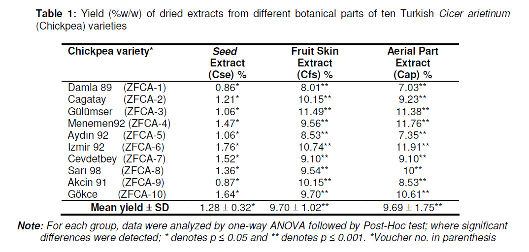
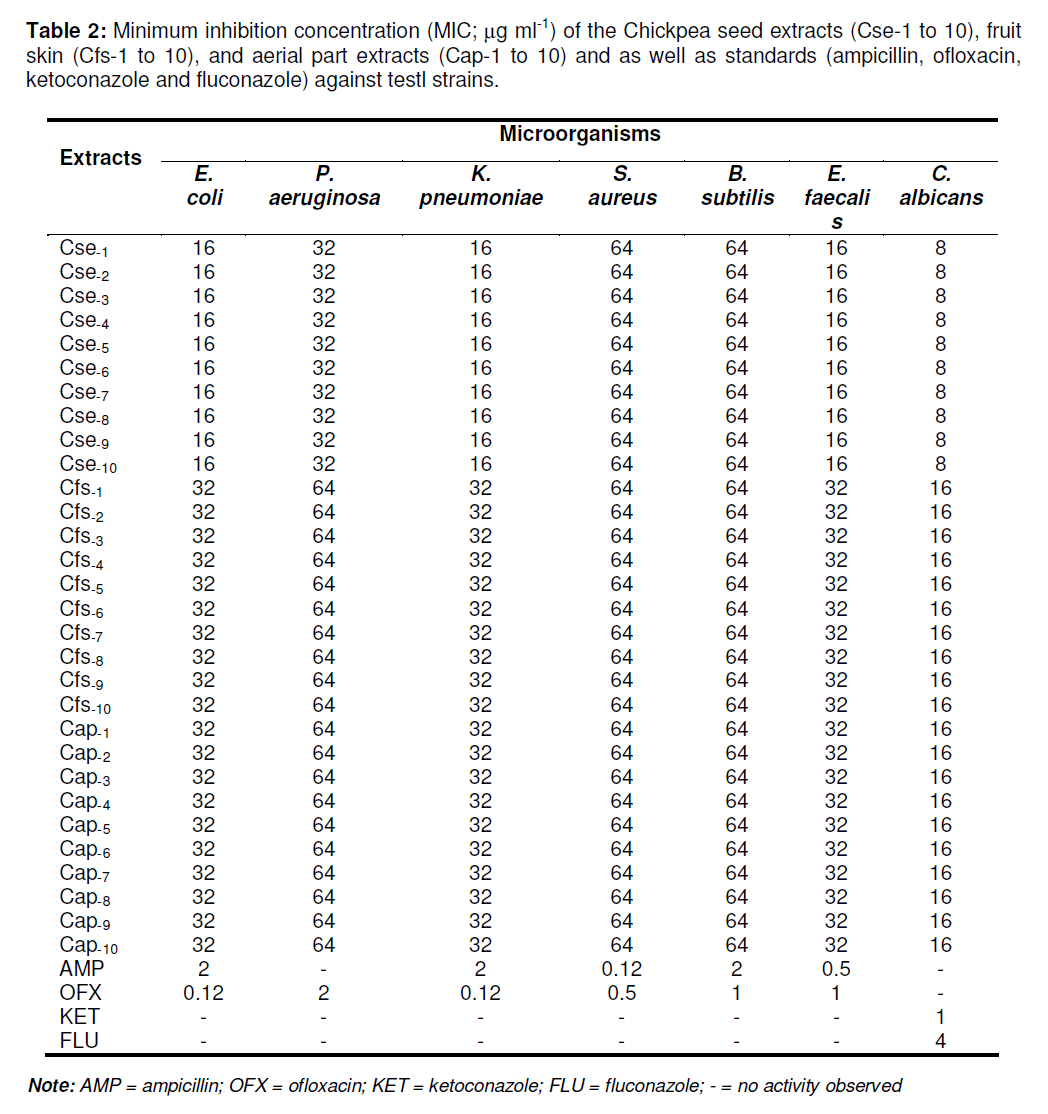
Tropical Journal of Pharmaceutical Research, Vol. 9, No. 5, September-October, 2010, pp. 475-481 Research Article In Vitro Antimicrobial Activities of Cicer arietinum L (Chickpea) A Kan1, B Özçelik2*, M Kartal3, ZA Özdemir4, and S Özgen2 1Programme for Food Technologies, Vocational School of Technical Sciences, University of Selcuk 42070, *Corresponding author: Email: berrin@gazi.edu.tr; microberr@yahoo.com Received: 11 December 2009 Revised accepted: 14 August 2010 Code Number: pr10057 AbstractPurpose: To evaluate the antibacterial and antifungal activities of the extracts
of the seed, fruit skin and aerial parts of ten registered varieties of Cicer
arietinum (Chickpea) Keywords: Antibacterial; Antifungal; Cicer arietinum; Chickpea INTRODUCTION Traditional healers have long used plants to prevent or cure infectious disease. Many of these plants have been investigated scientifically and shown to inhibit the growth of pathogenic/drug-resistant microorganisms. A number of these agents appear to have structures and modes of action that are distinct from those of antibiotics in current use. Therefore, it is worthwhile to study plants and plant products for activity against microorganisms. One approach that has been used for the discovery of antimicrobial agents from plants is based on the evaluation of traditional medicinal plant extracts [1-9]. Food legumes are crops of the family Leguminosae, also called Fabacae. They are mainly grown for their edible seeds and thus are also named grain legumes [10]. Based on world production estimates, Cicer arietinum L. (Chickpea) is the third most important coldseason food legume after the common bean (Phaseolus vulgaris L.) and pea (Pisum sativum L.) [10]. Chickpea is generally consumed as a seed food, being a good source of protein and other essential human nutrients. However, young chickpea leaves are also cooked and eaten as green vegetable in certain parts of the world and could be a useful source of dietary nutrients, especially in malnourished populations [5]. In this study, we aimed to investigate the antimicrobial and antifungal properties of edible and non-edible parts of Cicer arietinum L. (Chickpea) using the susceptibility testing method of the Clinical and Laboratory Standards Institute [11, 12]. EXPERIMENTAL Plant material Ten registered varietes (Damla 89, Cagatay, Gülümser, Menemen 92, Aydın 92, Izmir 92, Cevdetbey, Sarı-98, Akçin 91, Gökce) of chickpea were grown at Bahri Dagdas International Agricultural Research Institute in Central Anatolian Region of Turkey. This research was conducted to randomized block design with three replications in 2005. Chickpea seeds were sown on 30 March 2005 and harvested on 10 July 2005. The plants were identified by Prof Dr CY Ciftci of the Department of Field Crops, Faculty of Agriculture, Ankara University, Ankara, Turkey. The voucher specimens (Table 1) were preserved at the Herbarium of the Faculty Agriculture, Selcuk University, Konya, Turkey. The seed, fruit skin and aerial part of the plant were dried under shade and used for the study. Preparation of extractsSix grams of the powdered plant material (seed, fruit skin or aerial parts) were mixed with 50 ml methanol and extracted twice with an hour in a 100 ml Erlenmeyer flask with a magnetic stirrer at 40 °C. The extracts were combined and evaporated to dryness at a temperature not exceeding 40 °C. Dried and methanol-free extracts were used for testing the antibacterial and antifungal activities. The yields of the crude extracts obtained are given in Table 1. Microorganisms and inoculaStandards of Gram-negative bacteria used in the test were Escherichia coli (ATCC 35218), Pseudomonas aeruginosa (ATCC 10145) and Klepsiella pneumoniae (RSKK 574) while Gram-positive bacteria used were Staphylococcus aureus (ATCC 25923), Bacillus subtilis (ATCC 6633) and Enterococcus faecalis (ATCC 29212). For antifungal test, Candida albicans (ATCC 10231) was used. Mueller-Hinton Broth (MHB; Oxoid) and Mueller-Hinton Agar (MHA; Oxoid) were applied for growing and diluting the bacteria while Sabouraud liquid medium (SLM; Oxoid) and Sabouraud dextrose agar (SDA; Oxoid) were used for growing and diluting the fungus. The medium, RPMI-1640 with L-glutamine, was buffered to pH 7 with 3-{N-morpholino}-propansulfonic acid (MOPS). Prior to the test, the strains of fungus and bacteria were cultured on media and passaged at least twice to ensure purity and viability at 35 oC for 24 to 48 h. The bacterial suspensions used for inoculation were diluted to a concentration of 105 cfu ml-1 from fresh cultures with McFarland 0.5 density (108 cfu ml-1). The fungus suspension was diluted by the spectrophotometric method of inoculum preparation to a final culture suspension concentration of 2.5x10³ cfu ml-1 [11,12]. Preparation of extract and reference solutions The extracts were dissolved in dimethylsulphoxide (DMSO) to a final concentration of 512 µg ml-1, sterilized by filtration using a 0.22 µm filter (Millipore, USA) and used as stock solutions. Standard antibacterial powders of ampicillin (AMP; Faco) and ofloxacin (OFX; Hoechst Marion Roussel) as well as standard antifungal powders of ketoconazole (KET; Bilim) and flukonazole (FLU; Pfizer), were dissolved in phosphate buffer solution (AMP; pH: 8.0, 0.1 mol L-1), DMSO (KET), in water (FLU, OFX). The stock solutions were prepared in medium according to Clinical and Laboratory Standards Institute requirements [11,12]. Antibacterial and antifungal tests The microdilution method was employed for the antibacterial and antifungal tests. Susceptibility tests were performed according to the requirements of the Clinical and Laboratory Standards Institute [6,7] as previously reported, and the inhibition endpoint for the determination of the minimum inhibition concentrations was applied. Media were placed into each 96 wells of the microplates. Extract solutions were added into the first rows of the microplates and two-fold dilutions of the test antimicrobial materials (256 -0.0312 µg ml-1) and dispensed into the remaining wells. Culture suspensions (10 µl) were inoculated into all the wells. The sealed microplates were incubated at 35 ºC for 24 and 48 h in a humid (moist) chamber. All the tests were carried out in triplicate. The lowest concentration of the extracts that completely inhibited macroscopic growth was taken as the minimum inhibitory concentration (MIC) as previously reported [5-9]. Statistical analysisStatistical analysis of results was performed with Statistical Package for Social Sciences (SPSS), version 10.0 for analysis of variance (ANOVA) followed by Post-Hoc test. Prior to this, the homogeneity of each group was tested using Levene statistics. Tamhane test statistics was applied where required. Statistically difference was set at p < 0.05. RESULTSThe extract yields of Chickpea (Cicer arietinum L.) varieties, expressed as % of the seed (Cse%), fruit skin (Cfs%) and aerial part (Cap%) are listed in Table 1. The data show varying yield. The results of statistical analyses show that the extracts of the seed, fruit skin and aerial part have normal distribution at 5 % significance level. Hence, in order to test whether there was statistical difference between the yields of fruit skin and aerial part, analysis of variance (ANOVA) was applied. Post-hoc test results suggest that yield variances were not homogenous. Subsequently, application of Tamhane test statistics showed that the difference between the yield of the seed extract was different from those of the fruit skin and aerial part at 1 % significance level. The results of the antibacterial activities of the extracts obtained from Cicer arietinum L. varieties (seed extract, fruit skin extract and aerial part extract) are presented in Table 2. Chickpea seed extracts (Cse) showed varying antibacterial activity against Gramnegative strains (E. coli, P. aeruginosa, K. pneumoniae) in the MIC range 16–64 µg ml-1 but were less active against gram-positive (S. aureus, B. subtilis, E. faecalis) strains with MIC of 64 µg ml-1. Statistically different MICs were observed between the extracts of the fruit skin (Cfs) and the aerial part (Cap) (p < 0.05). The antibacterial activity of Chickpea fruit skin (Cfs1-10) and Chickpea aerial parts (Cap1- 10) extracts were not statistically different (p≥ 0.05) as they showed the same degree of inhibition against Gram-negative (E. coli and K. pneumoniae) bacteria and the grampositive bacterium, E. faecalis at the concentration of 32 µg/ml Additionally, they were both less effective against P. aeruginosa, S. aureus, and B. subtilis at a concentration of 64 µg/ml. Of all the Chickpea extracts, Chickpea seed extract (Cse; p ≤ 0.05). exhibited the stringest antifungal activity against C. albicans at a concentration of 8 µg/ml. Even at a concentration of 16 µg/ml, fruit skin (Cfs) and aerial part (Cap) extracts showed lowere antifungal activity than the seed extract. This is the first report showing that C. arietinum extracts had substantial antifungal activity at a concentration as low as 8 µ/ml. DISCUSSION As seen in Table 2, Chickpea seed extracts (Cse1-10) showed antibacterial activity on Gram-negative strains between the ranges of 16–32 µg/ml concentrations but were less active against Gram-positive strains even at a concentration of 64 µg/ml. Among Chickpea seed extracts (numbered 1-10), antibacterial activity against E. faecalis was greater than against the other two Gram-positive bacteria at a concentration of 16 µg/ml. Moreover, Cse1-10 extracts showed clear antifungal activity at a concentration of 8 µg/ml, which is close to that of the standard (FLU; 4 µg/ml). Although the antibacterial, and antifungal effects of Cicer arietinum L have been examined in some earlier studies, but its activity against commonly encountered human pathogens has not been addressed, to the best of our knowledge. In a previous study, several proteins, including a glucanase, a chitinase, an antifungal cyclophyllin-like protein, and three antifungal peptides designated cicerin, arietin, and cicearin were isolated from the chickpea (Cicer arietinum L) by Chu et al [3]. The antifungal protein, designated ‘chickpea cyclophilin-like antifungal protein’, has been isolated from seeds of the chickpea [4]. It possesses a molecular weight of 18kDa and an N-terminal sequence with resemblance to cyclophilin. It displayed an antifungal activity against a number of fungi including Mycosphaerelle arachidicola, Botrytis cinerea, and Rhizoctonia solani, to varying degrees. It was reported that probable structural differences further along the protein sequence may account for its varying antifungal activity. On the other hand, the protein was also capable of inhibiting human immunodeficiency virus-1 (HIV-1; RNA viruses) reverse transcriptase [4]. Aslam et al reported recently that stilbene compounds isolated from the roots of Chickpea inhibited not only the growth of the Gram-positive bacterium, Bacillus subtilis, but also that of the Gram-negative species, Pseudomonas syringae, and four species of filamentous fungi [13]. The results showed similar antimicrobial activity against the microorgaanisms with MICs in the range of 25 -100 µg/ml. The nine stilbenes evaluated in their studies inhibited the growth of both Gram-positive and Gram-negative bacteria, whereas a stilbene-3 compound was only active against the Gram-negative bacterium, P. syringae, at a concentration of 25 µg/ml. Aslam et al [13] also observed that stilbene compounds 3, 8, and 9 in the Chickpea root extract were active against four species of filamentous fungi; Aspergillus niger, Botrytis cinerea, Cladasporium herbarum and Monilinia aucupariae with MIC in the range of 25 -50 µg/ml. Ye et al reported the isolation of two antifungal peptides with novel N-terminal sequences from chickpea [4]. Although the two chickpea peptides, cicerin and arietin, were similar in molecular weight (5 -8 kDa), they differed somewhat in antifungal activity. Arietin was more potent against M. arachidicola, B. cinerea, and F. oxysporum while cicerin exhibited a higher cell-free translation-inhibiting activity than arietin [4]. Cicerarin, a novel peptide with potent antifungal activity, was isolated from the green chickpea and has been reported to exert antifungal activity against Botrytis cinere, Mycosphaerelle arachidicola and Physalospora piricola [3]. In chickpea, the role of isoflavonoid phytoalexins in fungal resistance to A. rabiei and F. oxysporum f.sp ciceri has been well reported, but there is yet no report on the antifungal activity of the flavonoids in chickpea. However, based on previous reports showing the effect of flavonoids on fungal resistance in other species and the differential patterns of flavanone 3-hydroxlase in chickpea in fungal infection, some degree of antifungal activity of flavonoids in chickpea should be expected [14]. The main phenolic constituents of chickpea seed are formononetin-7-Oglucoside-6’’-malonate, biochanin A-7-Oglucoside-6’’-malonate and biochanin A-7-Oglucoside [15,16]. Cicer arietinum contains only one major saponin, belonging to the soyasaponin group B, which is characterized by a reducing sugar 2, 3-dihydro-2, 5 dihydroxy-6-methyl-4H-pyran-4-one (DDMP) moiety on C-22. The pure chickpea saponin was said to have exhibited significant inhibitory activity against Penicillum digitatum and filamentous fungi [17]. CONCLUSIONThe results of the present study suggest that Chickpea extracts presumably possess compound(s) with antimicrobial properties against Gram-negative (E. coli, P. aeruginosa and K. pneumoniae), gram-positive bacteria (B. subtilis and E. faecalis) and the fungus, Candida albicans. It is probable that peptides and proteins in the Chickpea extracts along with phenolic compounds may be responsible for these activities but further studies are required to clearly elucidate the components responsible for antimicrobial activity as well as any pharmacological or toxicological properties that such extracts might have. REFERENCES
Copyright 2010 - Tropical Journal of Pharmaceutical Research The following images related to this document are available:Photo images[pr10057t2.jpg] [pr10057t1.jpg] |
| |||||||||
