 |
| About Bioline | All Journals | Testimonials | Membership | News |
|
||||||
|
||||||
Tropical Journal of Pharmaceutical Research, Vol. 9, No. 6, November-December, 2010, pp. 516-524 Research Article Development and In vitro Evaluation of Betahistine Adhesive-Type Transdermal Delivery System Ashish A Heda1*, Aravind R Sonawane1, Gautam H Naranje1, Vijay G Somani2 and Prashant K Puranik1 Department of Pharmaceutics, Government College of Pharmacy, Osmanpura, Aurangabad (M.S.), Themis Laboratories, R&D Centre, Thane, Mumbai, India. *Corresponding author: E-mail: aaheda@rediffmail.com; Tel: +91-9890360133. Received: 26 January 2010 Revised accepted: 16 September 2010 Code Number: pr10062 AbstractPurpose: To develop a transdermal betahistine (BTH) delivery system using
different pressure sensitive
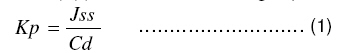
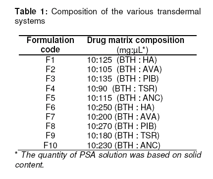
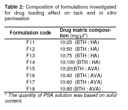
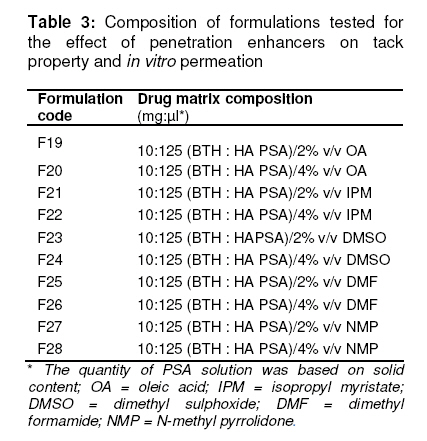
adhesives (PSAs) including acrylics, polyisobutylene and styrenic rubber solution. Keywords: Meniere’s syndrome, Transdermal delivery, Betahistine, Pressure-sensitive adhesives, Penetration enhancers. INTRODUCTION Betahistine (BTH) is an orally active histamine analogue known to improve the microcirculation of labyrinth, resulting in reduced endolymphatic pressure, and is therefore, used in diseases characterised by impaired peripheral circulation (Meniere’s syndrome). Peroral administration exhibit extensive first-pass metabolism and gastric irritation in patients suffering from peptic ulcer. As the disease is mostly observed in elderly patients, frequent dosing due to short biological half life may lead to noncompliance [1,2]. Transdermal drug delivery (TDD) can overcome these problems and improve treatment protocols by maintaining plasma levels over the dosing interval. Amongst various approaches to formulation development, drug-in-adhesive represents the simplest formulation design whereby a pressure-sensitive adhesive (PSA) not only fulfils the adhesion-to-skin function but also serves as a formulation matrix incorporating the drug and all the excipients. Commonly used PSAs include polyisobutylenes (PIB), silicones, acrylics and styrenic rubber solution [3]. Since only a few drugs can be administered percutaneously due to their low permeability, the formulation strategy often involves incorporation of chemical permeation enhancers. Penetration enhancers generally act by partitioning into the skin and interacting with different skin constituents to elicit temporary and ideally reversible reduction of barrier properties. However, permeation rate, PSA compatibility and skin adhesion must be considered before selection of an enhancer [4,5]. In the present study, an attempt was made to develop an adhesive-type TDD system for BTH using five PSAs of varying functionalities. The effects of penetration enhancers on the tack and in vitro permeation of the formulations were also evaluated. The most suitable formulation was further assessed for in vivo patch adhesion and preliminary stability. EXPERIMENTAL Materials Betahistine dihydrochloride was received as a gift from Geno Pharma, Goa, India. HPLC grade tetramethyl ammonium hydrogen sulphate and sodium heptane sulphonate (Merck Chemicals, Mumbai, India); HPLC grade methanol (Ranbaxy Fine Chemicals, New Delhi, India); N-methyl pyrrolidone (NMP), Isopropyl myristate (IPM), Dimethyl sulphoxide (DMSO), Dimethyl formamide (DMF) and Oleic acid (OA) – all from Lobachem, Mumbai, India -were purchased. Pressure-sensitive adhesives Duro Tak® 3872287A (a non-curing PSA containing 50.5 % acrylate-vinylacetate and with hydroxyl functionality, AVA), Duro Tak® 87-202A (a self-curing PSA containing 40 % hydrophilic acrylate and with hydroxyl functionality, HA), Duro Tak® 87-901A (an acrylic non-curing PSA containing 44 % solid content and without any functionality, ANC), Duro Tak® 87-608A (polyisobutylene rubber solution containing 38 % solid content, PIB), and Duro Tak87-611A (tackified styrenic rubber solution containing 57.5 % solid content, TSR) were all received (as organic solvent solutions) as gifts from National Starch and Chemicals Ltd, USA. CoTran9720 polyethylene backing film and Scotch pack9741 SBOPP release liner were received as gifts from 3M, USA. All other chemicals were used as received, without further purification/treatment. Preparation of betahistine (BTH) base and selection of PSABTH free base was liberated from its dihydrochloride salt by neutralization with strong sodium hydroxide solution followed by extraction with chloroform. The resultant extract was dried at room temperature to remove excess chloroform, leaving a viscous, dark yellow liquid [6]. Since selection of PSA matrix is a very important step in the design of adhesive-type TDD [4,7], it was necessary to select appropriate PSA by testing the miscibility of BTH with the PSAs under consideration (i.e., AVA, HA, ANC, PIB and TSR). Various formulations containing different ratios of BTH and PSAs were prepared and the resultant patches were subjected to microscopic examination. Miscibility was studied throughout the whole patch using a light microscope (Olympus camera SP-350, model CX 31, with Magnus PRO 3.0 image analysis software, 40 x magnification). BTH TDD was prepared by solvent evaporation method and fabricated by adhesive transfer technique. BTH (10 mg) was dissolved in sufficient quantity of chloroform and added to PSA solution. The proportion of BTH and PSA was varied (see Table 1 and 2) to obtain formulations F1 to F18. To formulations F19 to F28, various enhancers, namely, oleic acid (OA), isopropyl myristate (IPM), dimethyl sulphoxide (DMSO), dimethyl formamide (DMF) and N-methyl pyrrolidone (NMP) were added in different concentrations (see Table 3). The adhesive matrix was formed by casting the mixture on a release liner, using a casting knife to achieve the desired film thickness. It was set at room temperature for 20 min and subsequently oven-dried at 60 0C for 30 min to remove residual organic solvents. The dried film was then laminated onto a backing film using a standard 2 kg roller (Sri Sai Precision Instruments & R. C., Nasik, India) [8,9]. Evaluation of the effect of drug loading on tack properties and in vitro permeation Based on miscibility study, only adhesives with alcohol functionality (HA and AVA) were considered suitable for the preparation of the adhesive-type TDD system incorporating the drug. Since increase in drug loading is generally associated with increase in flux [4], further formulations containing higher BTH loading were prepared, as in Table 2. Tack parameter for all the formulations was subjectively evaluated by pressing a thumb briefly on the matrix [10]. Two formulations selected (F1 and F2), which showed adequate tack, were evaluated for in vitro permeation across guinea pig skin. Formulation F12 was also assessed to determine the effect of increase in drug loading on BTH permeation. In vitro skin permeation experimentThe animal experiments were approved by Institutional Animal Ethics Committee (IAEC) of Government College of Pharmacy, Aurangabad, India (ref. no. GCPA/IAEC/ 2008/330) and carried out as per the guidelines of the committee. Guinea pigs were housed in stainless steel cages and maintained under standard condition (12 h light/dark cycle; 25 ° C, 45 % RH). The animals were fed normally with green leafy vegetables and nutritional grains. They also freely received water and were acclimatized to laboratory conditions for one week before commencement of the experiment. The day before the experiment, hairs from the abdominal region of the guinea pigs were removed by a hair removing spray (Smooth skin, Regrace Ink, Mumbai). On the next day, full-thickness abdominal skin was excised and subcutaneous fats removed with scissors and scalpel. The excised skin was mounted on a modified Keshary Chien diffusion cell. Phosphate buffer (pH 7.4) was used as the receptor medium (15 ml, maintained at 32 ± 1 ° C) and it was stirred continuously with a magnetic stirrer (Whirlmatic-Mega, Spectra lab). The transdermal patch (1 cm2 ) was applied to skin on the donor side of the diffusion cell. Aliquots of 1 ml were withdrawn from the receptor medium at sampling intervals of 1, 2, 3, 6, 12, 24, 48 h and sink condition was maintained by replenishing the medium with 1 ml of phosphate buffer. The withdrawn samples were centrifuged (Spinwin, Tarsons) for 10 min. at 1000 rpm [1] and analysed by high performance liquid chromatography (HPLC) as described below. The experiment was performed in triplicate. HPLC determinationBTH was assayed by an in-house developed and validated RP-HPLC method. The HPLC system (Dionex, Germering, Germany) consisting of Chromeleon 6.70 acquisition software equipped with P680 HPLC pump, ASI-100 automated sample injector and UVD 170 UV detector. HPLC column Micra-NPS RP18 (length × OD × ID, 33 × 8.0 × 4.6 mm, 1.5 µm), was used. Isocratic elution was performed using a mobile phase consisting of a pH 3.5 buffer (20 mM tetramethyl ammonium hydrogen sulphate and 9 mM sodium salt of heptane sulphonic acid) and methanol in a ratio of 97:3 with a flow rate of 0.4 ml/min. A sample solution (05 µl) was injected into the column and analysed at a wavelength of 260 nm. Cumulative amounts of BTH (µg) permeated per unit area were plotted against time. Lag time (tL, h) was determined by extrapolating the linear portion of each curve to time axis. Steady state flux (Jss, µg/h/cm2) was calculated from the slope while permeability coefficient (Kp) was calculated using Eq 1.
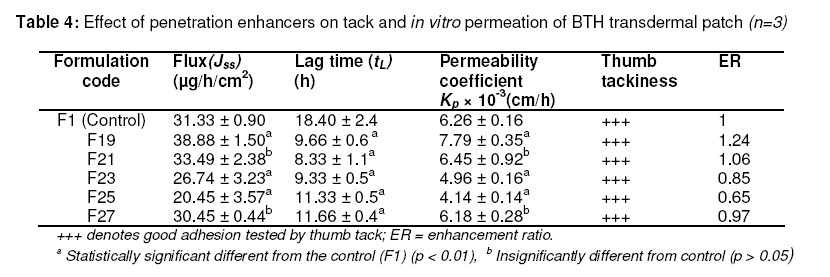
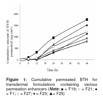
where Cd denotes the concentration of BTH in the donor cell [11,12]. Evaluation of the effect of penetration enhancers on tack property and in vitro permeationSince formulation F1 provided the highest flux as well as optimum tack, it was necessary to further increase the permeation rate using suitable penetration enhancers. Five different enhancers, viz, NMP, IPM, DMSO, DMF or OA were added to F1 at concentrations of 2 and 4 %w/w (see Table 3). The miscibility of enhancers with adhesive solution was tested by a previously reported method [13]. All the formulations were also evaluated by thumb tack test. The selected formulations (F19, F21, F23, F25 and F27) were then subjected to in vitro permeation study across guinea pig skin and enhancement ratio (ER) was calculated as the ratio between BTH flux from formulations with and without enhancers. In vivo patch adhesion performance test and stability studiesSince in vitro conditions do not necessarily represent the performance of TDD systems under in vivo conditions, the adhesion performance of the developed formulations was tested in vivo on guinea pig skin [14]. Also, since the transdermal system was intended to be applied over a period of at least one day based on lag time considerations, 5 cm2 patch of F19 was applied to the guinea pig skin and observed every 12 h for 36 h. Patch adhesion performance to skin was scored from 0 to 4 with 0 indicating that the patch was stuck firmly to the skin and 4 indicating that the patch completely peeled off the skin. Samples of F19 were wrapped in an aluminium foil, pouched in a self-sealing polybag and subjected to a 3-week accelerated stability condition by storing at 50 ° C in an oven. Stability was evaluated in terms of tack and in vitro permeation profile. Statistical analysis The data were analyzed using GraphPad InState Demo (version 3.10) and Microsoft Excel 2007. The permeation parameters obtained were compared by one-way analysis of variance (ANOVA). Tukey-Kramer test and Dunnett test were performed for multiple comparisons amongst the different formulations, respectively. The probability level, p < 0.001, was considered statistically significant. RESULTS Selection of PSA matrix for development of drug-in-adhesive TDD Microscopic examination of F1, F2, F6, and F7 showed complete miscibility of BTH with two of the PSAs (HA and AVA). Prominent yellow-coloured spots in F3, F4, F5, F8, F9 and F10 indicated immiscibility of BTH in ANC, PIB and TSR. Effect of drug loading on in vitro permeation and tack propertiesMaximum drug loading in HA and AVA was 1:2 and 1:5, respectively. Increased drug loading in HA reduced tack significantly. Significant differences were observed between flux through F1, F2 and F12 formulations (p < 0.001). F1 provided significantly higher flux (31.33 ± 0.90µ g/h/cm2) than F2 (4.27 ± 1.05 µg/h/cm2). F12, containing high BTH loading, showed significantly higher flux (39.24 ± 1.02µ g/h/cm2) than F1. However, significant loss of adhesion property was observed with increase in drug loading for F2 and F12. Effect of penetration enhancers on tack property and in vitro permeation All the five enhancers evaluated for miscibility dissolved homogenously in the adhesive solution. Thumb tack test data indicate that F20, F22, F24, F26, and F28 showed significant loss of adhesion property. Since these formulations (which contained high levels of enhancers) exhibited extremely poor tack, they were not considered suitable for the in vitro skin permeation studies. Formulations F19, F21, F23, F25 and F27 exhibited good adhesion. Microscopic examination did not show any evidence of immiscibility, separation or oozing out of the enhancers. Their in vitro permeation data are shown in Table 4 while the cumulative amount of BTH permeated per unit area for these formulations are compared in Figure 1. F1 (without enhancer) served as control. F19 containing OA as an enhancer exhibited the highest flux with an enhancement ratio 1.24 which was significantly different from that of the control (F1, p < 0.01). F21 and F27 containing 2 % IPM and NMP showed enhancement ratio of 1.06 and 0.97, respectively. This was not significantly different from that of the control. However, significant reduction in lag time was noted for F21 and F27 (p < 0.01). Enhancers DMSO and DMF (at a level of 2 %) produced significant reductions in flux, permeability coefficient and lag time compared with control (p < 0.01). In vivo patch adhesion and stabilityIn the in vivo adhesion test on the most suitable formulation (F19), an estimated 90 % of the patch area remained adhered to the skin and was, therefore, scored 0 as it did not peel off the guinea pig skin for a period of 36 h. Stability evaluation of F19 showed steady state flux of 37.3 ± 1.2 µg/cm2/h with a lag time of approx. 10 h. Statistically, no significant difference (p > 0.05) was observed between flux values before and after the storage period. DISCUSSION PSA matrix, drug loading, permeation and tack properties A suitable PSA is necessary in the design of TDD to ensure adequate drug release and intimate contact of the transdermal patch with the skin. However, these polymeric adhesives exert their own effect on the solubility and permeability of the drug incorporated. The acrylic adhesives, HA and AVA (which have a hydroxyl functional group) showed complete miscibility with BTH which might be due to the chemical nature of these PSAs. The hydrophilic acrylic component of HA might have enhanced the solubility of BTH in line with the reported effect of HA on the solubility of hydrophilic drugs [15]. It has also been reported that the chemical nature of a transdermal adhesive can affect the solubility of the drug in the PSA matrix [16]. Jeans et al demonstrated that adhesives containing acidic side-chains are not compatible with the basic form of primaquine base; on the other hand, adhesives containing OH group produced homogenous and stable blends, possibly due to hydrogen bond formation with primaquine base [9]. Similar observations were reported for granisetron base in that while adhesives comprising electronegative groups such as COOH groups could not be used to manufacture satisfactory transdermal patches of granisetron free base, an adhesive containing an OH functional group (Duro Tak® 387-2287A) was significantly better than non-nucleophilic, electroneutral adhesives [17]. When tested for in vitro permeation, F1 provided significantly higher flux than F2. Also, F12, which had high BTH loading, showed higher flux than F1; this supports the assumption that increase in drug loading increases drug permeation. Subsequently, however, there was a significant loss of adhesion by F12. Overall, drug loading, permeation and tack data suggest that HA was the most suitable PSA and was therefore employed for further evaluation. Furthermore, these results suggest that the chemical nature of PSA need to be considered before selecting an adhesive matrix (PSA). Effect of penetration enhancers on tack property and in vitro permeationTo develop matrix-type transdermal delivery system for a drug, an appropriate vehicle is often required to enhance the permeation rate and/or to increase the solubility of the drug in the adhesive. A vehicle can also act as a plasticizer in the adhesive, increasing the mobility of the drug in the adhesive. To further increase the permeation rate of BTH and to reduce the lag time, the effect of some vehicles on the permeation of BTH from HA matrix was investigated. The amount of each vehicle tested was 2 and 4% of the weight of acrylic adhesive polymer, respectively. Since 4% weight of permeation enhancer caused significant loss of adhesive force, the permeation rate was not determined for these formulations. Incorporation of OA into the acrylic adhesive matrix significantly enhanced permeation rate and shortened lag time. The enhancement effect of OA may be explained by the formation of permeable interfacial defects within the stratum corneum lipid bilayers which effectively decreased permeation resistance without necessarily invoking the formation of pores [18]. This might have increased the BTH permeation from HA matrix. The reported mechanism of permeation enhancement of IPM is by fluidization of intercellular lipids [19] whereas NMP rely on improving drug partitioning into the skin to promote permeation [20]. However, no such enhancing effects by these solvents were observed when tested for in vitro permeation of BTH across guinea pig skin. The permeation enhancement mechanism of sulphoxides and formamides (e.g., DMSO and DMF, respectively) is reported to be complex. They effectively promote permeation by reducing skin resistance to drug molecules or by promoting drug partitioning in the delivery system [22,22]. In the present work, DMSO and DMF also did not produce any significant increase in BTH permeation. Overall, 2 % OA proved to be the most effective and suitable penetration enhancer for HA -based transdermal BTH formulation since it resulted in the highest flux across the guinea pig skin while also showing the shortest lag time. In vivo patch adhesion and stabilitySeveral in vitro techniques, such as peel adhesion, tack and shear strength, have been used to monitor the adhesive performance of patches in vivo [23]. However, there is lack of evidence for a relationship between adhesion results obtained in vitro and those generated in vivo TDD systems [24]. F19 possessed adequate tack properties and remained adhered to skin for 36 h, a long enough period for TDD. The formulation was also stable for 3 weeks at 50° C, an important requirement for adhesive systems Previously reported bioavailability studies of BTH indicate that maximum plasma concentrations after oral dosings (50 mg/kg) to dogs were 2.6 -4.8 ng/ml [1]. After a single dose of 24 mg betahistine mesylate to 20 healthy Chinese male volunteers, the plasma concentration of the parent drug was less than 0.5 ng/ml [2]. Hence, the effective plasma concentration in man (oral dose 8-16 mg once) would be at ng/ml level. Although the effective plasma concentration range of BTH in man has not been evaluated, the overall results of the present study suggest that BTH reasonably permeated guinea pig skin. CONCLUSIONAcrylic acid-derived PSA containing hydroxyl functionality provided the most suitable matrix for BTH. The optimized formulation, F19, provided the highest flux with a lag time of approx 10 h. This patch showed adequate tack. These attributes warrants that further evaluation of this betahistine adhesive-type transdermal delivery system should be carried out. ACKNOWLEDGMENTThe authors are thankful to Geno Pharma, Goa, India for providing betahistine dihydrochloride free of charge. Thanks are also due to National Starch and Chemicals Ltd and 3M, both of USA, for generous provision of the PSAs and the other formulation components, respectively. Aravind R Sonawane is grateful to All India Council of Technical Education for providing financial assistance for the work. REFERENCES
Copyright 2010 - Tropical Journal of Pharmaceutical Research The following images related to this document are available:Photo images[pr10062t4.jpg] [pr10062t3.jpg] [pr10062t1.jpg] [pr10062t2.jpg] [pr10062f1.jpg] |
| |||||||||
