 |
| About Bioline | All Journals | Testimonials | Membership | News |
|
||||||
|
||||||
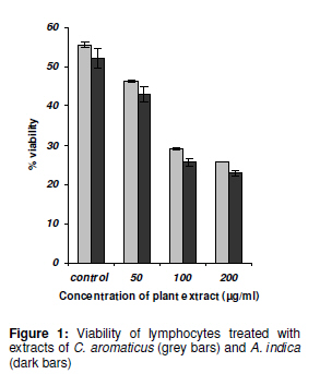
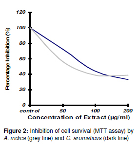
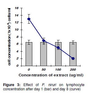
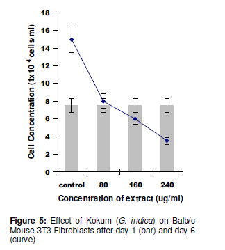
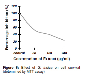
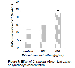
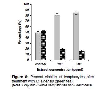
Research Article *Corresponding author: E-mail: varalakshmikn@yahoo.co.in, varalakshmikn.cpgs@jaingroup.info; Tel: +91-8043226500; Fax: +91-80-43226500 Received: 3 May 2010 Revised accepted: 2 December 2010 Code Number: pr11006 Abstract Purpose: To evaluate, using ethnomedical data approach, five Indian plants used in traditional medicine for cancer and other diseases for their safety and cytotoxic activity on human lymphocytes.Methods: The antiproliferative effect of the 50, 100 and 200 µg/ml aqueous extracts of five plants (leaves of Phyllanthus niruri, Coleus aromaticus, Azadirachta indica and Camellia sinensis, and dried fruit rind of Garcinia indica) were evaluated in vitro using trypan blue dye exclusion and clonogenic assay methods on two cell lines -Balb/c 3T3 mouse fibroblasts and human peripheral lymphocytes. Results: Among the five plants used traditionally to treat cancer and other infections, two of them (A. indica and C. aromaticus) exhibited cytotoxic effects on lymphocytes. Two other plants (G. indica and P. niruri) exhibited pronounced cytotoxic effects. Another plant (Camellia sinensis) exhibited cytostimulatory effect (> 50 % cell proliferation). For the plants that are traditionally used in anticancer therapy, there was a correlation between the reported use of these plants and their cytotoxic activity on lymphocytes. Conclusion: The plant extracts of the leaves of P. niruri, C, aromaticus and A. indica, and the dried fruit rind of G. indica are cytotoxic to lymphocytes and this lends some credence to their traditional use for cancer treatment. However, green tea (C. sinensis) is cytostimulatory and safe for consumption. Keywords: Balb/c mouse fibroblasts, Cytostimulatory, Cytotoxic, Lymphocytes, Traditional medicine. Introduction The medicinal use of plants is probably as old as mankind itself. Plants have continued to be a valuable source of natural products for maintaining human health, as studies on natural therapies have intensified. More than 150,000 plant species have been studied, and several of them contain therapeutic substances [1,2]. The use of plant compounds for pharmaceutical purposes has gradually increased. According to the World Health Organization [3], medicinal plants are probably the best source of a variety of drugs. About 80 % of individuals in developed countries use traditional medicine containing compounds derived from medicinal plants [4]. The use of plant extracts and phytochemicals can be of great significance in therapeutic treatments [5]. However, medicinal plants, and indeed plants in general, synthesize toxic substances which in nature act as a defense against infections, insects and herbivores but which often affect the organisms that feed on them. Thus, an assessment of their cytotoxic potential is necessary to ensure relatively safe use of medicinal plants [6]. The species Azadirachta indica (neem), Phyllanthus niruri, Garcinia indica and Coleus aromaticus (doddapatre) are examples of plants commonly used in popular medicine. Neem has been successfully used for centuries to reduce tumors by herbalists throughout Southeast Asia. [7]. Phyllanthus niruri has been found to exhibit marked inhibitory effect on hepatitis B virus which is evident by its wide application in cases of chronic jaundice. Phyllanthus niruri Linn has been used in Ayurvedic medicine for over 2,000 years in the therapy of a wide range of ailments such as jaundice, gonorrheoa, ‘frequent menstruation’ and diabetes, as well as topically as a poultice for skin ulcers, sores, swelling, and itchiness [8]. The plant is said to play a role in liver disorders due to its febrifuge, antiseptic, astringent, stomachic, deobstruent and diuretic actions [9] and has also been used to correct GIT problems such as dyspepsia, colic, diarrheoa and dysentery, and to tone the GIT tract back to functionality [10]. The young shoots of the plant are administered in the form of an infusion for the treatment of chronic dysentery [11]. Garcinol, a polyisoprenylated benzophenone from the Garcinia indica (kokum) fruit rind, has been claimed to be an anti-inflammatory and anticancer agent [12]. Rao et al [13] reported the antioxidant, anticlastogenic and radioprotective effect of Coleus aromaticus on Chinese hamster fibroblast cells (V79). The use of plant compounds for pharmaceutical purposes has gradually increased. Such plants should be investigated to better understand their properties, safety and efficiency. The objective of this study was to investigate five plants for acute cytotoxicity in vitro on lymphocytes from human peripheral blood. The plants evaluated were Azhadiracta indica (neem), Garcinia indica (kokum), Phyllanthus niruri, Camellia sinensis (green tea) and Coleus aromaticus (doddapatre). Fresh plant materials were collected from Lalbagh, Bangalore, India during the month of March 2009, and identified by Professor SB Sullia. A voucher specimen (no. JUH 125) was deposited at the herbarium of Jain University Research Center, Bangalore, India. The leaves of the plants and the fruit rind of Garcinia indica were shade-dried and coarsely powdered with a grinder. Cell lines Balb/c Mouse 3T3 fibrobalst was procured from National Center for Cell Sciences (NCCS), Pune, India while human peripheral lymphocytes were isolated from peripheral blood in our laboratory. Treatment of lymphocytes with plant extracts The flasks containing lymphocytes were inoculated with either 50, 100 or 200 µg/ml of the plant extracts. One flask was kept as control. All the treatments were performed in triplicate and the mean (± standard error) was calculated in each case. The flasks were labelled appropriately and kept in a CO2 incubator at 37 0C for one week and monitored regularly during the period. Thereafter, the cells were counted with a haemocytometer and cell viability determined by trypan blue dye exclusion method. In this procedure, an aliquot of the cell suspension (500µl) was mixed with 500µl of 0.4 % trypan blue (in phosphate buffered saline). This mixture (20µl) was introduced into a haemocytometer and observed under a light microscope. Viable cells were transparent and dead cells were blue. Antiproliferation test on Balb/c 3T3 mouse fibroblasts The fibroblasts were grown in 25 cm2 flasks containing DMEM with 10 % foetal bovine serum and streptomycin (1 %) and penicillin (1 %) in a 5 % CO2 incubator at 37 °C. The cells were harvested from the flasks after brief trypsinization which was carried out as follows[2]. The culture medium was removed and the cells were washed in a balanced salt solution. Thereafter, 2ml of trypsin-EDTA solution was added to the cells and the vessel placed in the incubator for 2 min. The cells were observed under a light microscope. Clonogenic assays using logarithmically growing cells were performed according to the published methods [16–17]. Briefly, approximately 400 cells obtained from subconfluent culture flasks were seeded per tissue culture dish in 5 ml of medium (five dishes per point). Twenty-four hours after seeding the cells, test compounds were added (at two selected concentration levels) to the medium. Control cultures received only solvent in the place of test chemical. Dimethyl sulphoxide (DMSO) served as the vehicle to solubilize compounds at a final concentration of 0.4 %v/v in the culture medium. Preliminary experiments showed that 0.4 %v/v DMSO has no effect on cell survival. The dishes were returned to the incubator for 7 to 8 days and surviving cells were allowed to form colonies. When the colonies became discrete and well defined, the dishes were washed with PBS solution, fixed with methanol, stained with Giemsa and allowed to dry. The number of colonies per dish was counted. The assays were carried out in >triplicate. MTT [3-(4,5-dimethylthiazol-2yl)-2,5diphenyl tetrazolium bromide)] assay MTT assay was performed to assess the cytotoxicity of the plant extracts. (MTT is an yellow dye which is reduced into purple colored formazan crystals by the activity of mitochondrial succinate dehydrogenase enzyme in viable cells). The cells were cultured in 96-well microtitre plates, treated with varying concentrations of different plant extracts for 6-7 days and incubated. At the end of the treatment period, 20µl of MTT was added to each well and the plates incubated (C. aromaticus) or neem (A. indica) fell as the concentration of the extracts increased (Fig 1). Treatment with 100 µg/ml of either of these two extracts resulted in 50 % inhibition of the cells (Fig 2). 60 Statistical analysis The results are presented as mean ± standard error (SE). Statistical significance was determined using one-way analysis of variance (ANOVA) and Student’s t-test. P < 0.05 was set as the level of significance. RESULTS Effect of plant extracts on cell viability The effect of the plant extracts on cell viability is illustrated in Figs 1 – 4. The viability of A. indica (grey line) and C. aromaticus (dark line) lymphocytes treated with either doddapatre (C. aromaticus) or neem (A. indica) fell as the concentration of the extracts increased (Fig.1.). Treatment with the 100ug/ml of either of the two extracts resulted in 50% inhibition of the cells (Fig 2). Figure 2: Inhibition of cell survival (MTT assay) by A. indica (grey line) and C. armocaticus (dark line) indica fruit rind extract, cell concentration also decreased as extract concentration increased (Fig 5). Cell concentration declined to nearly 50 % of the control after 6 days when treated with 80 µg/ml of the extract while the 240 µg/ml extract produced nearly 80 % inhibition within the same period (Fig 6). Discussion A. indica (neem) extract treatment resulted in decreased cell concentration in normal lymphocytes. According to Roy [18], treatment of leukemia and melanoma cells with 0.5 -5.0 µM concentration of nimbolide resulted in moderate to very strong growth inhibition in human leukaemic lymphoma (U937), human leukemia (HL-60 and murine melanoma (B16) cell lines.The ethanol extract of neem has been shown to cause death of prostate cancer cell lines [19]. Nimbolide, a triterpenoid isolate from the flowers of the neem tree, exhibits antiproliferative activity against some cancer cell lines [19] although their side effects on prolonged usage were not reported. In the present work, we found that neem extract was cytotoxic to normal lymphocytes just like other chemotherapeutic agents at higher concentrations. Our results indicate that they exerted deleterious side effects on normal lymphocytes. The extract of C. aromaticus also lowered cell concentration. Rao et al [13] reported the antioxidant, anticlastogenic and radioprotective effects of this extract on Chinese hamster fibroblast cells (V79) exposed to gamma radiation but there was no data on their effects on normal healthy cells. However, our results show that the extract is cytotoxic at high doses. P. niruri extract was also cytotoxic to normal peripheral human lymphocytes thus indicating that cytotoxicity was probable upon continued and unregulated consumption at high doses. Literature reports indicate that this extract primarily contains lignans (e.g.phyllanthine and hypophyllanthine), alkaloids, and bioflavonoids (e.g., quercetin) [8]. Phyllanthus blocks DNA polymerase, the enzyme required for hepatitis B virus (HBV) to reproduce. It has also been found to inhibit the DNA polymerase of HBV and to bind to HbsAg in vitro [20]. These earlier workers cautioned against the indiscriminate use of this extract by the populace. G. indica fruit rind extract reduced the cell concentration and viability of Balb/c mouse fibroblasts. These results are in agreement with those of Hong et al [12] who investigated the effect of an isolate of this plant, garcinol and its oxidative derivatives, cambogin, garcim-1, and garcim-2 on the growth of HT- 29 and HCT-116 colon cancer cells, as well as on IEC-6 and INT-407 normal immortalized intestinal cells. Garcinol and its derivatives showed potent growth-inhibitory effects on all intestinal cells with IC50 of 3.2 – 21.4 μM after a 3-day treatment. Yamiguchi[21] reported that orally administered garcinol prevented acute ulceration induced with indomethacin in rats and water immersion stress caused by radical formation. The results of Liao et al [22] suggest that garcinol has neuroprotective effects. Several epidemiologic studies have suggested an association between green tea (C. sinensis) consumption and reduced risk of different kinds of human cancer [23-25]. Hsu et al [26], tested exponentially growing and aged primary human epidermal keratinocytes in response to EGCG or a mixture of the four major green tea polyphenols. EGCG elicited cell differenttiation and proliferation with associated induction of p57/KIP2 within 24 h in growing keratinocytes. However, the effect of green tea on normal lymphocytes of the human body has not, to the best of our knowledge, been investigated in cell culture systems until now. Our data indicate that it promotes the proliferation of lymphocytes. Of the herbal extracts tested on normal healthy lymphocytes, this plant (green tea) is the only one which was not cytotoxic to lymphocytes. This property should be further explored for the treatment of cell deficiencies of lymphoid origin. Interestingly, this finding supports the reported benefit of green tea consumption in reducing the risk of different types of human cancer. Conclusion The findings of our study do not support the routine consumption of the plant extracts evaluated except for C. sinensis (green tea). Should they, however, be consumed, it is recommended that the extracts should be prepared exactly according to the recipe and taken in the prescribed doses, bearing in Acknowledgement The authors are grateful to the management of Jain Group of Institutions for the financial assistance provided to carry out the work. References
© Pharmacotherapy Group, Faculty of Pharmacy, University of Benin, Benin City, 300001 Nigeria. The following images related to this document are available:Photo images[pr11006f7.jpg] [pr11006f1.jpg] [pr11006f2.jpg] [pr11006f5.jpg] [pr11006f4.jpg] [pr11006f6.jpg] [pr11006f8.jpg] [pr11006f3.jpg] [pr11006f9.jpg] |
| |||||||||
