 |
| About Bioline | All Journals | Testimonials | Membership | News |
|
||||||
|
||||||
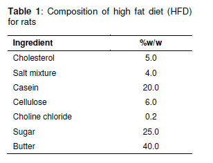
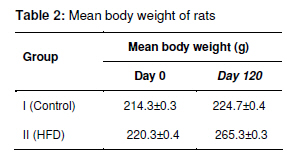
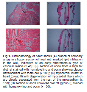
Tropical Journal of Pharmaceutical Research February 2011; 10 (1): 41-45 Research Article Role of C-Reactive Protein in the Development of Atherosclerosis in Diet-induced Lipidemia in Albino Rats RP Kalsait1, PB Khedekar2, AN Saoji2 and KP Bhusari1 1Sharad Pawar College of Pharmacy, Wanadongri, Hingna Road, Nagpur – 441 110, 2University Department of Pharmaceutical Sciences, Amravati Road, Nagpur, India *Corresponding author: E-mail: kalsait.ravi@gmail.com; Tel: (07104) 236352, 9960099852; Fax: (07104) 235084 Received: 2 July 2010 Revised accepted: 12 December 2011 Code Number: pr11007 Abstract Purpose: Blood C-reactive protein level serves as a useful biomarker of generalized injury and inflammation. Vast amounts of clinical data suggest its serum level predicts risk of cardiovascular disease. In the present study, the role of C-reactive protein in the development of atherosclerosis in albino rats was investigated. Introduction There is increasing evidence that complement activation may play an important role in atherogenesis. Thus activation products of the complement cascade have been demonstrated in atherosclerotic lesions of experimental animals and humans. Creactive protein (CRP) is the prototype acutephase protein in humans and numerous animals and it is widely used as an indicator of the activity of bacterial infection and various other diseases. In the acute-phase response, its plasma concentration can be elevated up to 500 times its normal level [1]. It is a marker of inflammation, and is synthesized mainly in the liver and regulated by circulating interleukin (IL-6). Emerging evidence suggests that elevated plasma levels of CRP have become one of the strongest independent predictors of coronary heart disease [2,3]. Mild elevations in CRP even when within the clinically ‘normal’ range are independently predictive of future cardiovascular events [4,5]. CRP directly participates in the process of atherogenesis by modulating endothelial function and its concentration known to predict cardiovascular events [6]. Recently, elevated level of CRP has been associated with the feature of the insulin resistance syndrome [7]. For clinical purposes, it is the most promising inflammatory biomarker, a classical acute-phase marker and a member of the pentraxin family of innate immune response protein [8]. It has a long half-life, affording stability of levels with no observable circadian variation [9]. Further, it is easily measured in usual outpatient settings, and standardized high sensitivity assays commercially available provide similar results in fresh, stored, and frozen plasma [10]. In this study, atherosclerosis was induced in animals using high fat diet. The main objective was to assess the role of CRP in the development atherosclerosis in albino rats. Experimental Animals Adult male albino rats of Wistar strain, weighing between 180 and 250 g, were used for the study. They were procured from the National Institute of Nutrition, Hyderabad, India. Approval of the institutional animal ethical committee was obtained for the animal studies and Committee for the Purpose of Control and Supervision of Experiments on Animals (CPCSEA) guidelines were followed throughout the experiment. The animals were housed in polypropylene cages at 25 ± 2º C and 12h/12h light/dark cycle, and fed with standard pellet diet and water ad libitum. The animals were divided into two groups of 12 rats each. Group I rats served as control and were fed normal pellet diet. Group II was fed on high fat diet (HFD) for 120 days to develop atherosclerosis. The composition of high fat diet is depicted in Table 1. Next, i.e., after 120 days, the rats were fasted for 12 h before collection of blood samples. Each rat was anesthetized with diethyl ether and blood was withdrawn from the retroorbital plexus, transferred to a centrifuge tube and allowed to clot. The separated serum was assayed for C-reactive protein by latex agglutination method [11] using Accucare creactive protein reagent latex test diagnostic kit (Labcare Diagnostics India Pvt Ltd). Total cholesterol, HDL, VLDL, and LDL were assayed using Span diagnostic kits. Histopathological studies The animals were sacrificed, the heart and aorta were removed as quickly as possible, fixed in 10 % formalin and prepared for histological examination [12]. The tissues were embedded in gelatin to prepare frozen sections using microtome. The aortae were first stained (gross staining) and examined for lesions with the aid of a magnifying lens. They were then cut longitudinally on the microtome, mounted and examined for lesions under a light microscope. The frozen sections of the aortae were stained with oil red O in triethylphosphate. Heart and frozen sections of the tissue were embedded in paraffin and stained routinely with hematoxylin and eosin [13-15]. Statistical analysis Data are expressed as mean ± SEM. Significant difference between means was tested by Students t-test using GraphPad Prism software, version 5.0. Differences were considered statistically significant at p < 0.001. Results As Table 2 shows, the animals on HFD showed a more rapid increase in weight than control. Though not quantified, most of them showed varying degrees of hair loss. The weight gain was significantly higher for group II. The consumption of diet by the animals was 15 to 20 g/day. C-reactive protein and lipid profile data are shown in Table 3. It is clear from the data that in all the lipid determination, there was a marked difference between the group I (control) and group II (HFD) group. Total cholesterol increased about 5-fold from an average normal level in group II after 120 days. The LDL, VLDL Cholesterol showed significant difference, an increase of about 4fold in group II as compared to control group I. Triglyceride concentration in HFD group II showed significant increase in its level. On the other hand, HDL cholesterol which is good cholesterol showed decrease in its level in group II. In this study, HFD group II showed marked increase in lipid profile but the concentration of C-reactive protein observed within normal range even after 120 days. Discussion Rats, which usually have omnivorous habits, but in which atherosclerosis has been experimentally induced, has been of great interest [16,17]. Induction of atherosclerosis in albino rat using a diet that included cholesterol, sodium cholate, and thiouracil has been reported by Fillios as well as by Hartroft and Thomas. The latter added a large amount of butter to the basic diet used by Fillos in their study and reported a high percentage of thrombosis and myocardial infarction in albino rats [18,19]. The development of atherosclerosis is a complex multi-factorial process involving a variety of environmental and genetic interactions. The data obtained in the present study allowed us to address the question of whether the lipid profile and the period of exposure to high fat diet are indeed important determinants of atherosclerotic lesion and whether c-reactive protein played a role. Our data show that exposure of the animals to high fat diet led to the development of atherosclerosis as evidenced by the lesion found in the intima and also the significantly increased serum lipid levels in group II (HFD) animals. However, the level of c-reactive protein in serum was within normal range. C-reactive protein has been recommended as the marker of choice to monitor cardiovascular risk, being a stronger predictor of atherosclerosis than even plasma LDL concentration. It is postulated that as an acute-phase protein, elevation of plasma CRP may signal underlying atherosclerosis process [20]. Fig 1A shows lipomatous changes in group II (HFD) animals, which consisted mostly of extracellular lipid infiltrations of the coronary vessels without proliferation. Fig 1B shows atheromatous lesion in the aorta of group II animals. In Fig 1C, the myocardial infarcts observed (group II animals) were small and irregular in outline. These infarcts were characterized by degeneration of myocardial fibers which are clearly separated from the rest of the myocardium. Fig 1D shows normal aorta (group I) with no cardiovascular lesions encountered. Sun and colleagues used well established animal atherosclerosis models, i.e., both cholesterol fed and Watanabe heritable hyperlipidemic rabbits, to study the role of CRP in atherogenesis and generated elaborate results. First, CRP level was significantly elevated in hypercholesterolemic rabbits. Second, elevated CRP levels strongly correlated with the extent of atherosclerosis in these animals [21]. These findings indicate the need for further investigation of human CRP concentration in cardiovascular disease. Conclusion We confirm that C-reactive protein did not play any role in the development of atherosclerosis in Wistar breed albino rat and therefore this animal model can’t be used to extrapolate C-reactive protein data. However, this animal model would useful for study of antihyperlipidemic drugs and atherosclerosis development. References
The following images related to this document are available:Photo images[pr11007t3.jpg] [pr11007f1.jpg] [pr11007t2.jpg] [pr11007t1.jpg] |
| |||||||||
