 |
| About Bioline | All Journals | Testimonials | Membership | News |
|
||||||
|
||||||
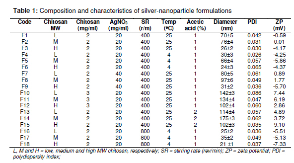
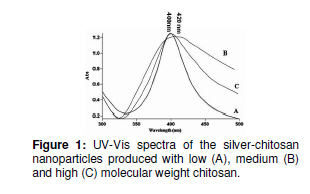
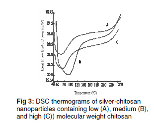
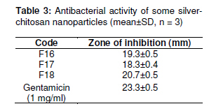
Tropical Journal of Pharmaceutical Research February 2011; 10 (1): 69-74 Research Article Preparation, Characterization and Antibacterial Properties of Silver-Chitosan Nanocomposites Using Different Molecular Weight Grades of Chitosan S Honary1*, K Ghajar1, P Khazaeli2 and P Shalchian2 1 Mazandaran University of Medical Sciences, School of Pharmacy& Pharmaceutical Sciences Research Center, Sari, Iran; 2Kerman University of Medical Sciences, School of Pharmacy, Kerman, Iran *Corresponding author: E-mail: shonary@yahoo.com, SHonary@mazums.ac.ir; Tel: +98 151 3543084, 151 3246491; Fax: +98 151 2224521 Received: 14 July 2010 Revised accepted: 10 January 2011 Code Number: pr11011 Abstract Purpose: To study the effect of chitosan molecular weight on the physicochemical and antibacterial properties of silver-chitosan nanoparticles. Introduction The antibacterial effects of silver (Ag) salts have been known for over 2000 years, but they have only been in common use as an antimicrobial since the 19th century [1]. Ag is currently used to control bacterial growth in a variety of applications, including dental work, catheters, and burn wounds [1]. Silver nitrate solution causes argyria (staining of the skin) and a burning sensation on application. Ag ions and Ag-based compounds are highly toxic to microorganisms. They show strong biocidal activity against as many as 12 species of bacteria including E. coli [2]. Today, other forms of silver are available which do not have the disadvantages of earlier solutions. Modern dressings are capable of delivering silver through a sustained-release mechanism. This helps to avoid toxicity and yet ensures delivery of a therapeutic dose of silver to the wound [3]. It has been shown that reducing the particle size of materials is an efficient and reliable tool for improving their actions; hence, silver nanoparticles in some modern dressings, such as Acticoat®, aids in burns management [4]. The antimicrobial properties of silver-containing wound dressings have been studied by a microcalorimetric technique. The results showed that this kind of dressing has the capacity to kill common wound pathogens -Staphylococcus aureus and Pseudomonas aeruginosa [5]. Different nano silver-coated barrier dressings were reported to exhibit antimicrobial activity and reduce infection in wounds [6]. The antibacterial activity of chitosan tripolyphosphate nanoparticles loaded with various metal ions was investigated by Du et al and the results showed that antibacterial activity was enhanced by the loaded metal ions [7]. An earlier work showed that accumulation of silver nanoparticles in bacterial membrane caused a significant increase in membrane permeability, resulting in the death of the cell [8]. Silver nanoparticles have been produced by different methods, one of which is the chemical reaction method [9]. In this method, Ag ions are reduced by UV or γ-irradiation, ultrasound, prolonged reflux, or chemicals to produce silver nanoparticles [10]. Two other methods used include nanoparticle colloid [11] and organic-inorganic composition film [12]. Hybridation of Ag nanoparticles with amphiphilic hyperbranched macromolecules has been shown to exhibit good antimicrobial properties [13]. Chitosan is the N-deacetylated derivative of chitin. The positive charge of chitosan affords the polymer numerous and unique physiological and biological properties. Several studies have demonstrated the effect of molecular weight and concentration of chitosan on antibacterial and antifungal activities [14]. The aim of this work was to formulate and evaluate the antibacterial activity of simple and cost-effective silverchitosan nanocomposite formulations made with different molecular weight grades of chitosan, for possible use as dressings. Experimental Materials Low, medium and high molecular weight (MW) grades of chitosan with MW of 100, 400 and 600 KD, respectively, were purchased from Fluka BioChemica, Japan. Their degree of deacetylation was 85 %. Silver nitrate (AgNO3) and sodium borohydride (NaBH4) were purchased from Merck, Germany. Preparation of silver-chitosan nanocomposites A solution of chitosan (1 -3 mg/ml) in acetic acid solution (1 -2 %) was first prepared. Due to the poor solubility of chitosan, the mixture was vortexed to achieve complete dissolution, and then kept overnight at room temperature. The solution was filtered through a 0.22 µm millipore syringe filter to remove any impurity before use. Silver-chitosan nanocomposites were obtained by chemical reduction of the silver salt to yield the corresponding zero valent silver nanoparticles with NaBH4. To ensure complete reduction, the concentration of NaBH4 was 10 times that of the silver salt. Table 1 shows the composition of the various formulations prepared and the process conditions. The silver nanoparticles were separated by centrifugation at 15000 rpm and dried at 60 ºC for 24 h on a Petri dish, yielding a thin layer. Characterization of nanoparticles Scanning electron microscope (model 2360, Leo Oxford, England) was used to evaluate the surface and shape characteristics of the particles after prior coating with gold. UV-Vis absorption spectra of the samples were recorded in the wavelength range of 300 to 500 nm with Genesys 2 spectrophotometer, while size, polydispersity and zeta potential of the particles were determined using Zetasizer 3600 (Malvern Instruments, UK) at 25 °C and scattering angle of 90°. Differential scanning calorimetric studies (Perkin Elmer, Pyris 6, Germany) on the nanoparticles were carried out with the sample (≈10 mg) placed in a conventional aluminium pan with aluminium lid and heated from 50 to 250 °C at a rate of 10 °C/min under a nitrogen atmosphere. An empty, loosely covered aluminium pan served as the reference. Antibacterial test The antibacterial activity of the nanoparticles was evaluated against Staphylococcus aureus (PTCC 1112) by the agar diffusion method with soybean-casein digest agar (SCDA) as the medium. An aliquot of silvernanoparticle dispersion (50 µl) was added into each of six wells in a plate, and then incubated for 24 h at 40 °C. Antibacterial activity was measured, in triplicate, as the diameter of the inhibitory zones in the plates; the mean of three measurements was taken. Gentamicin solution (50 µl, 1 mg/ml) was used as reference standard. Control experiments with polymers and solvents were carried out and the control inhibitory zone was subtracted from inhibitory zone for the test sample to arrive at the actual value of the inhibitory zone [15]. Statistical Analysis The results were expressed as mean ± standard deviation (SD). Student’s t-test and one-way analysis of variance (ANOVA) were applied to ascertain significant differences in antibacterial activity and particle size for various formulations using Sigma Stat software, version 2. Differences were considered to be statistically significant at p < 0.05. Results Size and zeta potential of nanoparticles Table 1 shows, amongst others the particle size of the formulations. It indicates that the size of silver nanoparticles depends on various process factors including chitosan MW, temperature, zeta potential and stirring rate. Alteration of these process variables had a major effect on the size of the nanoparticles. In terms of chitosan molecular weight (MW), the rank order of particle size was medium MW > low MW > high MW. Nanoparticle size decreased significantly with increase in stirrer speed and also as temperature decreased from 25 to 4 °C(p < 0.05). The smallest size (21.9 nm) was achieved for F18 when high MW chitosan was used at 4 °C and 800 rev/min. All the formulations showed low zeta potential (Table 1) and significant variation in particle size after storage at 4, 25 or 45 °C for 10 days in dispersion form (p < 0.05). There was a direct correlation between particle size and temperature of preparation. Although the nanoparticles showed aggregation and physical instability in liquid form, leading to the formation of larger particles, they nevertheless exhibited good physical stability in the solid state. Spectral properties Fig 1 shows the UV-Vis spectra of the silverchitosan nanocomposites. All the spectra exhibited an absorption band at around 400 420 nm, which is a typical plasmon band, suggesting the formation of silver nanoparticles. The width of the absorption band for the nanoparticles containing medium molecular weight chitosan is greater than those containing low and high molecular weight chitosan. Furthermore, the center of absorption band also varied slightly with chitosan molecular weight, a factor that operates as a controller of nucleation as well as a stabilizer [16]. Figure 1: UV-Vis spectra of the silver-chitosan nanoparticles produced with low (A), medium (B) and high (C) molecular weight chitosan. Scanning electron microscopy (SEM) The results of SEM, shown in Fig 2, indicate that the silver-chitosan nanoparticles were spherical with particle size in the range of 20 -120 nm. Differential scanning calorimetry (DSC) Fig 3 indicates that the thermograms of the nanoparticles containing low, medium and high MW chitosan exhibited an endothermic peak below 100 ºC, which is probably related to water loss, and a slight exothermic transition above 200 ºC, which likely corresponds to chitosan decomposition. Antimicrobial activity Table 3 shows that the diameter of the zone of inhibition increased as the size of the particles decreased. Table 3: Antibacterial activity of some silverchitosan nanoparticles (mean±SD, n = 3) Code Zone of inhibition (mm) F16 19.3±0.5 F17 18.3±0.4 F18 20.7±0.5 Gentamicin 23.3±0.5 (1 mg/ml). Discussion When AgNO3 was mixed with chitosan solution, Ag+ ions probably bound to chitosan macromolecules via electrostatic interaction between the electron–rich oxygen atoms of the polar hydroxyl and ether groups of chitosan and the electropositive transition cations (Ag+). However, a study of the mechanism of the reaction process of silver nitrate with chitosan by Wei et al using FTIR [17] also showed possible interaction between silver salts and chitosan molecules, which may account for the reduction of Ag ions and stabilization of silver-chitosan nanoparticles. Thus the attachment of silver to the nitrogen atoms in chitosan reduced the vibration intensity of the N-H bond due to increased molecule weight after silver binding. It has been shown that the stiffness and conformations of small molecular weight chitosan were greater and more extended, respectively, than higher molecular weight chitosan [18]. Furthermore, high molecular weight chitosan is more flexible [19]. Generally, high MW chitosan would be a more effective stabilizer for Ag-chitosan nanoparticles owing to its greater flexibility and viscosity than lower MW grades. Also, production of the nanoparticles at 4 °C would result in smaller particles than at 25 °C due probably to increase in medium viscosity. The inhibitory effect of silver on microorganisms tested is effected via two possible mechanisms First, is the electrostatic attraction between the negatively charged cell membrane of the microorganisms and the positively charged Ag, and second, is the formation of ‘pits’ in the cell wall of bacteria related to Ag concentration [20]. In this study, since the zero valent metal nanoparticles were obtained by chemical reduction of metal salts, it seems the latter mechanism would have been mooted. A previous study showed that 25 nm diameter particles exhibited maximum antibacterial activity [18]; therefore, the method used in this study would be suitable for producing 25 nm silver-chitosan particles. The smaller the size of the nanoparticles the larger the surface area in contact with the bacterial cells and hence, the greater the interaction with the cells. Conclusion Although chitosan has often been used as a stabilizer in silver nanoparticles production process, there is no report, to the best of our knowledge, about the effect of its MW in this process. This study shows that the size of silver nanoparticles can be controlled by varying chitosan MW as well as process conditions such as temperature and stirring speed. One of the most important properties of silver nanoparticles is their antimicrobial property. This study demonstrates that the antimicrobial effect of silver nanoparticles strongly depends on their size. Smaller particles showed greater antimicrobial activity than bigger one.
References
© Pharmacotherapy Group, Faculty of Pharmacy, University of Benin, Benin City, 300001 Nigeria. All rights reserved. Available online at http://www.tjpr.org The following images related to this document are available:Photo images[pr11011t1.jpg] [pr11011f1.jpg] [pr11011t3.jpg] [pr11011f2.jpg] [pr11011f3.jpg] |
| |||||||||
