 |
| About Bioline | All Journals | Testimonials | Membership | News |
|
||||||
|
||||||
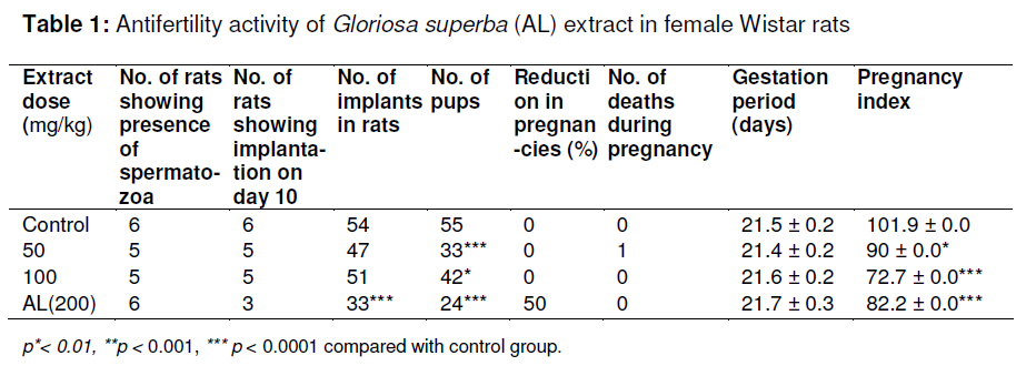
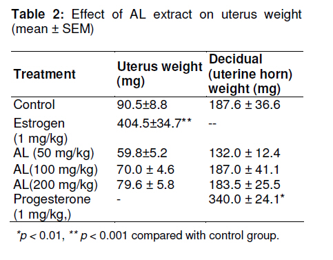
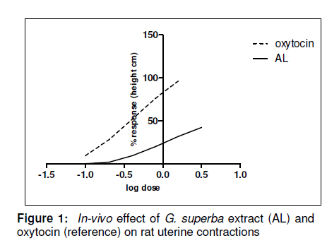
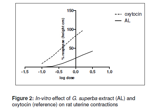
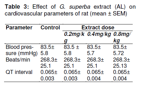
Tropical Journal of Pharmaceutical Research, Vol. 10, No. 2, April, 2011, pp. 169-176 Research Article Effect of the Aqueous Extract of Gloriosa superba Linn (Langli) Roots on Reproductive System and Cardiovascular Parameters in Female Rats Arati A Malpani1, Urmila M Aswar2, Shiv K Kushwaha2, GN Zambare2 and SL Bodhankar2* 1Department of Pharmacology, HKES’s College of Pharmacy, Sedam Road, Gulbarga 585105, Received: 20 July 2010 Revised accepted: 22 February 2011 Code Number: pr11024 Abstract Purpose: Gloriosa superba Linn (liliaceae) has been used to induce labor in the traditional Indian system of medicine. The objective of the study was to evaluate the activity of the aqueous extract of Gloriosa superba (AL) root on the female reproductive system of rat. Keywords: Abortificient activity; Blood pressure; Gloriosa superba; Oxytocic activity INTRODUCTION Most of the plants claimed to be oxytocics are used to induce and maintain labour, aid the removal of retained placenta, regulate post-partum bleeding and as abortifacient. The plant extracts increase the spontaneous activity of the uterus causing increase in contractions [1]. Some animal products have also been used to induce labour and removal of retained placenta, e.g., Hippopotamus amphibious (skin and meat) and Panthera leo (fats and faeces) which are boiled and the cooled decoction administered orally. Medicinal plants used to speed the birth process are usually administered towards the end of the gestation period or at the onset of labour labor pains. Plants that produce uterine contractions have a similar action to that of oxytocin which stimulates the uterus, causing strong contractions, and thus producing labour [2]. Traditional birth attendants, mothers-in-law, mothers and the expectant mother mostly prescribe these herbal remedies to induce labour. Some of these medicinal plants are also fed to cows and goats in labour. Gloriosa superba L., (Langli), is widely distributed in the tropical jungles of Africa and in many parts of tropical Asia, including India, Burma, Malaysia and Srilanka [10]. In India, it is mainly found in Nasik, Ratnagiri and Savanthwadi in Maharashtra state; Uttar Kannada, Hassan, Chikmangalur, Coorg and Mysore in Karnataka state; Cannanore, Palakkad and Trivandrum in Kerala state; as well as in Tamil Nadu and Goa. It is an erect, perennial, climbing herb. Tribesmen of Patalkot apply the rhizome extract over the navel and vagina to induce labour and facilitate normal delivery. According to them, 250 to 500 mg of the extract may lead to abortion if given to a lady with a pregnancy of 1 -2 months [3]. It is also used for the treatment of ulcers, leprosy, piles, inflammations [4], intestinal worm infestations, thirst, bruises, skin problems and snakebite [5,6]. Although some pharmacological studies have been carried out on this plant [7], to the best of our knowledge, there is no report on its effect on the reproductive system. In view of its folkloric use, the first objective of this study was to evaluate the effect of the aqueous extract of the root of Gloriosa superba L. on the reproductive system of rat. Since hypertension is one of the most common medical problems encountered during pregnancy, causing complications in 2 – 3 % of pregnancies [8], the second objective was to determine the effect of the extract on some cardiovascular parameters. EXPERIMENTAL Materials Histamine and estrogen benzoate (1mg/ml, Himedia Labs, India); hydroxyprogesterone caproate (German Remidies, India); oxytocin injection (Ranbaxy Laboratories Limited, India), adrenaline bitartarate and atropine sulphate injection (T. Walker’s Pharmaceuticals Pvt. Ltd, Pune, India); and De-Jalons solution (comprising 9.0 g of NaCl, 4.2 ml of 10 % KCl, 0.27 ml of 1M CaCl2, 0.5 g of glucose and 0.5 g of NaHCO3 in 1 L of distilled water) were the main materials used. Collection and identification of plant material The roots of Gloriosa superba L. were collected from a local market in Pune City, Maharashtra, India and authenticated by Dr A M Mujumdar a taxonomist and Head, Plant Sciences Division, Agharkar Research Institute, Pune, Maharashtra, India. The plant specimen was deposited in the herbarium of Agharkar Research Institute, Pune, India (voucher no. ARI/PG/897). Animals All the animal experiments were performed on in-bred adult female Wistar rats (weighing 250 -300g) . They were maintained at a temperature of 25 ± 1°C and relative humidity of 45 - 55 % under 12h light/12h dark cycle. The animals had free access to food pellets (Chakan Oil Mills, Pune, India) and water. The experimental protocol was approved by the Institutional Animal Ethics Committee (IAEC) of Poona College of Pharmacy, Pune, India, (approval ref no. CPCSEA/32/2008) and followed the guidelines of India’s Committee for the Purpose of Control and Supervision of Experiments on Animals (CPCSEA) [22]. Preparation of extract Gloriosa superba root powder (100 g) was defatted with 150 ml of petroleum ether (b.p. 40 - 60ºC). Cold maceration by addition of distilled water (150 ml) to dried, defatted Gloriosa superba root powder was carried out for 24 h in a flask using a mechanical shaker. The supernatant was removed, the marc dried and again macerated with 150 ml of distilled water for 24 h. The extract was filtered through Whatman filter paper no. 44 and dried in a tray dryer at 30ºC. The yield was 6%. The extract was reconstituted in distilled water prior to pharmacological tests. Preliminary phytochemical tests The extract was analyzed for phytochemicals including steroids using Salkowski test [9], flavonoids and tannins using ferric chloride test [10], alkaloids by Mayer’s test [11], and proteins and glycosides by Biuret and Legal tests, respectively [12,13]. Acute oral toxicity study For acute oral toxicity and LD50 determination, OECD (Organization for Economic Cooperation and Development) guideline AOT-425 was followed [14]. Healthy adult Swiss mice of either sex weighing between 20 -25 g were used for acute oral toxicity study. Food, but not water, was withheld for 4 h before the drug was administered orally. The test drug was given in doses of 5000, 1750, 550 mg/kg p.o.; the number of mice per dose was three. The mice were observed for 2 h for behavioural, neurological and autonomic profiles and for any lethality or death over the next 48 h. Assessment of antifertility activity of extract Adult female Wistar rats were kept overnight with male rats of proven fertility (in the ratio of 2 females to 1 male) and the vaginal smear of the female rats was examined for spermatozoa in the morning. The day on which the spermatozoa were observed in the smear was considered as day zero of pregnancy (Day 0) and such female rats were divided into four groups of 5 rats each. Group-I (Control) received the vehicle (distilled water), while the other groups received extract as follows: Group-II (50 mg/kg), Group-III (100 mg/kg), and Group-IV (200 mg/kg). The rats received these treatments daily for 7 days. On day 10, all the animals were laprotomized under light ether anesthesia and the number of implantation sites in both horns of the uteri was recorded. Pregnancy parameters such as length of pregnancy (days), pregnancy index (i.e., no. of pups delivered/no. of implants, expressed as a percentage), no. of pups on day 7 and teratogenicity were recorded [15]. Determination of estrogenic activity of extract Immature female rats (50 – 80 g) were bilaterally ovariectomized under light ether anesthesia [16]. The animals were classified into different groups (6 each). Group-I (control) was control while groups II -IV received the extract - 50, 100 and 200 mg/kg, respectively; Group-V received 0.1 mg estradiol benzoate in olive oil (subcutaneously) biweekly. The treatment period was 28 days. On day 29, the animals were sacrificed and dissected, their uteri removed and freed from surrounding tissues, blotted on filter paper and then weighed quickly on an electronic balance. Determination of progestogenic activity The deciduoma model was used for this experiment [17]. Adult female Wistar rats weighing 200 to 250 g were ovariectomized. The animals were classified, according to the extract (unless otherwise specified) dose given, as follows: Group-I (Control, vehicle, i.e., distilled water), Group-II (hydroxyprogesterone caproate (reference, 0.04 mg/animal, s.c.), Group-III (AL-50 mg/kg), Group-IV (AL-100 mg/kg), Group-V (AL-200 mg/kg). One week later, all the animals were treated with 0.5 µg estradiol/animal once daily s.c. for 4 days, followed by progesterone (Group-II) or extract (Groups-III to V in doses of 50, 100 and 200 mg/kg, respectively) and vehicle (control group) for 9 days. The animals were anaesthetized, the uterus of each rat exposed on day 5 of the preceding treatment, and then 1 mg histamine dihydrochloride injected into the lumen of the left horn. The animals were sacrificed thereafter and the left uterine horn from each animal removed and weighed. The degree of deciduoma formation was evaluated as percent increase in the weight of the histamine-injected uterine horn, compared with the control horn. In-vivo assay of uterotonic activity of extract Microballoon technique was used for this assay. Adult female Wistar rats weighing 250 to 300 g were divided into two groups each containing six animals, and primed with 100 µg of estradiol benzoate s.c. 24 h prior to the experiment. The rats showing estrous phase were selected for the experiment. Each animal was anaesthetized with urethane (1.25 g/kg) and the jugular vein cannulated for drug administration. A small incision was made on the lower abdominal skin and the uterus exposed. Another small incision was made near the midrostral end of the left uterine horn. A saline-filled catheter with a microlatex balloon on its end (approx 2 cm diameter) was inserted into the lumen of the uterine horn and secured to it by stitching [18]. The catheter was connected to a pressure transducer and normal uterine contractions were recorded using an 8-channel recorder (Power Lab, model 8/30, AD Instruments, Australia). The extract (0.2, 0.4 or 0.8 mg/kg, i.v) and oxytocin (0.2, 0.4 or 0.8 µg/kg, i.v) were administered to two different groups of female rats. The dose responses of oxytocin (reference) and the extract were recorded until a ceiling effect was observed. In-vitro assay of uterotonic activity of extract Adult female Wistar rats weighing 120 to 200 g were primed with 100 µg of estradiol benzoate s.c. 24 h prior to the experiment. The rats showing estrous phase were selected for the experiment, sacrificed by cervical dislocation and the uterus was removed. One horn of the uterus was mounted on an organ bath containing DeJelon’s solution and allowed to stabilize for 45 min during which the tissue was washed at least 4 times with fresh De-Jelon’s solution. Dose response was recorded for oxytocin (0.05 – 0.8 µg) and the extract (0.1 6.4mg) until a ceiling (peak) effect was observed. The response of the extract was compared with that of oxytocin (reference) response [19]. Effect of extract on blood pressure (BP) and electrocardiogram (ECG) of female rat Adult female Wistar rats weighing 200 to 250 g were anesthetized with urethane (1.25 g/kg) and the jugular vein cannulated for administration of the extract. The carotid artery was cannulated for the recording of blood pressure (BP) while electrocardiogram (ECG) electrodes were attached to the right and left forelimb and right hindlimb for recording of ECG. ECG, BP and heart rate were recorded using an 8-channel recorder (Powerlab model 8/30, AD Instruments, Australia). Statistical analysis The arithmetic means of the data were calculated for each experiment. The results were subjected to statistical analysis using one-way analysis of variance followed by Dunnett’s test. The level of significance used was p < 0.05. The statistical software used was Graph Pad Prism, USA, versions 4 and 5. RESULTS Preliminary phytochemical assessment showed that the aqueous extract of roots of Gloriosa superba contains flavonoids, tannins, alkaloids and glycosides, while acute toxicity studies indicate that the extract was safe up to a dose level of 550 mg/kg of body weight. Antifertility activity of extract The antifertility data for the extract are shown in Table 1. The reduction in pregnancies, based on the number of rats showing implants on day 10 of laprotomy was 0 % for control and all extract doses except for 200 mg/kg which was 50 % (p < 0.05). The extract reduced the number of pups born when compared to the number of implants present in the uterus on day 10 of laprotomy. The reduction was significant for all three doses of the extract. However, none of the test animals from days 1 -22 of pregnancy irrespective of the treatment received. Gestation period was approximately 21 days for all the animal groups and all the pups born survived up to the 7th post-natal day, the number being, 8 or 9 per group. Pregnancy index (PI) of the control group was 101.9 % while for extract groups, it was 90 (50 mg/kg, p < 0.01), 72.7 (100 mg/kg, (p < 0.0001). and 82.2 % (200 mg/kg, p < 0.0001), respectively. Effect of G. superba (AL) extract on uterus weight Table 2 shows the effect of the extract on the uterus weight of immature female rats. The administration of estradiol benzoate provoked a significant increase (p< 0.001) in uterus weight compared with control while extract administration (all doses) did not increase uterus weight significantly. Similarly, while administration of hydroxyprogesterone caproate significantly increased (p < 0.01) the weight of uterine horn, the extract, at all doses, did not; although the 50 mg/kg appeared to have reduced the weight of the uterine horn, this was not statistically significant. Effect of G. superba extract (AL) on rat uterine contractility Figure 1 shows the spasmogenic activity (i.e., contraction) of the rat uterus of the extract in estrogen-primed non-pregnant uterus in-vivo while Figure 2 indicates the same parameter in vitro. The extract treatments produced increase in uterine contractility both in vivo and in vitro but height of contractions produced by the extract was slightly less than that produced. Effect of G. superba (AL) extract on rat cardiovascular parameters Table 3 indicates that the extract had no significant effect on rat blood pressure and heart rate when compared with the control group. DISCUSSION The persistent use of plants by pregnant women and traditional birth attendants for the induction of labor suggests that some herbs might be potent uterine stimulants. Therefore, studies of such herbs could provide a helpful guide to the discovery of new oxytocics. Gloriosa superba is used for labour induction by traditional birth attendants in India. Phytochemical analysis of the extract showed presence of flavonoids, tannins, alkaloids, and glycosides in the aqueous extract of Gloriosa superba. The extract (AL) was safe up to a dose of 550 mg/kg which is approx. 10 times higher than the minimum dose at which the extract effectively reduced the number of pups produced. While the extract had significant anti-implantation and early abortifacient activities, gestation period and pregnancy index were not altered, the extract did not show any apparent teratogenicity as the pups born seemed normal and no death occurred during the 7-day observation period. It is well known that for implantation to take place, the exact equilibrium of estrogen and progesterone must obtain, and hence any disturbance in the level of these hormones may cause abortion or infertility. In order to determine the mechanism of abortifacient activity of the extract, two hormones, progesterone and estrogen, which have the capacity for termination/reduction of pregnancy were used in the study to ascertain whether the extract exhibits estrogenic and progestogenic activities. The failure of the extract to increase the uterine weight suggests absence of estrogenic activity. Similarly, progestogenic activity was not manifest as the extract did not increase decidual (uterine horn) weight. The uterine contractility of the extract may be due to its oxytocic effects. The effect was fast in onset and could be totally eliminated by washing with extract-free Dejalon solution, thus suggesting the presence of low molecular weight active compound(s) in the extract, which may have penetrated rapidly to the site of action. However, the extract demonstrated spasmogenic activity, which was not as strong as that of oxytocin, Thus, its anti-implantation and early abortifacient activity may be due to its oxytocic property. The ability to cause uterine contractility raises the possibility of its being developed as a medicine for induction of labour. Since many pregnancies are associated with gestational hypertension, it is worth noting that the extract had no effect on blood pressure and other cardiovascular parameters measured. CONCLUSION The tests carried out on G. superba extract indicate that its mechanism of action was neither estrogenic nor progesterone like. However, its early abortifacient activity appears to suggest that its activity is oxytocic. Nonetheless, further studies are required to determine which phytochemical constituents are responsible for the plant’s abortifacient and oxytocic activities. The absence of any effects on the cardiovascular parameters enhances the plant extract’s safety profile in pregnancy. Overall, the study lends some credence to the folkloric use of Gloriosa superba Linn. (Langli) in labour induction. ACKNOWLEDGEMENT The authors are thankful to the Vice Chancellor, Dr. SS Kadam, and Principal, Dr KR Mahadik, of Bharati Vidyapeeth University, Poona College of Pharmacy, Pune for providing resources and facilities for this research work. REFERENCES
Copyright © 2011 - Pharmacotherapy Group, Faculty of Pharmacy, University of Benin, Benin City, 300001 Nigeria. The following images related to this document are available:Photo images[pr11024t2.jpg] [pr11024t3.jpg] [pr11024f1.jpg] [pr11024f2.jpg] [pr11024t1.jpg] |
| |||||||||
